. 2021 Feb 1;94(1118):20200703.
doi: 10.1259/bjr.20200703. Epub 2020 Dec 9.

COVID-19 mimics on chest CT: a pictorial review and radiologic guide

Affiliations

COVID-19 mimics on chest CT: a pictorial review and radiologic guide

Bruno Hochhegger et al. Br J Radiol. .

Abstract

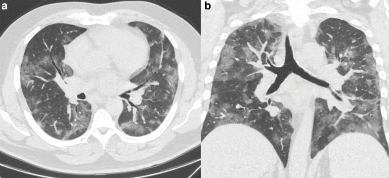
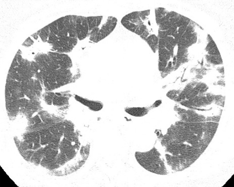
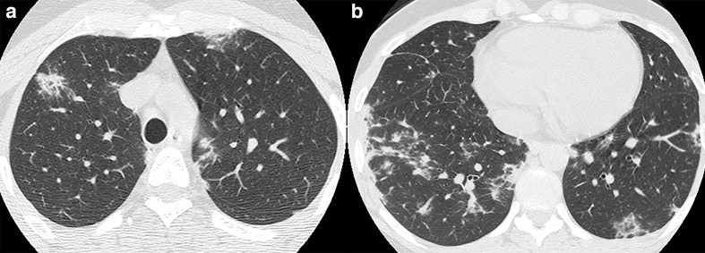
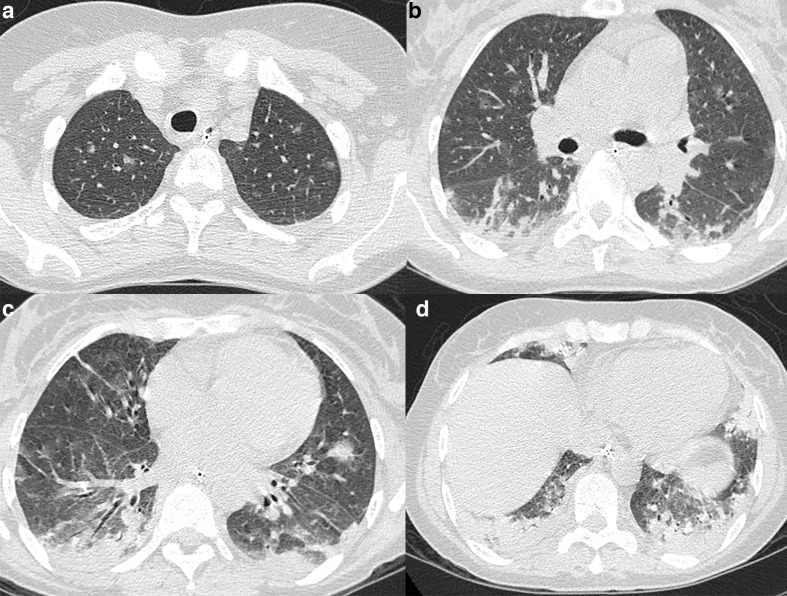
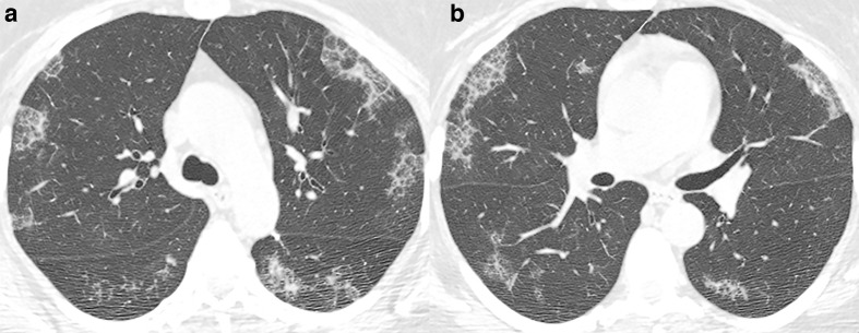
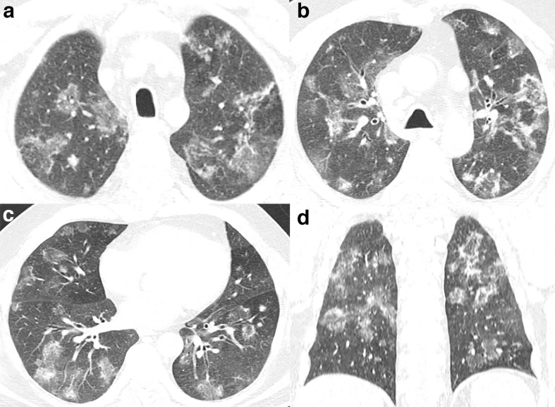
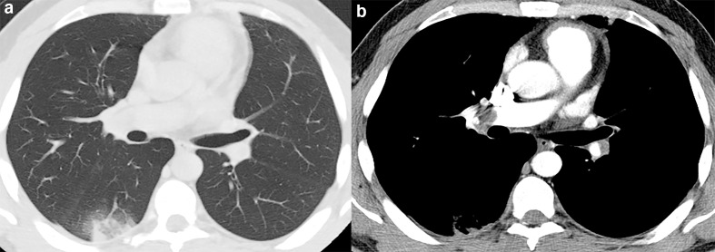
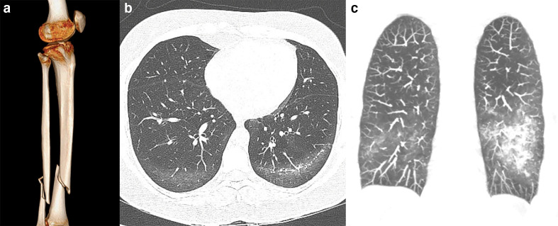
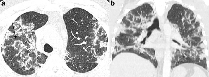
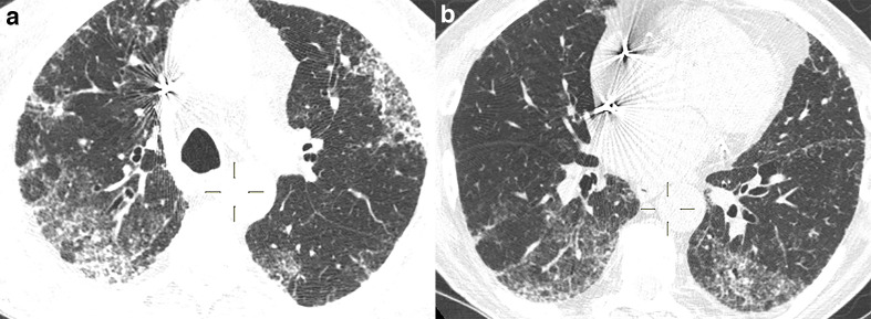
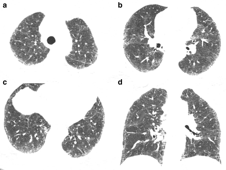
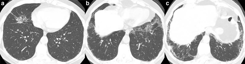
Chest imaging is often used as a complementary tool in the evaluation of coronavirus disease 2019 (COVID-19) patients, helping physicians to augment their clinical suspicion. Despite not being diagnostic for COVID-19, chest CT may help clinicians to isolate high suspicion patients with suggestive imaging findings. However, COVID-19 findings on CT are also common to other pulmonary infections and non-infectious diseases, and radiologists and point-of-care physicians should be aware of possible mimickers. This state-of-the-art review goal is to summarize and illustrate possible etiologies that may have a similar pattern on chest CT as COVID-19. The review encompasses both infectious etiologies, such as non-COVID viral pneumonia, Mycoplasma pneumoniae, Pneumocystis jiroveci, and pulmonary granulomatous infectious, and non-infectious disorders, such as pulmonary embolism, fat embolism, cryptogenic organizing pneumonia, non-specific interstitial pneumonia, desquamative interstitial pneumonia, and acute and chronic eosinophilic pneumonia.


Figures

Figure 1.
Figure 1.
51-year-old patient presenting with CT findings of COVID-19 confirmed by RT-PCR. (a) Axial chest CT in lung windows demonstrates bilateral predominantly peripheral ground-glass attenuation without consolidation. (b) Coronal reformatted CT shows bilateral ground-glass attenuation without a significant gradient. RT-PCR, reverse transcription polymerase chain reaction.
Figure 2.
Figure 2.
58-year-old male with Influenza A H1N1 pneumonia. Axial CT image demonstrates a predominant pattern of consolidation with diffuse distribution, air bronchograms, and sparse ground-glass opacities.
Figure 3.
Figure 3.
67-year-old male with adenovirus pneumonia. (a, b) Axial CT images demonstrate areas of consolidation with peripheral distribution, lower lobe predominance, and interlobular septal thickening.
Figure 4.
Figure 4.
48-year-old female with non-Hodgkin lymphoma and Mycoplasma pneumoniae infection. (a–d) Axial CT images demonstrate random centrilobular nodules with ground-glass and solid attenuation, bronchial wall thickening and consolidations predominantly in the lower lobes.
Figure 5.
Figure 5.
32-year-old male with acquired immunodeficiency syndrome and Pneumocystis jiroveci pneumonia. (a, b) Axial CT images demonstrate ground-glass opacities and intralobular septal thickening in the periphery of the upper lobes and the superior segments of the lower lobes.
Figure 6.
Figure 6.
58-year-old male with paracoccidioidomycosis. (a–c) Axial and (d) coronal CT images demonstrate diffuse ground-glass lesions in a random distribution, some with a peripheral rim of consolidation yielding the reversed halo sign and some solid nodules.
Figure 7.
Figure 7.
48-year-old male with pulmonary embolism. (a) Axial CT image demonstrate a wedge-shaped peripheral lung opacity in the right lower lobe with a ground-glass core and a peripheral rim of consolidation yielding the reversed halo sign. (b) CT angiography evidencing the thrombus in the right interlobar artery.
Figure 8.
Figure 8.
27-year-old male victim of a motorcycle accident with multiples injuries and developed respiratory distress a couple of days after the trauma due to fat embolism. (a) Three-dimensional rendering reconstruction of CT images demonstrates tibial and fibular fracture. (b) Axial CT image and (c) coronal maximum intensity projection reconstruction demonstrate patchy ground-glass opacities in the lower lobes with subpleural sparing.
Figure 9.
Figure 9.
57-year-old female with cryptogenic organizing pneumonia. (a) Axial and (b) coronal CT images shows bilateral perilobular opacities, interlobular septal thickening, and the reversed halo sign (arrow).
Figure 10.
Figure 10.
61-year-old male with amiodarone-induced organizing pneumonia. (a, b) Axial CT images with peripheral and bilateral areas of ground-glass lesions in random distribution and with some reticular opacities.
Figure 11.
Figure 11.
49-year-old female with non-specific interstitial pneumonia. (a–c) Axial and (d) coronal CT images demonstrate reticulations in the subpleural zones, predominantly in the lower lobes, with some ground-glass opacities and bronchiectasis.
Figure 12.
Figure 12.
44-year-old male, smoker, with desquamative interstitial pneumonia. (a–c) Axial CT images demonstrate ground-glass and reticular opacities in the periphery of the lower lobes and the middle lobe.
Figure 13.
Figure 13.
40-year-old male with acute eosinophilic pneumonia. Axial CT demonstrates bilateral and peripheral ground-glass opacites with interlobular septal thickening.

References

    1. World Health Organization . WHO Coronavirus Disease (COVID-19) Dashboard 2020. 2020. Available from: https://covid19.who.int/.
    1. Rubin GD, Ryerson CJ, Haramati LB, Sverzellati N, Kanne JP, Raoof S, et al. The role of chest imaging in patient management during the COVID-19 pandemic: a multinational consensus statement from the Fleischner Society. Chest 2020; 158: 106–16. doi: 10.1016/j.chest.2020.04.003 - DOI - PMC - PubMed
    1. Zu ZY, Jiang MD, Xu PP, Chen W, Ni QQ, Lu GM, et al. Coronavirus disease 2019 (COVID-19): a perspective from China. Radiology 2020; 296: E15–25. doi: 10.1148/radiol.2020200490 - DOI - PMC - PubMed
    1. Ding X, Xu J, Zhou J, Long Q. Chest CT findings of COVID-19 pneumonia by duration of symptoms. Eur J Radiol 2020; 127: 109009. doi: 10.1016/j.ejrad.2020.109009 - DOI - PMC - PubMed
    1. World Health Organization. Laboratory testing of 2019 novel coronavirus (‎‎‎‎‎‎‎‎‎‎‎‎2019-nCoV)‎‎‎‎‎‎‎‎‎‎‎‎ in suspected human cases: interim guidance, 17 January 2020. 2020. Available from: https://apps.who.int/iris/handle/10665/330676.

Publication types

MeSH terms