. 2020 Dec 7;8(12):578.
doi: 10.3390/biomedicines8120578.

Human Induced Pluripotent Stem Cell-Derived Cardiomyocytes, in Contrast to Adipose Tissue-Derived Stromal Cells, Efficiently Improve Heart Function in Murine Model of Myocardial Infarction

Affiliations

Human Induced Pluripotent Stem Cell-Derived Cardiomyocytes, in Contrast to Adipose Tissue-Derived Stromal Cells, Efficiently Improve Heart Function in Murine Model of Myocardial Infarction

Jacek Stępniewski et al. Biomedicines. .

Abstract

Cell therapies are extensively tested to restore heart function after myocardial infarction (MI). Survival of any cell type after intracardiac administration, however, may be limited due to unfavorable conditions of damaged tissue. Therefore, the aim of this study was to evaluate the therapeutic effect of adipose-derived stromal cells (ADSCs) and human induced pluripotent stem cell-derived cardiomyocytes (hiPSC-CMs) overexpressing either the proangiogenic SDF-1α or anti-inflammatory heme oxygenase-1 (HO-1) in a murine model of MI. ADSCs and hiPSCs were transduced with lentiviral vectors encoding luciferase (Luc), GFP and either HO-1 or SDF-1α. hiPSCs were then differentiated to hiPSC-CMs using small molecules modulating the WNT pathway. Genetically modified ADSCs were firstly administered via intracardiac injection after MI induction in Nude mice. Next, ADSCs-Luc-GFP and genetically modified hiPSC-CMs were injected into the hearts of the more receptive NOD/SCID strain to compare the therapeutic effect of both cell types. Ultrasonography, performed on days 7, 14, 28 and 42, revealed a significant decrease of left ventricular ejection fraction (LVEF) in all MI-induced groups. No improvement of LVEF was observed in ADSC-treated Nude and NOD/SCID mice. In contrast, administration of hiPSC-CMs resulted in a substantial increase of LVEF, occurring between 28 and 42 days after MI, and decreased fibrosis, regardless of genetic modification. Importantly, bioluminescence analysis, as well as immunofluorescent staining, confirmed the presence of hiPSC-CMs in murine tissue. Interestingly, the luminescence signal was strongest in hearts treated with hiPSC-CMs overexpressing HO-1. Performed experiments demonstrate that hiPSC-CMs, unlike ADSCs, are effective in improving heart function after MI. Additionally, long-term evaluation of heart function seems to be crucial for proper assessment of the effect of cell administration.

Keywords: adipose-derived stromal cells; heme oxygenase-1; human induced pluripotent stem cells; myocardial infarction; stromal cell-derived factor-1α.


Conflict of interest statement

The authors declare no conflict of interest.

Figures
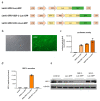
Figure 1
Figure 1
Generation of genetically modified adipose-derived stromal cells (ADSCs). (a) Schematic representation of lentiviral vectors used in the study. (b) GFP expression in ADSCs-Luc-GFP after cell sorting. Activity of luciferase (c) as well as expression of SDF-1 (d) and HO-1 (e) in transduced and purified ADSCs. Control—nontransduced ADSCs. Scale bar—50 µm.
Figure 2
Figure 2
Induction of myocardial infarction (MI) in athymic Nude mice. (a) Schematic representation of murine heart subjected to MI. Arrows indicate the site of ADSC injections. (b) Level of cTnI in plasma of mice subjected to surgery and cell administration confirming successful MI induction. N = 4–7 animals/group. (c) Kaplan–Meier survival curves for Nude mice subjected to MI and cell administration. Sham-treated animals served as a control. N = 4–7 animals/group (numbers in parentheses represent surviving mice/all mice used).
Figure 3
Figure 3
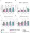
No improvement of heart function after administration of genetically modified ADSCs. Parameters of left ventricle (LV) function: ejection fraction (a), fractional shortening (b), end-systolic volume (c) and end-diastolic volume (d) in animals subjected to MI and administration of cells. N = 4–7 animals/group. * p < 0.05 vs. saline, one-way ANOVA with Bonferroni’s post hoc test.
Figure 4
Figure 4
Generation of genetically modified human induced pluripotent stem cell-derived cardiomyocytes (hiPSC-CMs). (a) Schematic representation of lentiviral vectors used to transduce hiPSCs. Secretion of SDF-1α (b) and expression of HO-1 (c) in transduced hiPSCs. (d) Cardiac differentiation efficiency of transduced and sorted hiPSCs measured as percentage of cells positive for cardiac troponin T (cTnT). Secretion of SDF-1α (e) and expression of HO-1 (f) in genetically modified hiPSC-CMs.
Figure 4
Figure 4
Generation of genetically modified human induced pluripotent stem cell-derived cardiomyocytes (hiPSC-CMs). (a) Schematic representation of lentiviral vectors used to transduce hiPSCs. Secretion of SDF-1α (b) and expression of HO-1 (c) in transduced hiPSCs. (d) Cardiac differentiation efficiency of transduced and sorted hiPSCs measured as percentage of cells positive for cardiac troponin T (cTnT). Secretion of SDF-1α (e) and expression of HO-1 (f) in genetically modified hiPSC-CMs.
Figure 5
Figure 5
Detection of human cells in murine hearts in vivo. Bioluminescent signal collected intravitally on days 7, 14, 28 and 42 after MI induction from the chest of all animals used in the experiment. N = 3 (ADSC) or 7–9 animals/group.
Figure 6
Figure 6
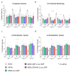
Administration of hiPSC-CMs improves heart function 42 days after MI. (a) LV ejection fraction (LVEF) measured 7, 14, 28 and 42 days after MI induction and administration of genetically modified hiPSC-CMs and ADSCs. N = 3 (ADSC) or 7–12 animals/group. *** p < 0.005 vs. sham group; ††† p < 0.005 vs. saline group; $$$ p < 0.005 vs. ADSC-Luc-GFP group; one-way ANOVA with Bonferroni’s post hoc test. (b) LV fractional shortening (LVFS) measured 7, 14, 28 and 42 days after MI induction and administration of genetically modified hiPSC-CMs and ADSCs. N = 3 (ADSC) or 7–12 animals/group. * p < 0.05, ** p < 0.01, *** p < 0.005 vs. sham group; †† p < 0.01, ††† p < 0.005 vs. saline group; one-way ANOVA with Bonferroni’s post hoc test. (c) LV end-systolic volume (LVVs) measured 7, 14, 28 and 42 days after MI induction and administration of genetically modified hiPSC-CMs and ADSCs. N = 3 (ADSC) or 7–12 animals/group. * p < 0.05, ** p < 0.01, *** p < 0.005 vs. sham group; p < 0.05 vs. saline group; one-way ANOVA with Bonferroni’s post hoc test. (d) LV end-diastolic volume (LVVd) measured 7, 14, 28 and 42 days after MI induction and administration of genetically modified hiPSC-CMs and ADSCs. N = 3 (ADSC) or 7–12 animals/group. * p < 0.05, ** p < 0.01, *** p < 0.005 vs. sham group; one-way ANOVA with Bonferroni’s post hoc test.
Figure 7
Figure 7
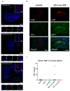
Immunofluorescent analysis of hearts subjected to MI and cell therapy. (a) Immunofluorescent analysis of human Ku80 (red) as well as Ki67 (green) in murine hearts subjected to MI and injected with hiPSC-CMs-Luc-GFP, hiPSC-CMs-SDF-1-Luc-GFP and hiPSC-CMs-HO-1-Luc-GFP. Nuclei stained with DAPI (blue). (b) Immunofluorescent analysis of Ku80 (red) and HO-1 expression (green) in hearts treated with either control hiPSC-CMs or hiPSC-CMs-HO-1-Luc-GFP. Nuclei stained with DAPI (blue). (c) Human SDF-1α-targeted ELISA assay performed on plasma collected from sham as well as saline-, hiPSC-CM-GFP-Luc-GFP-, hiPSC-CM-SDF-1-Luc-GFP- and hiPSC-CM-HO-1-Luc-GFP-treated animals 42 days after MI induction. N = 7–9 animals/group.
Figure 8
Figure 8
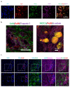
Expression of cardiac markers in human cells detected in murine myocardium. (a) Representative pictures for immunofluorescent analysis of human Ku80 (red), NKX2.5 (green) and GATA4 (purple) in murine heart subjected to MI and hiPSC-CM-SDF-1-Luc-GFP administration. Nuclei stained with DAPI (blue). (b) Representative pictures for immunofluorescent analysis of human Ku80 (red), connexin 43 (Cx43, green) and troponin T (purple) in the left panel and NKX2.5 (green) and α-actinin (purple) in the right panel in murine heart subjected to MI and hiPSC-CM-SDF-1-Luc-GFP administration. Nuclei stained with DAPI (blue). (c) Representative pictures for immunofluorescent analysis of human Ku80 (red), connexin 43 (Cx43, green) and troponin T (purple) in murine heart subjected to MI and hiPSC-CM-SDF-1-Luc-GFP administration. Nuclei stained with DAPI (blue). Similar results were obtained for hiPSC-CMs-Luc-GFP and hiPSC-CMs-HO-1-Luc-GFP (Supplementary Figure S9).
Figure 9
Figure 9
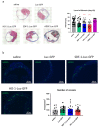
Histological analysis of hearts subjected to MI and cell therapy. (a) Masson’s trichrome analysis of heart sections obtained from animals subjected to MI and cell therapy—representative pictures. Level of fibrosis was calculated based on histological analysis of heart sections. N = 2 (ADSC) or 4 animals/group, each dot represents a single section, 3–23 sections/heart were analyzed. * p < 0.05, ** p < 0.01 vs. saline. (b) Representative pictures for immunofluorescent analysis of αSMA-positive vessels (green) in left ventricle of saline-, hiPSC-CM-GFP-Luc-GFP-, hiPSC-CM-SDF-1-Luc-GFP- and hiPSC-CM-HO-1-Luc-GFP-treated hearts. Nuclei stained with Hoechst33342 (blue). Data presented as number of vessels/mm2. N = 2–3 animals/group, each dot represents a single section, 5–12 sections/animal were analyzed.

References

    1. Bergmann O., Bhardwaj R.D., Bernard S., Zdunek S., Barnabé-Heider F., Walsh S., Zupicich J., Alkass K., Buchholz B.A., Druid H., et al. Evidence for cardiomyocyte renewal in humans. Science. 2009;324:98–102. doi: 10.1126/science.1164680. - DOI - PMC - PubMed
    1. Gerbin K.A., Murry C.E. The winding road to regenerating the human heart. Cardiovasc. Pathol. 2015;24:133–140. doi: 10.1016/j.carpath.2015.02.004. - DOI - PMC - PubMed
    1. Laflamme M.A., Murry C.E. Heart regeneration. Nature. 2011;473:326–335. doi: 10.1038/nature10147. - DOI - PMC - PubMed
    1. Langrzyk A., Nowak W.N., Stępniewski J., Jaźwa A., Florczyk-Soluch U., Józkowicz A., Dulak J. Critical View on Mesenchymal Stromal Cells in Regenerative Medicine. Antioxid. Redox Signal. 2018;29:169–190. doi: 10.1089/ars.2017.7159. - DOI - PubMed
    1. Eggenhofer E., Benseler V., Kroemer A., Popp F.C., Geissler E.K., Schlitt H.J., Baan C.C., Dahlke M.H., Hoogduijn M.J. Mesenchymal stem cells are short-lived and do not migrate beyond the lungs after intravenous infusion. Front. Immunol. 2012;3 doi: 10.3389/fimmu.2012.00297. - DOI - PMC - PubMed

LinkOut - more resources