Review
. 2020 Dec 7;9(12):2630.
doi: 10.3390/cells9122630.

Deciphering the Relevance of Bone ECM Signaling

Affiliations
Review

Deciphering the Relevance of Bone ECM Signaling

Natividad Alcorta-Sevillano et al. Cells. .

Abstract

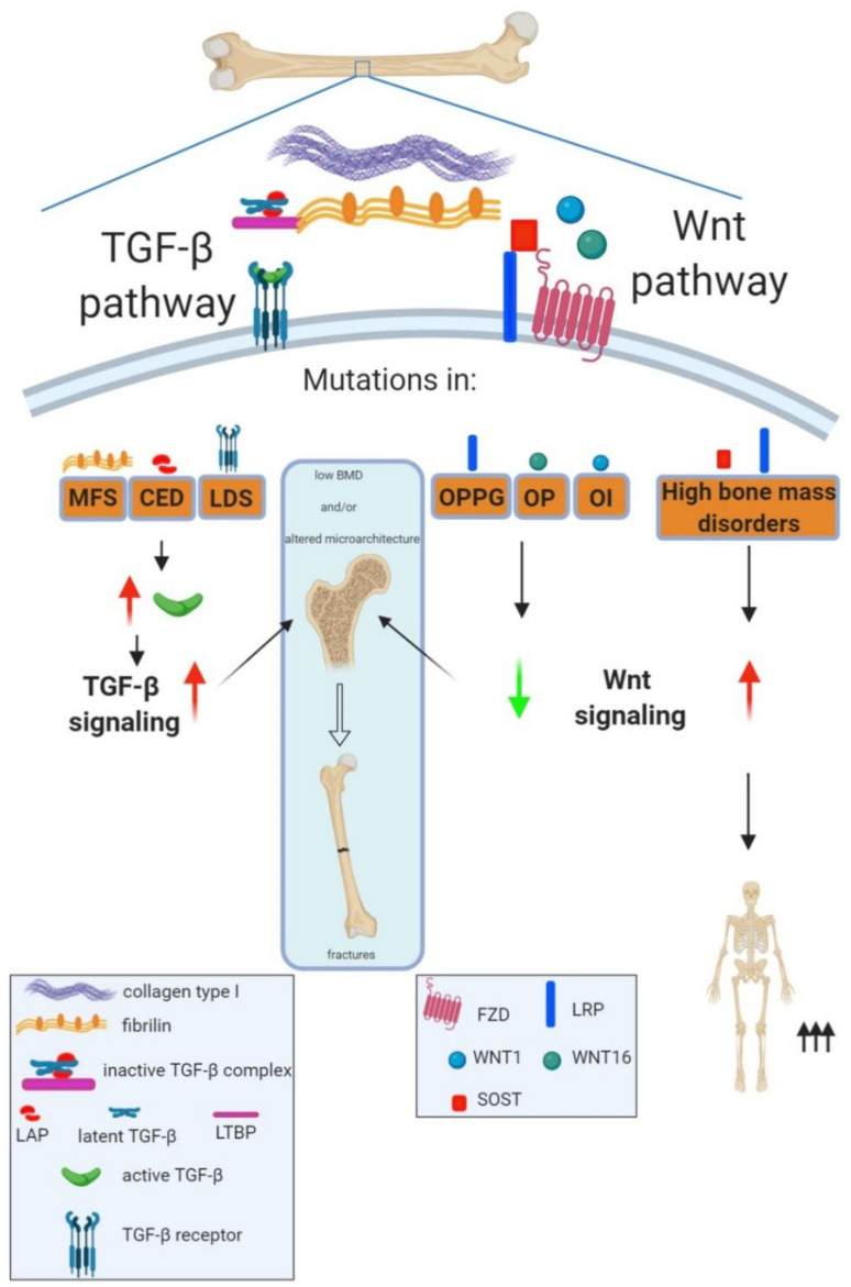
Bone mineral density, a bone matrix parameter frequently used to predict fracture risk, is not the only one to affect bone fragility. Other factors, including the extracellular matrix (ECM) composition and microarchitecture, are of paramount relevance in this process. The bone ECM is a noncellular three-dimensional structure secreted by cells into the extracellular space, which comprises inorganic and organic compounds. The main inorganic components of the ECM are calcium-deficient apatite and trace elements, while the organic ECM consists of collagen type I and noncollagenous proteins. Bone ECM dynamically interacts with osteoblasts and osteoclasts to regulate the formation of new bone during regeneration. Thus, the composition and structure of inorganic and organic bone matrix may directly affect bone quality. Moreover, proteins that compose ECM, beyond their structural role have other crucial biological functions, thanks to their ability to bind multiple interacting partners like other ECM proteins, growth factors, signal receptors and adhesion molecules. Thus, ECM proteins provide a complex network of biochemical and physiological signals. Herein, we summarize different ECM factors that are essential to bone strength besides, discussing how these parameters are altered in pathological conditions related with bone fragility.

Keywords: ECM; ECM signaling; bone disease; bone fragility; fracture risk.


Conflict of interest statement

The authors declare no conflict of interest. The funders had no role in the collection, analyses or interpretation of data; in the writing of the manuscript, or in the decision to publish the review.

Figures

Figure 1
Figure 1
Roles of transforming growth factor beta (TGF-β) and Wnt signaling in bone strength maintenance. Bone strength depends on bone mineral density and bone microarchitecture; disturbances in any of them lead to increased risk of fractures. Human musculoskeletal disorders with mutations in TGF-β or Wnt family members have revealed the key role of these pathways in regulating bone strength. Thus, mutations in different TGF-β family members leading to an increased pool of active TGF-β and therefore an increased TGF-β signaling give rise to rare disorders characterized by low bone mineral density, alterations in bone microarchitecture and increased risk of fractures. This is the case of Marfan (MFS) and Loeys–Dietz (LDS) syndromes and Camurati–Engelmann disease (CED), with mutations in FBN1 (Fibrillin-1), TGFβ receptors (TGFβR1 and TGFβR2) and LAP, respectively. In the case of the Wnt pathway, mutations leading to increased Wnt signaling can cause high bone mass disorders, whereas inactivating mutations of the Wnt pathway are associated to low bone mass disorders with different ranges of severities. Thus, heterozygous mutations in Wnt1 or SNPs in Wnt16 can lead to osteoporosis (OP) whereas homozygous mutations of Wnt1 cause osteogenesis imperfecta (OI). Moreover, autosomal recessive loss of function mutations in LRP5 are known to cause the rare osteoporosis pseudoglioma syndrome (OPPG) characterized by extremely severe childhood onset osteoporosis. For definitions of other abbreviations, please see the main text. Red arrow increased expression; green arrow decreased expression.

References

    1. Bonucci E. Bone mineralization. Front. Biosci. 2012;17:100. doi: 10.2741/3918. - DOI - PubMed
    1. Seeman E., Delmas P.D. Bone Quality—The Material and Structural Basis of Bone Strength and Fragility. N. Engl. J. Med. 2006;354:2250–2261. doi: 10.1056/NEJMra053077. - DOI - PubMed
    1. Fonseca H., Moreira-Gonçalves D., Coriolano H.-J.A., Duarte J.A. Bone Quality: The Determinants of Bone Strength and Fragility. Sports Med. 2014;44:37–53. doi: 10.1007/s40279-013-0100-7. - DOI - PubMed
    1. Murshed M. Mechanism of Bone Mineralization. Cold Spring Harb. Perspect. Med. 2018;8:a031229. doi: 10.1101/cshperspect.a031229. - DOI - PMC - PubMed
    1. Narisawa S., Yadav M.C., Millán J.L. In Vivo Overexpression of Tissue-Nonspecific Alkaline Phosphatase Increases Skeletal Mineralization and Affects the Phosphorylation Status of Osteopontin. J. Bone Miner. Res. 2013;28:1587–1598. doi: 10.1002/jbmr.1901. - DOI - PMC - PubMed

Publication types

MeSH terms

LinkOut - more resources