Comparative Study
. 2020 Dec 9;20(1):1211.
doi: 10.1186/s12885-020-07675-7.

Gene expression analysis of human prostate cell lines with and without tumor metastasis suppressor CD82

Affiliations
Comparative Study

Gene expression analysis of human prostate cell lines with and without tumor metastasis suppressor CD82

Pushpaja Dodla et al. BMC Cancer. .

Abstract

Background: Tetraspanin CD82 is a tumor metastasis suppressor that is known to down regulate in various metastatic cancers. However, the exact mechanism by which CD82 prevents cancer metastasis is unclear. This study aims to identify genes that are regulated by CD82 in human prostate cell lines.

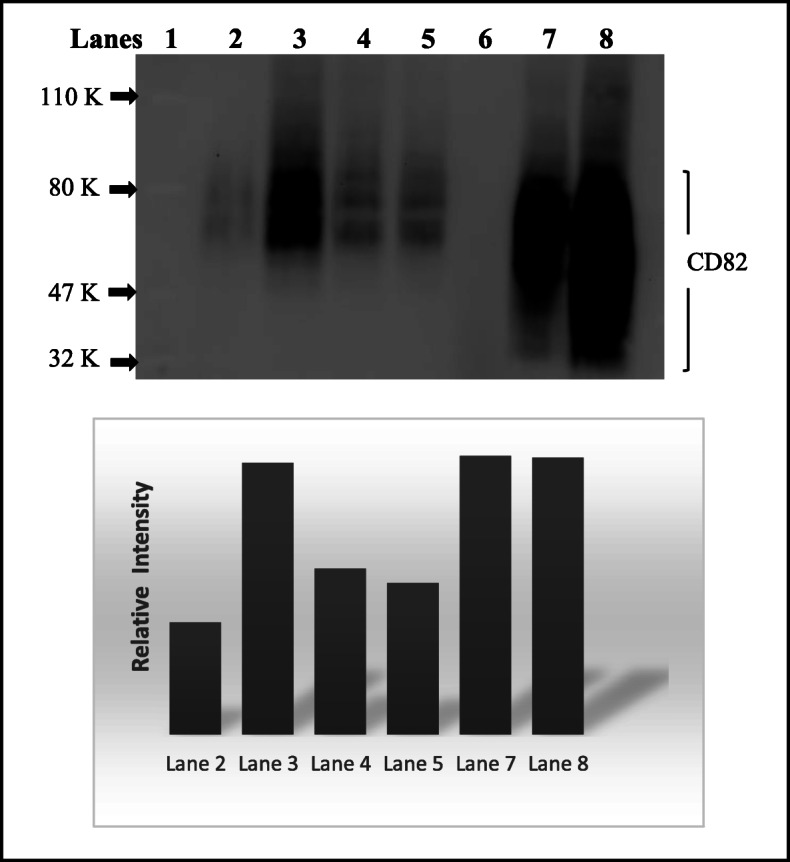
Methods: We used whole human genome microarray to obtain gene expression profiles in a normal prostate epithelial cell line that expressed CD82 (PrEC-31) and a metastatic prostate cell line that does not express CD82 (PC3). Then, siRNA silencing was used to knock down CD82 expression in PrEC-31 while CD82 was re-expressed in PC3 to acquire differentially-expressed genes in the respective cell line.

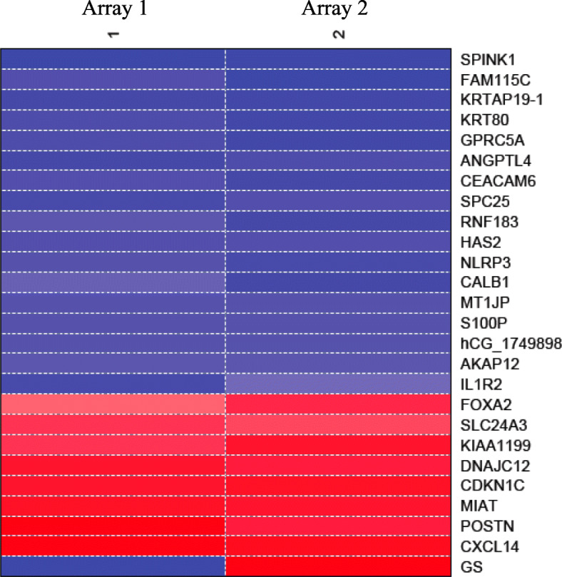
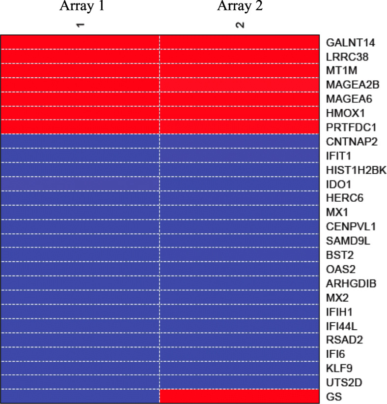
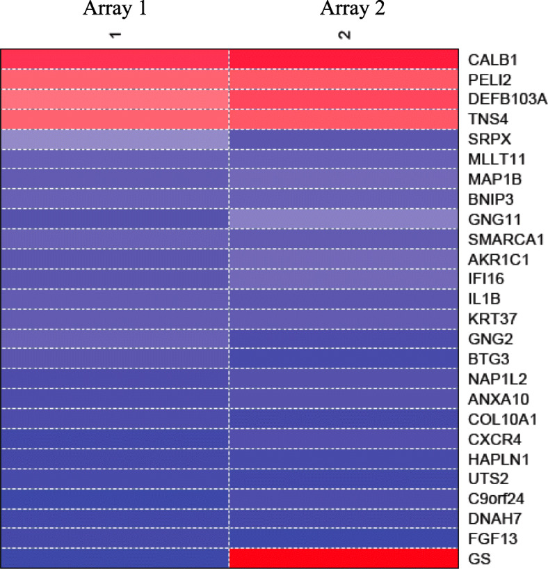
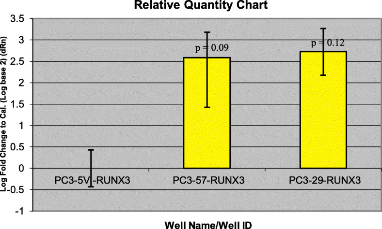
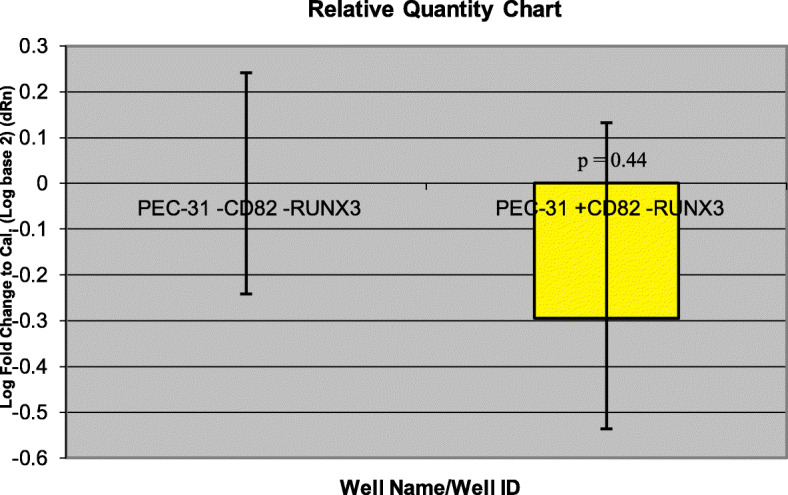
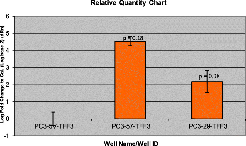
Results: Differentially-expressed genes with a P < 0.05 were identified in 3 data sets: PrEC-31 (+CD82) vs PrEC-31(-CD82), PC3-57 (+CD82) vs. PC3-5 V (-CD82), and PC3-29 (+CD82) vs. PC3-5 V (-CD82). Top 25 gene lists did not show overlap within the data sets, except (CALB1) the calcium binding protein calbindin 1 which was significantly up-regulated (2.8 log fold change) in PrEC-31 and PC3-29 cells that expressed CD82. Other most significantly up-regulated genes included serine peptidase inhibitor kazal type 1 (SPINK1) and polypeptide N-acetyl galactosaminyl transferase 14 (GALNT14) and most down-regulated genes included C-X-C motif chemokine ligand 14 (CXCL14), urotensin 2 (UTS2D), and fibroblast growth factor 13 (FGF13). Pathways related with cell proliferation and angiogenesis, migration and invasion, cell death, cell cycle, signal transduction, and metabolism were highly enriched in cells that lack CD82 expression. Expression of two mutually inclusive genes in top 100 gene lists of all data sets, runt-related transcription factor (RUNX3) and trefoil factor 3 (TFF3), could be validated with qRT-PCR.

Conclusion: Identification of genes and pathways regulated by CD82 in this study may provide additional insights into the role that CD82 plays in prostate tumor progression and metastasis, as well as identify potential targets for therapeutic intervention.

Keywords: CD82; Gene expression; KAI1; Metastasis tumor suppressor; Microarray; Prostate cancer.


Conflict of interest statement

The authors declare that they have no competing interests.

Figures

Fig. 1
Fig. 1
Western blot of CD82 protein expression in prostate cancer cell lines. Lane 1. Protein ladder with Phosphorylase B (110 K), BSA (80 K), Ovalbumin (47 K), and Carbonic Anhydrase (32 K). Lane 2. PC3-5 V metastatic prostate clonal cells with empty vector, Lane 3. PrEC-31 transfected with 40 nM of scrambled siRNA. Lane 4 and 5. PrEC-31 transfected with 30 nM and 40 nM of CD82 siRNA, respectively. Lane 6. empty. Lane 7 and 8. PC3–29 and PC3–57 clonal cells. Restored with CD82, respectively. A heavily glycosylated CD82 runs as a wide band between 30 and 90 KDa. Below is a graph that represents the relative intensity of the CD82 band in different lanes, based on the densitometric analysis of the blot. An uncropped full-length blot is presented in supplementary Fig. S9
Fig. 2
Fig. 2
Heat map of top 25 differentially expressed genes in PrEC-31 (+/−CD82) cells. GS: graphic scale for the array, where red represents downregulation and blue represents upregulation of a gene in the normal PrEC (+CD82) compared to siRNA treatment sample PrEC (−CD82). Columns 1, 2 represent the two arrays used i.e., array 1 and array 2 as a result of dye swapping
Fig. 3
Fig. 3
Heat map of top 25 differentially expressed genes in PC3–57 vs. PC3-5 V cells. GS: graphic scale for the array, where red represents upregulation and blue represents downregulation of a gene in the treatment PC3–57 (+CD82) compared to control PC3-5 V (−CD82). Columns 1, 2 represent the two arrays used i.e., array 1 and array 2 as a result of dye swapping
Fig. 4
Fig. 4
Heat map of top 25 differentially expressed genes in PC3–29 vs. PC3-5 V cells. GS: graphic scale for the array, where red represents upregulation and blue represents downregulation of a gene in the treatment PC3–29 (+CD82) compared to control PC3-5 V (−CD82). Columns 1, 2 represent the two arrays used i.e., array 1 and array 2 as a result of dye swapping
Fig. 5
Fig. 5
Comparative quantification for RUNX3 gene in PC3 cell lines using qRT-PCR. PC3-5 V cell line was used as the calibrator. Yellow bars represent the log-fold change for the PC3–57 and PC3–29 cell lines compared to PC3-5 V. Fold change were initially calculated for all the three cell lines by subtracting RUNX3 Ct values from the respective cell lines beta actin Ct values. The fold change for PC3-5 V cell lines was equaled to 0 and the values for PC3–57 and PC3–29 were calculated by comparing to PC3-5 V. A t-test performed on the final fold change values yielded p values of 0.12 (PC3–29) and 0.09 (PC3–57) respectively
Fig. 6
Fig. 6
Comparative quantification for RUNX3 gene in PrEC-31(+/− CD82) cells with qRT-PCR. PrEC-31-CD82 cell line was used as the calibrator. Yellow bars represent the log-fold change for the PrEC-31 + CD82 cell lines compared to PrEC-31-CD82. Fold change was initially calculated for both cell lines by subtracting RUNX3 Ct values from the respective cell lines beta actin Ct values. The fold change for PrEC-31-CD82 cell line was equaled to 0 and the values for PrEC-31 + CD82 were calculated by comparing to PrEC-31-CD82. A t-test performed on the final fold change value yielded a p value of 0.44
Fig. 7
Fig. 7
Comparative quantification for TFF3 gene in PC3 cells using qRT-PCR. PC3-5 V cell line was used as the calibrator. Yellow bars represent the log-fold change for the PC3–57 and PC3–29 cell lines compared to PC3-5 V. Fold change was initially calculated for all the three cell lines by subtracting TFF3 Ct values from the respective cell lines beta actin Ct values. The fold change for PC3-5 V cell line was equaled to 0 and the values for PC3–57 and PC3–29 were calculated by comparing to PC3-5 V. A t-test performed on the final fold change values yielded p values of 0.08 (PC3–29) and 0.18 (PC3–57) respectively
Fig. 8
Fig. 8
Comparative quantification for TFF3 gene in PrEC-31(+/− CD82) cells using qRT-PCR. PEC-31-CD82 cell line was used as a calibrator. Yellow bars represent the log-fold change for the PEC-31 + CD82 cell lines compared to PEC-31-CD82. Fold change was initially calculated for both cell lines by subtracting TFF3 Ct values from the respective cell lines beta actin Ct values. The fold change for PEC-31-CD82 cell line was equaled to 0 and the values for PEC-31 + CD82 were calculated by comparing to PEC-31-CD82. A t-test performed on the final fold change yielded a p value of 0.47

References

    1. Dong JT, Lamb PW, Rinker-Schaeffer CW, Vukanovic J, Ichikawa T, Isaacs JT, et al. KAI1, a metastasis suppressor gene for prostate cancer on human chromosome 11p11.2. Science. 1995;268:884–886. doi: 10.1126/science.7754374. - DOI - PubMed
    1. Bouras T, Frauman AG. Expression of the prostate cancer metastasis suppressor gene KAI1 in primary prostate cancers: a biphasic relationship with tumour grade. J Pathol. 1999;188:382–388. doi: 10.1002/(SICI)1096-9896(199908)188:4<382::AID-PATH365>3.0.CO;2-O. - DOI - PubMed
    1. Tricoli JV, Schoenfeldt M, Conley BA. Detection of prostate cancer and predicting progression: current and future diagnostic markers. Clin Cancer Res. 2004;10:3943–3953. doi: 10.1158/1078-0432.CCR-03-0200. - DOI - PubMed
    1. Ueda T, Ichikawa T, Tamaru J, Mikata A, Akakura K, Akimoto S, et al. Expression of the KAI1 protein in benign prostatic hyperplasia and prostate cancer. Am J Pathol. 1996;149:1435–1440. - PMC - PubMed
    1. Hinoda Y, Adachi Y, Takaoka A, Mitsuchi H, Satoh Y, Itoh F, et al. Decreased expression of the metastasis suppressor gene KAI1 in gastric cancer. Cancer Lett. 1998;129:229–234. doi: 10.1016/S0304-3835(98)00112-8. - DOI - PubMed

Publication types

MeSH terms