. 2020 Dec 9;12(1):113.
doi: 10.1186/s13073-020-00814-6.

A Klebsiella pneumoniae ST307 outbreak clone from Germany demonstrates features of extensive drug resistance, hypermucoviscosity, and enhanced iron acquisition

Affiliations

A Klebsiella pneumoniae ST307 outbreak clone from Germany demonstrates features of extensive drug resistance, hypermucoviscosity, and enhanced iron acquisition

Stefan E Heiden et al. Genome Med. .

Abstract

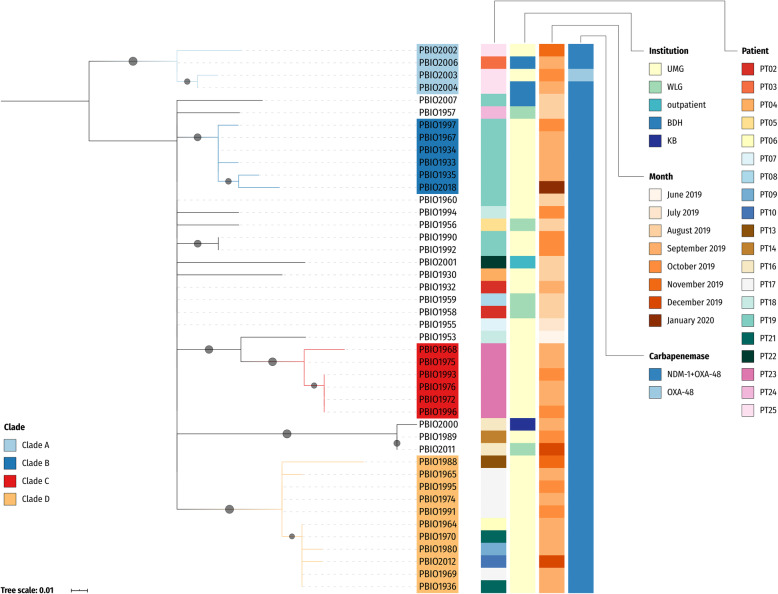
Background: Antibiotic-resistant Klebsiella pneumoniae are a major cause of hospital- and community-acquired infections, including sepsis, liver abscess, and pneumonia, driven mainly by the emergence of successful high-risk clonal lineages. The K. pneumoniae sequence type (ST) 307 lineage has appeared in several different parts of the world after first being described in Europe in 2008. From June to October 2019, we recorded an outbreak of an extensively drug-resistant ST307 lineage in four medical facilities in north-eastern Germany.

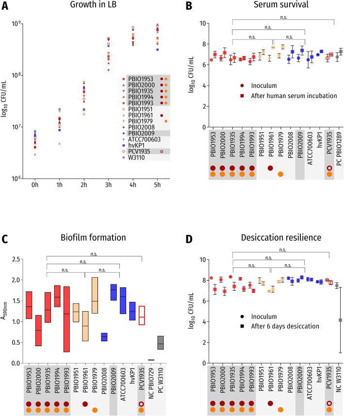
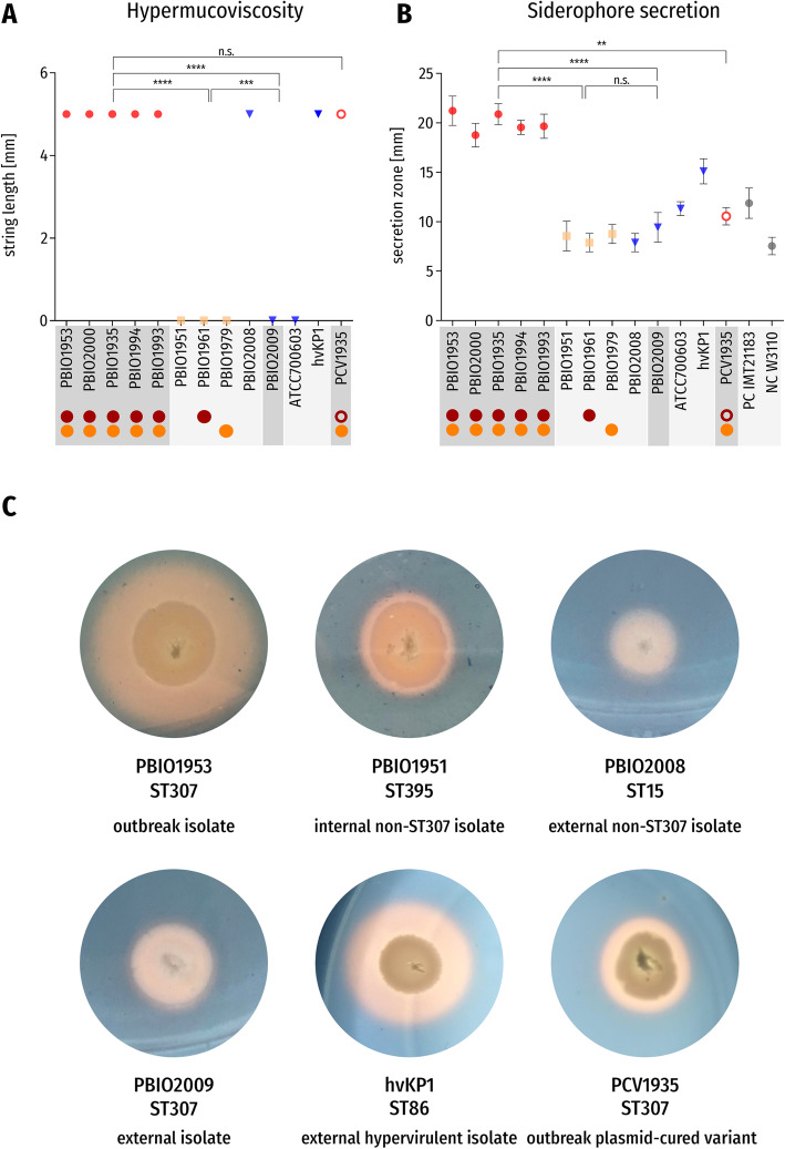
Methods: Here, we investigated these isolates and those from subsequent cases in the same facilities. We performed whole-genome sequencing to study phylogenetics, microevolution, and plasmid transmission, as well as phenotypic experiments including growth curves, hypermucoviscosity, siderophore secretion, biofilm formation, desiccation resilience, serum survival, and heavy metal resistance for an in-depth characterization of this outbreak clone.

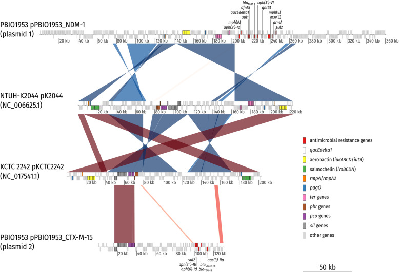
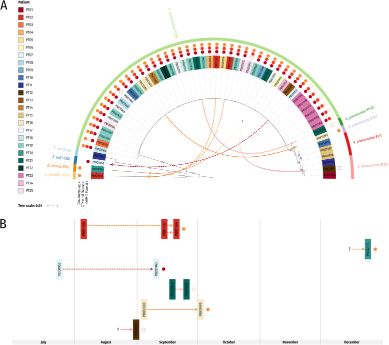
Results: Phylogenetics suggest a homogenous phylogram with several sub-clades containing either isolates from only one patient or isolates originating from different patients, suggesting inter-patient transmission. We identified three large resistance plasmids, carrying either NDM-1, CTX-M-15, or OXA-48, which K. pneumoniae ST307 likely donated to other K. pneumoniae isolates of different STs and even other bacterial species (e.g., Enterobacter cloacae) within the clinical settings. Several chromosomally and plasmid-encoded, hypervirulence-associated virulence factors (e.g., yersiniabactin, metabolite transporter, aerobactin, and heavy metal resistance genes) were identified in addition. While growth, biofilm formation, desiccation resilience, serum survival, and heavy metal resistance were comparable to several control strains, results from siderophore secretion and hypermucoviscosity experiments revealed superiority of the ST307 clone, similar to an archetypical, hypervirulent K. pneumoniae strain (hvKP1).

Conclusions: The combination of extensive drug resistance and virulence, partly conferred through a "mosaic" plasmid carrying both antibiotic resistance and hypervirulence-associated features, demonstrates serious public health implications.

Keywords: Hypervirulence; Outbreak; Plasmid transmission; XDR Klebsiella pneumoniae; “Mosaic” plasmid.


Conflict of interest statement

The authors declare that they have no competing interests.

Figures

Fig. 1
Fig. 1
Core SNP phylogeny of K. pneumoniae ST307 isolates. The phylogenetic tree was inferred with a maximum likelihood-based approach and is based on a core SNP alignment (83 sites). The (midpoint-rooted) tree is shown with bootstrap proportions for values ≥ 50% (1000 replicates; filled circles on branches) and additional metadata. The patient associated with isolate PBIO1964 (PT06) was initially present on a submission ward for bacterial isolation but later on transferred to a different ward. Abbreviations: UMG, University Medicine Greifswald; WLG, clinic in Wolgast; BDH, clinic in Greifswald; KB, clinic in Karlsburg
Fig. 2
Fig. 2
Synteny plot of hybrid resistance/virulence plasmid 1 (pPBIO1953_NDM-1) and resistance plasmid 2 (pPBIO1953_CTX-M-15). The plot depicts pairwise BlastN comparisons (E value 1e−10) between pPBIO1953 plasmid 1 and 2 and pK2044 and pKCTC2242. Alignment lengths were required to be at least 1% of the smaller replicon in the comparison to be included. Direct comparisons are colored with red hues whereas reverse comparisons are colored with blue hues. Boxes on top depict CDS on the forward strand, and those at the bottom depict CDS on the reverse strand. Boxes with a dotted outline are annotated as pseudo CDS. Plasmid 1 of PBIO1953 combines virulence features, such as aerobactin, with acquired antimicrobial resistance genes (e.g., blaNDM-1), which are missing from typical virulence plasmids of hypervirulent strains NTUH-K2044 and KCTC 2242. Note that PBIO1953 plasmid 2 carries several metal resistance genes (pco and sil genes) present on hvKP virulence plasmids but absent from plasmid 1
Fig. 3
Fig. 3
Putative plasmid transfer among K. pneumoniae isolates and between different bacterial genera. Solid arrows indicate putative intra-species transfer, whereas dashed arrows indicate inter-bacterial transfer. The putative inter-patient transfer is illustrated through a question mark. Note that from PT12 (PBIO1961), no additional carbapenem-resistant isolates were obtained and that no other ST307-positive patient was simultaneously present on the same ward whereas inter-patient transfer for PT20 (PBIO2014) is further supported by the epidemiologic data. Line colors match the colors of transferred virulence/resistance plasmids and are also shown next to the isolate names as circles. Closed circles—complete plasmid backbone (Megablast hits with identity ≥ 99%: coverage ≥ 99%). Open circles—incomplete plasmid backbone (Megablast hits with identity ≥ 99%: coverage > 77% and < 99% (Additional file 2: Table S4)). a The phylogenetic tree is based on distance-corrected MinHash dissimilarities between sequenced genomes and was inferred with FastME as part of JolyTree. Shown at the branches is the rate of elementary quartets (REQ) for values ≥ 0.75 (circle diameter). b Timeline figure of plasmid transfer between July and December 2019
Fig. 4
Fig. 4
Results of phenotypic experiments without significant differences. Red symbols: K. pneumoniae ST307 outbreak isolates; yellow symbols: K. pneumoniae non-ST307 internal isolates; blue symbols: K. pneumoniae external isolates. Red circle: occurrence of plasmid 1 in K. pneumoniae ST307 outbreak isolates and PBIO1961; orange circle: occurrence of plasmid 2 in K. pneumoniae ST307 outbreak isolates, PBIO1979, and PCV1935; red open circle: incomplete plasmid 1 in PCV1935. Isolates belonging to ST307 are highlighted in dark gray, and all other isolates in light gray. a Results of growth experiments in LB medium are given as mean values of CFU/mL for each isolate over 5 h. Abbreviations: n.s., not significant. b Results of serum survival experiments are given as mean values and standard deviation of CFU/mL for each isolate before (inoculum: circles) and after 4 h of incubation in human serum (squares). c Results of biofilm formation experiments are given as absorbance values (mean with coefficient of variation) of crystal violet at 590 nm for each isolate after 3 h of incubation. d Results of desiccation tolerance experiments are given as mean values and standard deviation of colony-forming units (CFU) per milliliter (CFU/mL) for each isolate before (inoculum: circles) and after 6 days of desiccation (squares)
Fig. 5
Fig. 5
Results of phenotypic experiments with significant differences. Red symbols: K. pneumoniae ST307 outbreak isolates; yellow symbols: K. pneumoniae non-ST307 internal isolates; blue symbols: K. pneumoniae external isolates. Red circle: occurrence of plasmid 1 in K. pneumoniae ST307 outbreak isolates and PBIO1961; orange circle: occurrence of plasmid 2 in K. pneumoniae ST307 outbreak isolates, PBIO1979, and PCV1935; red open circle: incomplete plasmid 1 in PCV1935. Isolates belonging to ST307 are highlighted in dark gray, and all other isolates in light gray. a Results of the hypermucoviscosity test are given as mean values and standard deviation of the string length in millimeters (mm) for each isolate. b Results of siderophore secretion tests are given as mean values and standard deviation of the secretion zone diameter in millimeters (mm) for each isolate. c Siderophore secretion experiment on CAS-Agar of six exemplary isolates: PBIO1953 (outbreak isolate [ST307]), PBIO1951 (internal control [ST395]), PBIO2008 (external control [ST15]), PBIO2009 (external control [ST307]), hvKP1 (external hypervirulent control [ST86]), and PCV1935 (outbreak plasmid-cured variant [ST307]). Yellow areas around colonies indicate siderophore secretion. Abbreviations and symbols: n.s., not significant; ****p value < 0.0001; ***p value < 0.001; **p value < 0.01

References

    1. Podschun R, Ullmann U. Klebsiella spp. as nosocomial pathogens: epidemiology, taxonomy, typing methods, and pathogenicity factors. Clin Microbiol Rev. 1998;11(4):589–603. doi: 10.1128/CMR.11.4.589. - DOI - PMC - PubMed
    1. Fazili T, Sharngoe C, Endy T, Kiska D, Javaid W, Polhemus M. Klebsiella pneumoniae liver abscess: an emerging disease. Am J Med Sci. 2016;351(3):297–304. doi: 10.1016/j.amjms.2015.12.018. - DOI - PubMed
    1. Villa L, Feudi C, Fortini D, Brisse S, Passet V, Bonura C, et al. Diversity, virulence, and antimicrobial resistance of the KPC-producing Klebsiella pneumoniae ST307 clone. Microb Genom. 2017;3(4):e000110. - PMC - PubMed
    1. Rossi B, Gasperini ML, Leflon-Guibout V, Gioanni A, de Lastours V, Rossi G, et al. Hypervirulent Klebsiella pneumoniae in cryptogenic liver abscesses, Paris, France. Emerg Infect Dis. 2018;24(2):221–229. doi: 10.3201/eid2402.170957. - DOI - PMC - PubMed
    1. Russo TA, Marr CM. Hypervirulent Klebsiella pneumoniae. Clin Microbiol Rev. 2019;32(3):e00001–19. - PMC - PubMed

Publication types

MeSH terms

LinkOut - more resources