Case Reports
. 2020 Dec 9;13(12):e236056.
doi: 10.1136/bcr-2020-236056.

Colonic inflammatory myofibroblastic tumour presenting as 'pyrexia of unknown origin': report of a rare disease and its unique presentation

Affiliations
Case Reports

Colonic inflammatory myofibroblastic tumour presenting as 'pyrexia of unknown origin': report of a rare disease and its unique presentation

Victor Vijay Coelho et al. BMJ Case Rep. .

Abstract

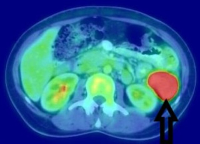
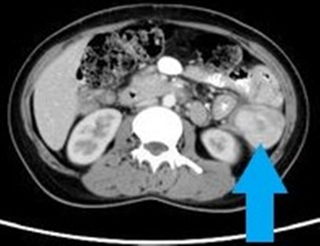
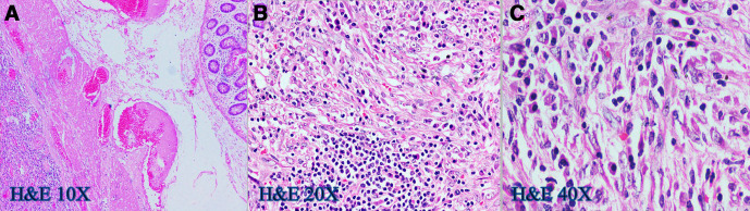
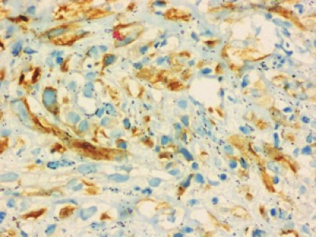
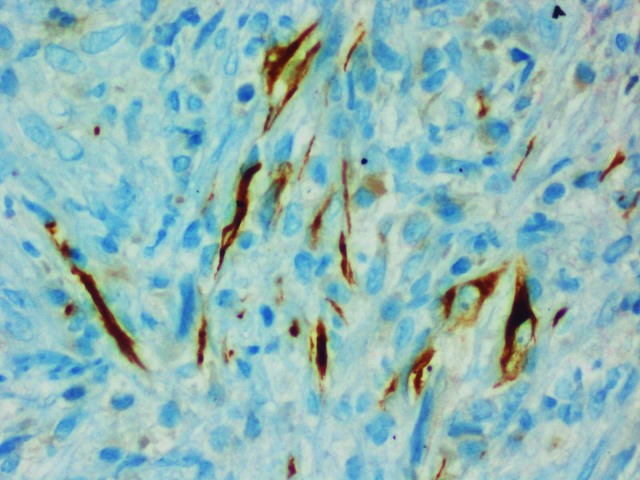
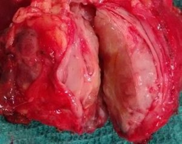
An intra-abdominal inflammatory myofibroblastic tumour (IMT) belongs to a rare group of diseases initially described as an inflammatory pseudotumour. Even though it is seen more often in children, its incidence in adults is even rarer. Clinical presentations can vary depending on its site and inherent tumour properties. The colon is an uncommon site for IMT and pyrexia of unknown origin (PUO) as its dominant clinical presentation is even rarer. A 27-year-old woman presented with PUO. She was evaluated under the department of internal medicine before undergoing an 18F-fluorodeoxyglucose positron emission tomography-computed tomography scan. This showed an intensely enhancing descending colon mass. An image-guided biopsy of this lesion was reported as IMT. She underwent a left hemicolectomy and complete excision of the tumour, following which her symptoms resolved completely. The patient has been disease-free at a 6-month follow-up and is asymptomatic at 1 year.

Keywords: colon cancer; pathology; surgery; surgical oncology.


Conflict of interest statement

Competing interests: None declared.

Figures

Figure 1
Figure 1
18F-fluorodeoxyglucose positron emission tomography scan showing an intensely enhancing retroperitoneal mass in the left lumbar region.
Figure 2
Figure 2
Contrast-enhanced computed tomography scan of the abdomen/pelvis showing a well-defined, intensely enhancing lesion in the left lumbar retroperitoneal region, indenting the proximal descending colon.
Figure 3
Figure 3
(A–C) H&E slides showing sheets of polygonal cells with moderate nuclear pleomorphism, vesicular chromatin, prominent nucleoli and moderate amounts of eosinophilic cytoplasm and admixed lymphocytes, plasma cells, eosinophils and foamy histiocytes.
Figure 4
Figure 4
Immunohistochemistry slide (40×) showing tumour cells with anaplastic lymphoma kinase-1 (ALK-1) positivity.
Figure 5
Figure 5
Immunohistochemistry slide (40×) showing tumour cells with desmin positivity.
Figure 6
Figure 6
A cut section of the surgical specimen showing a fleshy tumour.

References

    1. Shi H, Wei L, Sun L, et al. . Primary gastric inflammatory myofibroblastic tumor: a clinicopathologic and immunohistochemical study of 5 cases. Pathol Res Pract 2010;206:287–91. 10.1016/j.prp.2009.09.002 - DOI - PubMed
    1. Kim EY, Lee IK, Lee YS, et al. . Inflammatory myofibroblastic tumor in colon. J Korean Surg Soc 2012;82:45–9. 10.4174/jkss.2012.82.1.45 - DOI - PMC - PubMed
    1. Raffaeli E, Cardinali L, Fianchini M, et al. . Inflammatory myofibroblastic tumor of the transverse colon with synchronous gastrointestinal stromal tumor in a patient with ulcerative colitis: a case report. Int J Surg Case Rep 2019;60:141–4. 10.1016/j.ijscr.2019.06.012 - DOI - PMC - PubMed
    1. Park S-H, Kim J-H, Min BW, et al. . Exophytic inflammatory myofibroblastic tumor of the stomach in an adult woman: a rare cause of hemoperitoneum. World J Gastroenterol 2008;14:136–9. 10.3748/wjg.14.136 - DOI - PMC - PubMed
    1. Zhou RU, Xiang J, Chen Z, et al. . Fever of unknown origin as a presentation of colonic inflammatory myofibroblastic tumor in a 36-year-old female: a case report. Oncol Lett 2014;7:1566–8. 10.3892/ol.2014.1942 - DOI - PMC - PubMed

Publication types

MeSH terms

Substances