. 2020 Dec 9;5(6):e00943-20.
doi: 10.1128/mSphere.00943-20.

Amylases in the Human Vagina

Affiliations

Amylases in the Human Vagina

Kenetta L Nunn et al. mSphere. .

Abstract

Dominance of Lactobacillus species in vaginal communities is a hallmark of healthy conditions in the female genital tract. Key nutrients for lactobacilli include sugars produced when glycogen is degraded by α-amylase in the vagina. While α-amylase activity has been demonstrated in vaginal fluids, it is unclear whether α-amylases are produced solely by the host, bacteria in the vagina, or both. We screened cervicovaginal mucus from 23 reproductive-age women, characterized the species composition of vaginal communities, measured vaginal pH, and determined levels of amylase activity, glycogen, and lactic acid. Based on differences in these measured variables, one sample from each of four individual donors was selected for metagenomic and proteomic analyses. Of eight putative bacterial amylases identified in the assembled bacterial metagenomes, we detected four in vaginal fluids. These amylases were produced by various bacteria in different vaginal communities. Moreover, no two communities were the same in terms of which bacteria were producing amylases. Although we detected bacterial amylases in vaginal fluids, there was no clear association between the bacterial species that was dominant in a community and the level of amylase activity. This association was likely masked by the presence of human α-amylase, which was also detected in vaginal fluids. Finally, the levels of amylase activity and glycogen were only weakly associated. Our findings show, for the first time, that multiple amylases from both bacterial and human origins can be present simultaneously in the vagina. This work also suggests that the link between glycogen, amylase, and Lactobacillus in the vagina is complex.IMPORTANCE In this study, we show that multiple bacteria in the vaginal community produce amylases that hydrolyze glycogen into simpler sugars (i.e., maltose and maltotriose). These sugars serve as "common goods" that sustain bacterial populations in vaginal communities. Given the temporal changes that are observed in the human vaginal microbiome, we expect the kinds of bacterial amylases produced will also vary over time. These differences influence the pool of resources that are broadly shared and shape the species composition of the vaginal bacterial community.

Keywords: Lactobacillus; amylase; glycogen; vaginal microbiome.


Figures

FIG 1
FIG 1
Scatterplot of glycogen measurements (milligrams per milliliter) as a function of amylase activity (units per milligram of total protein). The points in the graph represent glycogen measurements for each sample, plotted with the corresponding amylase activity. The regression line was generated by fitting a generalized linear model to these data points. The dashed lines represent the 95% confidence interval for the model fit. The pseudo-R2 and the P value for the model are shown at the top right.
FIG 2
FIG 2
Amylase activity relative to bacterial species composition in the human vagina. Amylase activity (units per milligram total protein) in each sample was arranged in increasing order (A) along with the corresponding stacked bar (B). The dashed gray line in panel A represents the mean of amylase activity across samples (0.55 U/mg). The stacked bars represent the relative proportions of bacteria in each community. The colors for bacterial taxa are shown in the legend at the bottom.
FIG 3
FIG 3
Amylase enzymes in vaginal fluids collected from donors F02, F06, F08, and F12 separated by native PAGE. These columns represent image slices taken from Fig. S1 in the supplemental material (containing all of the images of native PAGE gels completed for all samples) for donor samples F02, F06, F08, and F12. A total of 20 μg of total protein was loaded for each sample. After the amylase proteins were separated in the gels, gels were incubated in 1% starch to enable starch hydrolysis and stained with iodine solution to observe where hydrolysis took place. The images that the slices were extracted from were inverted so that the dark background shows up white and the clear zones indicating hydrolysis of starch show up dark. Thus, the dark bands reflect amylase isozymes that were resolved in each donor sample. We were unable to resolve any amylase proteins in donor F12. PA (human pancreatic α-amylase) and GA (Rhizopus spp. glucoamylase) are two commercial amylases that were used as positive controls for the assay.
FIG 4
FIG 4
Human proteins expressed in vaginal fluids of donors F02, F06, F08, and F12. (Left) Heat map that reflects the relative abundances of human proteins detected in each sample. Relative abundances are indicated by the Z-scores as shown at the bottom. Blue values indicate low abundances based on the mean of all of the proteins in a sample, whereas yellow indicates high abundances. (Right) Clusters identified in the enrichment analysis that identified GO terms that were enriched in the proteome for each sample.
FIG 5
FIG 5
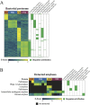
Bacterial proteins expressed in vaginal fluids from donors F02, F06, F08, and F12. (A) Heat map that reflects the relative abundances of bacterial proteins detected in each sample. Relative abundances are indicated by the Z-scores shown at the bottom. (Right) Green boxes indicate in which MAG (bacteria) the proteins were detected. (B) Putative bacterial and human amylases that were detected in vaginal fluids using LC-MS/MS. The relative abundance of the protein (listed on the left) is shown in individual heat maps below each sample. Relative abundances are indicated by the Z-scores shown at the bottom. Black squares indicate that the protein was not detected in a sample. Similar to that in panel A, the green boxes at the right show which MAG the protein sequence was found in.

References

    1. Boskey ER, Telsch KM, Whaley KJ, Moench TR, Cone RA. 1999. Acid production by vaginal flora in vitro is consistent with the rate and extent of vaginal acidification. Infect Immun 67:5170–5175. doi:10.1128/IAI.67.10.5170-5175.1999. - DOI - PMC - PubMed
    1. Boskey ER, Cone RA, Whaley KJ, Moench TR. 2001. Origins of vaginal acidity: high d/l lactate ratio is consistent with bacteria being the primary source. Hum Reprod 16:1809–1813. doi:10.1093/humrep/16.9.1809. - DOI - PubMed
    1. O'Hanlon DE, Moench TR, Cone RA. 2011. In vaginal fluid, bacteria associated with bacterial vaginosis can be suppressed with lactic acid but not hydrogen peroxide. BMC Infect Dis 11:200. doi:10.1186/1471-2334-11-200. - DOI - PMC - PubMed
    1. O'Hanlon DE, Moench TR, Cone RA. 2013. Vaginal pH and microbicidal lactic acid when lactobacilli dominate the microbiota. PLoS One 8:e80074. doi:10.1371/journal.pone.0080074. - DOI - PMC - PubMed
    1. Antonio MA, Hawes SE, Hillier SL. 1999. The identification of vaginal Lactobacillus species and the demographic and microbiologic characteristics of women colonized by these species. J Infect Dis 180:1950–1956. doi:10.1086/315109. - DOI - PubMed

Publication types

LinkOut - more resources