. 2020 Dec;8(2):e001591.
doi: 10.1136/jitc-2020-001591.

Mobilization of pre-existing polyclonal T cells specific to neoantigens but not self-antigens during treatment of a patient with melanoma with bempegaldesleukin and nivolumab

Affiliations

Mobilization of pre-existing polyclonal T cells specific to neoantigens but not self-antigens during treatment of a patient with melanoma with bempegaldesleukin and nivolumab

Joshua R Veatch et al. J Immunother Cancer. 2020 Dec.

Erratum in

Abstract

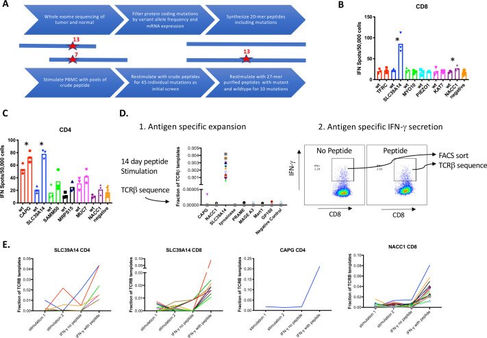
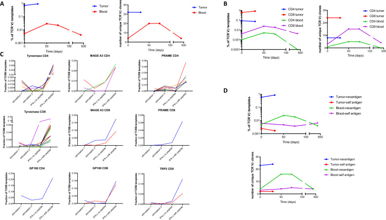
T cells that recognize self-antigens and mutated neoantigens are thought to mediate antitumor activity of immune checkpoint blockade (ICB) in melanoma. Few studies have analyzed self and neoantigen-specific T cell responses in patients responding to ICB. Here, we report a patient with metastatic melanoma who had a durable clinical response after treatment with the programmed cell death protein 1 inhibitor, nivolumab, combined with the first-in-class CD122-preferential interleukin-2 pathway agonist, bempegaldesleukin (BEMPEG, NKTR-214). We used a combination of antigen-specific T cell expansion and measurement of interferon-γ secretion to identify multiple CD4+ and CD8+ T cell clones specific for neoantigens, lineage-specific antigens and cancer testis antigens in blood and tumor from this patient prior to and after therapy. Polyclonal CD4+ and CD8+ T cells specific to multiple neoantigens but not self-antigens were highly enriched in pretreatment tumor compared with peripheral blood. Neoantigen, but not self-antigen-specific T cell clones expanded in frequency in the blood during successful treatment. There was evidence of dramatic immune infiltration into the tumor on treatment, and a modest increase in the relative frequency of intratumoral neoantigen-specific T cells. These observations suggest that diverse CD8+ and CD4+ T cell clones specific for neoantigens present in tumor before treatment had a greater role in immune tumor rejection as compared with self-antigen-specific T cells in this patient. Trial registration number: NCT02983045.

Keywords: T-lymphocytes; antigens; melanoma; neoplasm.


Conflict of interest statement

Competing interests: JRV, BJ and SR have equity interest in Lyell Immunopharma, and JZ and EE are employees of Nektar therapeutics, and this paper discusses use of an investigational drug owned by Nektar therapeutics.

Figures

Figure 1
Figure 1
Treatment response and transcriptome analysis. (A) RECIST clinical disease burden following initiation of treatment. (B) Lung nodule on CT scan that underwent needle biopsy at day 19 of treatment. (C) Log2 fold change of immune-related genes between on-treatment and pretreatment biopsy by whole transcriptome sequencing. (D) Log2 fold change of melanocytic lineage-specific and cancer-testis antigens between on-treatment and pre-treatment tumor biopsy.
Figure 2
Figure 2
Identification of neoantigen-specific T cell clones. (A) Schematic of approach for identifying T cell responses to neoantigens. (B–C) Peripheral blood mononuclear cells (PBMCs) were stimulated with pools of crude peptides and then magnetically enriched into CD8+ or CD4+ T cell subsets. CD8+ (B) and CD4+ (C) T cells were restimulated with single purified wild-type or mutant peptides and interferon (IFN)-γ secretion was quantitated by ELISpot. (D) PBMCs were stimulated with purified peptides containing mutations and TCRVβ clonotypes were quantitated by sequencing (left panel). IFN-γ secreting cells were identified following stimulation with peptide or no peptide (control) and sorted by fluorescence activated cell sorting (FACS) and subjected to TCRVβ sequencing. (E) Multiple CD4+ and CD8+ clonotypes were enriched after IFN-γ secretion after stimulation with the peptide containing the mutation in SLC39A14, CAPG or NACC1.
Figure 3
Figure 3
Isolation of patient-derived neoantigen-specific CD8 and CD4 T cells. (A) Schematic of strategy for isolating neoantigen-specific T cells: stimulation of peripheral blood mononuclear cell (PBMC) with peptides followed by isolation of interferon (IFN)-γ secreting cells, expansion of T cell lines and screening of T cell lines for antigen specificity and TCRVβ sequencing of reactive T cell lines. (B–D) Clonotype analysis of six reactive CD8+ T cell lines with each color representing an individual clone (B) and three CD4+ T cell lines (C) specific for mutation in SLC39A14 and four CD8+ T cell lines specific for a mutation in NACC1 (D). (E–G) Expanded CD8+ T cell lines were incubated with different concentrations of mutant and wild-type peptides containing mutations in SLC39A14 (E) and NACC1 (F) and CD4+ T cell lines were incubated with peptides containing mutant and wild-type SLC39A14. (G) Sequences and IFN-γ secretion was measured by ELISA assay. (H) Consensus sequence of nine NACC1-specific TCRVβ CDR3 amino acid sequences of length 12 were generated using weblogo.
Figure 4
Figure 4
Neoantigen, but not self-antigen-specific T cell clones localize to tumor and expand in the blood following treatment. (A–B) Cumulative frequency (left) or number of distinct neoantigen-specific TCRVβ clones (right) in tumor and blood samples at the indicated timepoints following treatment for total clones (A) and separated into CD4+ and CD8+ clones (B). (C) Peripheral blood mononuclear cell (PBMC) was stimulated with purified peptides containing mutations or peptide pools of self-antigens and TCRVβ clonotypes were quantitated by sequencing in two independent replicates. These cultures were restimulated with peptides or no peptide controls, interferon (IFN)-γ secreting cells were sorted by fluorescence activated cell sorting (FACS) and TCRVβ clonotypes were quantitated by sequencing. The frequency of putative antigen-specific clones is shown for CD4+ and CD8+ T cells for the indicated antigens. (D) Cumulative frequency (top panel) and number (bottom panel) of distinct neoantigen and self-antigen-specific TCRVβ clones in tumor and blood samples following treatment.

References

    1. Schumacher TN, Schreiber RD. Neoantigens in cancer immunotherapy. Science 2015;348:69–74. 10.1126/science.aaa4971 - DOI - PubMed
    1. Lennerz V, Fatho M, Gentilini C, et al. The response of autologous T cells to a human melanoma is dominated by mutated neoantigens. Proc Natl Acad Sci U S A 2005;102:16013–8. 10.1073/pnas.0500090102 - DOI - PMC - PubMed
    1. Gubin MM, Zhang X, Schuster H, et al. Checkpoint blockade cancer immunotherapy targets tumour-specific mutant antigens. Nature 2014;515:577–81. 10.1038/nature13988 - DOI - PMC - PubMed
    1. Gilboa E. The makings of a tumor rejection antigen. Immunity 1999;11:263–70. 10.1016/S1074-7613(00)80101-6 - DOI - PubMed
    1. Anagnostou V, Smith KN, Forde PM, et al. Evolution of neoantigen landscape during immune checkpoint blockade in non-small cell lung cancer. Cancer Discov 2017;7:264–76. 10.1158/2159-8290.CD-16-0828 - DOI - PMC - PubMed

Publication types

Associated data