Review
. 2020 Dec 9;8(1):42.
doi: 10.1038/s41413-020-00119-9.

Neurological heterotopic ossification: novel mechanisms, prognostic biomarkers and prophylactic therapies

Affiliations
Review

Neurological heterotopic ossification: novel mechanisms, prognostic biomarkers and prophylactic therapies

Ker Rui Wong et al. Bone Res. .

Abstract

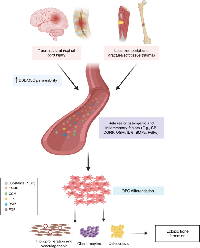
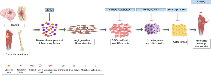
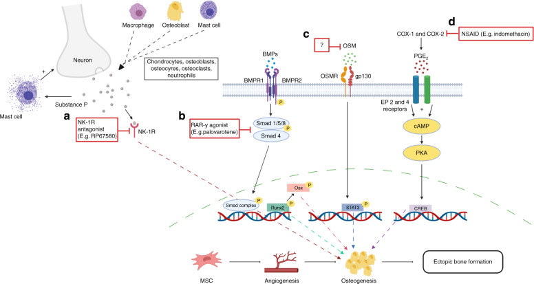
Neurological heterotopic ossification (NHO) is a debilitating condition where bone forms in soft tissue, such as muscle surrounding the hip and knee, following an injury to the brain or spinal cord. This abnormal formation of bone can result in nerve impingement, pain, contractures and impaired movement. Patients are often diagnosed with NHO after the bone tissue has completely mineralised, leaving invasive surgical resection the only remaining treatment option. Surgical resection of NHO creates potential for added complications, particularly in patients with concomitant injury to the central nervous system (CNS). Although recent work has begun to shed light on the physiological mechanisms involved in NHO, there remains a significant knowledge gap related to the prognostic biomarkers and prophylactic treatments which are necessary to prevent NHO and optimise patient outcomes. This article reviews the current understanding pertaining to NHO epidemiology, pathobiology, biomarkers and treatment options. In particular, we focus on how concomitant CNS injury may drive ectopic bone formation and discuss considerations for treating polytrauma patients with NHO. We conclude that understanding of the pathogenesis of NHO is rapidly advancing, and as such, there is the strong potential for future research to unearth methods capable of identifying patients likely to develop NHO, and targeted treatments to prevent its manifestation.


Conflict of interest statement

The authors declare no competing interests.

Figures

Fig. 1
Fig. 1
Proposed mechanism for NHO development. Simultaneous injury to the CNS and peripheral sites triggers the release of osteogenic and inflammatory factors including; SP, CGRP, OSM, IL-6, BMPs and FGFs. The influx of osteogenic and inflammatory factors, initiates the differentiation of OPCs into fibroblasts which is mediated by fibroblast growth factors (FGFs). This influx also elicits angiogenesis, which results in an increase in oxygen tension, triggering the differentiation of OPCs into chondrocytes which undergo hypertrophy and form a cartilage matrix. This cartilaginous matrix provides a structural framework for the formation of blood vessels, osteoblast proliferation and differentiation and formation of ectopic bone (created with BioRender.com)
Fig. 2
Fig. 2
Current treatments targeting specific pathways of NHO. NHO development is triggered by a cascade of inflammatory factors. Presently, the preferred prophylactic treatment for NHO/HO involves NSAIDs (e.g. Indomethacin) to downregulate the inflammatory response and prevent OPC differentiation. Radiotherapy is thought to prevent the formation and development of ectopic bone specifically by inhibiting the differentiation of OPCs. RAR-γ agonists have been shown to prevent chondrogenesis and therefore subsequent mineralisation. While, nitrogen-containing bisphosphonates (e.g. sodium etidronate) have been used to inhibit mineralisation, and the formation of ectopic bone. Finally, when bone is completely mineralised, surgical resection is the only remaining intervention. This invasive procedure, however, is accompanied by the risk of recurrence and is associated with complications which include incomplete resection, functional and physiological impairment (created with BioRender.com)
Fig. 3
Fig. 3
Key pathways/mechanisms implicated in the development of NHO highlighting potential and existing therapeutic targets to mitigate NHO. a Substance P receptor (NK-1R) antagonists (e.g. RP67580) have been found to reduce NHO volume in murine models. b Downstream of the BMP pathway, RAR-γ agonists such as palovarotene have been reported to prevent early stages of NHO development by disrupting OPC differentiation, chondrogenesis and osteogenesis by downregulating mRNA expression of SOX-9 and RUNX2. c OSM is a potential therapeutic target and may serve as a biomarker for NHO. Blocking OSM has been found to reduce NHO likely by inhibiting downstream transcription factor, STAT3, which is known to trigger bone formation. d NSAID, indomethacin is currently the preferred prophylaxis for NHO/HO. It targets COX-1 and COX-2 non-selectively, inhibiting the production of prostaglandins and osteogenesis (created with BioRender.com)

References

    1. Garland, D. E. A clinical perspective on common forms of acquired heterotopic ossification. Clin. Orthop. Relat. Res.263, 13–29 (1991). - PubMed
    1. Hoyt BW, Pavey GJ, Potter BK, Forsberg JA. Heterotopic ossification and lessons learned from fifteen years at war: a review of therapy, novel research, and future directions for military and civilian orthopaedic trauma. Bone. 2018;109:3–11. doi: 10.1016/j.bone.2018.02.009. - DOI - PubMed
    1. Citta-Pietrolungo TJ, Alexander MA, Steg NL. Early detection of heterotopic ossification in young patients with traumatic brain injury. Arch. Phys. Med. Rehabil. 1992;73:258–262. - PubMed
    1. Mavrogenis AF, Soucacos PN, Papagelopoulos PJ. Heterotopic ossification revisited. Orthopedics. 2011;34:177. doi: 10.3928/01477447-20110124-08. - DOI - PubMed
    1. Adiguzel E, et al. Knee pain relief with genicular nerve blockage in two brain injured patients with heterotopic ossification. Brain Inj. 2015;29:1736–1739. doi: 10.3109/02699052.2015.1075171. - DOI - PubMed