. 2020 Dec 9;11(1):6303.
doi: 10.1038/s41467-020-20132-0.

Condensation of Rubisco into a proto-pyrenoid in higher plant chloroplasts

Affiliations

Condensation of Rubisco into a proto-pyrenoid in higher plant chloroplasts

Nicky Atkinson et al. Nat Commun. .

Abstract

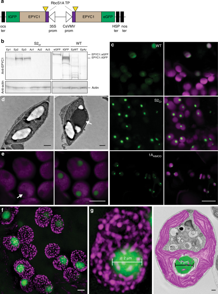
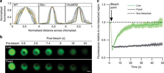
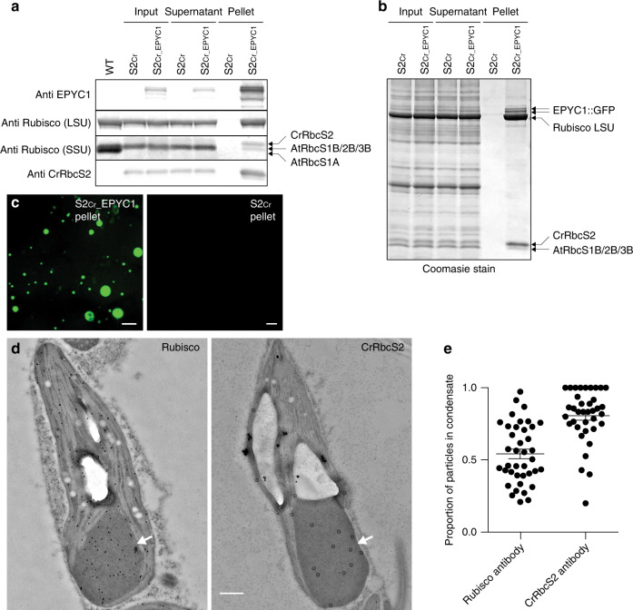
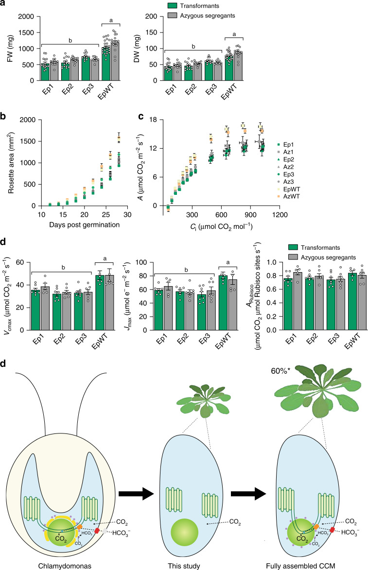
Photosynthetic CO2 fixation in plants is limited by the inefficiency of the CO2-assimilating enzyme Rubisco. In most eukaryotic algae, Rubisco aggregates within a microcompartment known as the pyrenoid, in association with a CO2-concentrating mechanism that improves photosynthetic operating efficiency under conditions of low inorganic carbon. Recent work has shown that the pyrenoid matrix is a phase-separated, liquid-like condensate. In the alga Chlamydomonas reinhardtii, condensation is mediated by two components: Rubisco and the linker protein EPYC1 (Essential Pyrenoid Component 1). Here, we show that expression of mature EPYC1 and a plant-algal hybrid Rubisco leads to spontaneous condensation of Rubisco into a single phase-separated compartment in Arabidopsis chloroplasts, with liquid-like properties similar to a pyrenoid matrix. This work represents a significant initial step towards enhancing photosynthesis in higher plants by introducing an algal CO2-concentrating mechanism, which is predicted to significantly increase the efficiency of photosynthetic CO2 uptake.


Conflict of interest statement

The authors declare no competing interests.

Figures

Fig. 1
Fig. 1. Expression of EPYC1 in the Arabidopsis line S2Cr results in condensate formation.
a Schematic representation of the dual GFP expression system (EPYC1-dGFP) for EPYC1 truncated at amino acid residue 27 (as indicated by yellow triangles) and fused at the N-terminus to the chloroplastic transit peptide (TP) sequence of the Arabidopsis Rubisco small subunit RbcS1A. EPYC1 was further fused at the C-terminus to either enhanced GFP (eGFP) or turboGFP (tGFP), and driven by the 35S CaMV promoter (35S prom) or CsVMV promoter (CsVMV prom), respectively. For the latter expression cassette, a dual terminator system was used to increase expression (Supplementary Fig. 1). b EPYC1 protein levels in Arabidopsis plants as assessed by immunoblot analysis with anti-EPYC1 antibodies. Shown are three T2 S2Cr transgenic plants expressing EPYC1-dGFP (Ep1−3) and azygous segregants (Az1−3), and S2Cr plants transformed with only EPYC1::tGFP (55.4 kDa) or EPYC1::eGFP (63.9 kDa). Also displayed are a T2 EPYC1-dGFP WT transformant (EpWT) and azygous segregant (AzWT). Anti-actin is shown as a loading control underneath. c Expression of EPYC1-dGFP in WT, S2Cr and 1AAtMOD backgrounds. Green and purple signals are GFP and chlorophyll autofluorescence, respectively. Overlapping signals are white. Scale bar = 10 µm for all images. d TEM images of chloroplasts from S2Cr plants with (right) and without (left) expression of EPYC1. A white arrowhead indicates the dense dark grey area of the EPYC1 condensate. The large white structures are starch granules. Scale bars = 0.5 µm. A representative chloroplast from a wild-type plant expressing EPYC1-dGFP is shown for comparison in Extended Data Fig. 3. e Chlorophyll autofluorescence is reduced at the site of EPYC1-dGFP accumulation (white arrow). Scale bar = 5 µm. f SIM microscopy showing EPYC1-dGFP condensates inside the chloroplast (see also Supplementary Movie 1). The magenta puncta show the position of grana stacks. Light magenta puncta indicate grana stacks behind the condensate. Scale bar = 2 µm. g Example comparison of condensate size (left, 2 µm) with that of a pyrenoid in Chlamydomonas (right, representative TEM image highlighting the pyrenoid in green and chloroplast in purple). Scale bar for TEM image = 0.5 µm. Images of EPYC1-dGFP condensates in the S2Cr background are from line Ep3 (c, e, f, g). Confocal images are representative of multiple imaging sessions (c, e). SIM microscopy (f, g) imaging was performed once. The immunoblots shown were derived from the same experiment and gels/blots were processed in parallel. Immunoblots results were representative of six gels/blots (a), TEM images are representative of 26 and 12 images from S2Cr plants with and without EPYC1, respectively (d) and 55 images from Chlamydomonas (g). Source data are provided as a Source Data file.
Fig. 2
Fig. 2. In planta condensates behave like liquid−liquid phase-separated microcompartments.
a Fluorescence distribution plots of EPYC1-dGFP across the chloroplast. The intensity of the GFP fluorescence signal over the cross-section of a chloroplast is shown in WT (n = 28), S2Cr (line Ep3, n = 17) and 1AAtMOD (n = 22) backgrounds. Both GFP fluorescence and cross-section values have been normalised to 1 (as indicated by the dashed line), with the highest value in the centre. b Fluorescence recovery after photobleaching (FRAP) assays. Condensates are shown from live (top) and fixed (bottom) leaf tissue from S2Cr transgenic line Ep3 expressing EPYC1-dGFP. Scale bar = 1 µm for both images. c Fluorescence recovery of the bleached area in relation to the non-bleached area of condensates. The mean ± SEM are shown for live (n = 13) and fixed (n = 16) chloroplasts. Source data are provided as a Source Data file.
Fig. 3
Fig. 3. Condensates contain EPYC1 and plant-algal hybrid Rubisco.
a EPYC1 and Rubisco protein levels in whole leaf tissue (input), the supernatant following condensate extraction and centrifugation (supernatant) and the pellet (pellet) as assessed by immunoblot analyses with anti-EPYC1, anti-Rubisco (LSU and SSU shown) or anti-CrRbcS2 antibodies. Samples are shown for WT plants (WT), and S2Cr mutants not expressing EPYC1 (S2Cr) and expressing EPYC1-dGFP (S2Cr_EPYC1). The pellet is 40× more concentrated than the input and supernatant. Molecular weights: LSU, 55 kDa; RbcS1B, RbcS2B and RbcS3B, 14.8 kDa; AtRbcS1A, 14.7 kDa; CrRbcS2 15.5 kDa. The immunoblots shown were derived from the same experiment and gels/blots were processed in parallel. Immunoblots results were representative of four gels/blots for EPYC1, two for Rubisco, and one for CrRbcS2. b Coomassie-stained SDS-PAGE gel showing the composition of the pelleted condensate. Images are representative of three gels. c The condensates in the S2Cr_EPYC1 pellet coalescence to form large liquid droplets. Scale bars = 25 µm. d Representative immunogold labelling of Rubisco in chloroplasts of an S2Cr transgenic line Ep3 expressing EPYC1-dGFP probed with polyclonal anti-Rubisco (left) or anti-CrRbcS2 (right) antibodies (dots are highlighted for the latter). The condensates are marked by a white arrowhead. Large white structures are starch granules. Scale bar = 0.5 µm for both images. e Proportion of gold nanoparticles inside the condensate compared to the chloroplast for each antibody. The mean ± SEM are shown for n = 37 individual chloroplast images. Source data are provided as a Source Data file.
Fig. 4
Fig. 4. EPYC1-mediated condensation of Rubisco has no negative impact on growth and photosynthesis.
a Fresh and dry weights of three T2 EPYC1-dGFP S2Cr transgenic lines (Ep1−3) and an EPYC1-dGFP WT transformant (EpWT) (both in green) with their respective azygous segregants (Az1−3 and AzWT) (in grey). Plants were measured after 32 days of growth under 200 μmol photons m−2 s−1 light. The mean ± SEM are shown for n = 10−26 individual plants for each line. b Rosette expansion of S2Cr and WT lines in (a). c Net CO2 assimilation (A) based on intercellular [CO2] (Ci) under saturating light (1500 μmol photons m−2 s−1). Values show the mean ± SEM of measurements made on individual leaves from individual rosettes (n = 5−8). d Variables derived from gas exchange data include maximum rate of Rubisco carboxylation (Vcmax), maximum electron transport rate (Jmax), stomatal conductance (Gs), mesophyll conductance (Gm) and the net CO2 assimilation rate at ambient concentrations of CO2 normalised to Rubisco (ARubisco). Letters indicate significant difference (p < 0.05) of EpWT lines compared to Ep lines as determined by one-way ANOVA followed by Tukey’s honestly significant difference (HSD) post-hoc tests (a, d). e Algal CCM components required for enhancing photosynthesis. Generating a pyrenoid-like condensate in a plant chloroplast provides a platform for introducing bicarbonate (HCO3) channels/pumps at the chloroplast envelope (e.g. LCIA, shown in red) and thylakoid membrane (e.g. BST1−3, shown in orange), a lumenal carbonic anhydrase to convert HCO3 to CO2 for release into the surrounding Rubisco condensate (CAH3, shown in blue), mechanisms to capture CO2 as HCO3 (LCIB and LCIC, shown in purple), and traversing thylakoid membranes. Current models suggest that introduction of a functional biophysical CCM into a C3 plant could lead to productivity gains of up to 60% . Source data are provided as a Source Data file.

References

    1. Andersson I, Backlund A. Structure and function of Rubisco. Plant Physiol. Bioch. 2008;46:275–291. doi: 10.1016/j.plaphy.2008.01.001. - DOI - PubMed
    1. Salesse-Smith CE, et al. Overexpression of Rubisco subunits with RAF1 increases Rubisco content in maize. Nat. Plants. 2018;4:802–810. doi: 10.1038/s41477-018-0252-4. - DOI - PubMed
    1. Flamholz AI, et al. Revisiting trade-offs between Rubisco kinetic parameters. Biochem.-Us. 2019;58:3365–3376. doi: 10.1021/acs.biochem.9b00237. - DOI - PMC - PubMed
    1. Galmes J, et al. Expanding knowledge of the Rubisco kinetics variability in plant species: environmental and evolutionary trends. Plant Cell Environ. 2014;37:1989–2001. doi: 10.1111/pce.12335. - DOI - PubMed
    1. Young JN, et al. Large variation in the Rubisco kinetics of diatoms reveals diversity among their carbon-concentrating mechanisms. J. Exp. Bot. 2016;67:3445–3456. doi: 10.1093/jxb/erw163. - DOI - PMC - PubMed

Publication types

MeSH terms