Review
. 2020 Nov 23;5(1):22.
doi: 10.1038/s41536-020-00107-x.

Mitochondrial dysfunction in neurological disorders: Exploring mitochondrial transplantation

Affiliations
Review

Mitochondrial dysfunction in neurological disorders: Exploring mitochondrial transplantation

Pedro Norat et al. NPJ Regen Med. .

Erratum in

Abstract

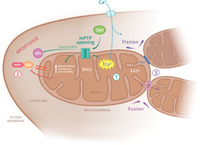
Mitochondria are fundamental for metabolic homeostasis in all multicellular eukaryotes. In the nervous system, mitochondria-generated adenosine triphosphate (ATP) is required to establish appropriate electrochemical gradients and reliable synaptic transmission. Notably, several mitochondrial defects have been identified in central nervous system disorders. Membrane leakage and electrolyte imbalances, pro-apoptotic pathway activation, and mitophagy are among the mechanisms implicated in the pathogenesis of neurodegenerative diseases, such as Alzheimer's, Parkinson's, and Huntington's disease, as well as ischemic stroke. In this review, we summarize mitochondrial pathways that contribute to disease progression. Further, we discuss pathological states that damaged mitochondria impose on normal nervous system processes and explore new therapeutic approaches to mitochondrial diseases.


Conflict of interest statement

M.R.L. owns stocks of eLoupes, Inc., and Cerebrotech Medical Systems, Inc. The rest of the authors declare that there are no competing interests.

Figures

Fig. 1
Fig. 1. Mitochondrial damage contributes to a wide range of pathologies.
The main pathways leading to mitochondria-associated cellular dysfunction include (1) calcium overload in the matrix and mPTP pore opening, (2) cytochrome c release and activation of apoptosis, and (3) defects in mitochondrial fission and fusion affecting mitochondrial longevity. APAF1 apoptotic protease activating factor 1, ATP adenosine triphosphate, CypD cyclophilin D, cytc cytochrome c, Drp1 dynamin-related protein 1, IAPs inhibitor of apoptosis proteins, Mfn mitofusin, mPTP mitochondrial permeability transition pore, OPA optic atrophy mitochondrial fusion protein, ROS reactive oxygen species, Smac/DIABLO pro-apoptotic mitochondrial protein.
Fig. 2
Fig. 2. Stereotactic injection facilitates efficient delivery of mitochondria into the brain parenchyma.
Injected brains were harvested, cryo-sectioned and stained with nuclear stain DAPI (blue). ac Control brain injected with PBS, df Experimental brain injected with exogenous mitochondria labeled with MitoTracker Red CMXRos (Invitrogen). Both mice were injected directly into the striatum and striatal sections were imaged with confocal microscopy. The images were acquired at various distances from the injection site: a, d 200 μm, b, e 400 μm, and c, f 800 μm. Mitochondria were found to diffuse in the parenchyma and detected over 1 mm from the injection site. Scale bar, 25 μm.
Fig. 3
Fig. 3. Intra-arterial injection can deliver mitochondria in the ischemic brain parenchyma.
Mice were stroked with middle cerebral artery occlusion, injected intraarterially and their brains were cryo-sectioned and stained with DAPI (blue). a and b Stroked brain injected with exogenous mitochondria pre-labeled with MitoTracker (red), c Stroked brain injected with PBS. Both animals were injected in the common carotid artery following middle cerebral artery occlusion. Images were acquired in the striatum with confocal microscopy. Mitochondria crossed the blood–brain barrier and localized throughout the parenchyma. Occasionally, mitochondria were found deposited in the blood vessels (white arrow). b Higher magnification of a mitochondrial deposit shown in a. a Scale bar, 50 μm. b, c Scale bar, 25 μm.

Similar articles

Cited by

References

    1. Frey TG, Mannella CA. The internal structure of mitochondria. Trends Biochem. Sci. 2000;25:319–324. - PubMed
    1. Picard M, Taivassalo T, Gouspillou G, Hepple RT. Mitochondria: isolation, structure and function. J. Physiol. 2011;589:4413–4421. - PMC - PubMed
    1. Rose J, et al. Mitochondrial dysfunction in glial cells: Implications for neuronal homeostasis and survival. Toxicology. 2017;391:109–115. - PMC - PubMed
    1. Liu F, Lu J, Manaenko A, Tang J, Hu Q. Mitochondria in ischemic stroke: new insight and implications. Aging Dis. 2018;9:924–937. - PMC - PubMed
    1. Briston T, et al. Mitochondrial permeability transition pore: sensitivity to opening and mechanistic dependence on substrate availability. Sci. Rep. 2017;7:10492. - PMC - PubMed