. 2020 Nov 12;5(1):105.
doi: 10.1038/s41541-020-00255-7.

Mucosal delivery of a multistage subunit vaccine promotes development of lung-resident memory T cells and affords interleukin-17-dependent protection against pulmonary tuberculosis

Affiliations

Mucosal delivery of a multistage subunit vaccine promotes development of lung-resident memory T cells and affords interleukin-17-dependent protection against pulmonary tuberculosis

Claudio Counoupas et al. NPJ Vaccines. .

Abstract

The development of effective vaccines against bacterial lung infections requires the induction of protective, pathogen-specific immune responses without deleterious inflammation within the pulmonary environment. Here, we made use of a polysaccharide-adjuvanted vaccine approach to elicit resident pulmonary T cells to protect against aerosol Mycobacterium tuberculosis infection. Intratracheal administration of the multistage fusion protein CysVac2 and the delta-inulin adjuvant Advax™ (formulated with a TLR9 agonist) provided superior protection against aerosol M. tuberculosis infection in mice, compared to parenteral delivery. Surprisingly, removal of the TLR9 agonist did not impact vaccine protection despite a reduction in cytokine-secreting T cell subsets, particularly CD4+IFN-γ+IL-2+TNF+ multifunctional T cells. CysVac2/Advax-mediated protection was associated with the induction of lung-resident, antigen-specific memory CD4+ T cells that expressed IL-17 and RORγT, the master transcriptional regulator of Th17 differentiation. IL-17 was identified as a key mediator of vaccine efficacy, with blocking of IL-17 during M. tuberculosis challenge reducing phagocyte influx, suppressing priming of pathogen-specific CD4+ T cells in local lymph nodes and ablating vaccine-induced protection. These findings suggest that tuberculosis vaccines such as CysVac2/Advax that are capable of eliciting Th17 lung-resident memory T cells are promising candidates for progression to human trials.


Conflict of interest statement

N.P. is the research director for Vaxine P/L. E.L.S. was an employee of Vaxine P/L. The remaining authors declare that they have no competing interests.

Figures
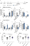
Fig. 1
Fig. 1. Pulmonary vaccination with CysVac2/AdvaxCpG demonstrates improved protection against M. tuberculosis infection compared to parenteral administration.
C57BL/6 mice (n = 5–6) were vaccinated by either the i.m. or i.t. route with CysVac2(CV2)/AdvaxCpG (three times, 2 weeks apart). One week after last vaccination mice were bled for vaccine immunogenicity assessment. Six weeks after last immunization mice were challenged with H37Rv by aerosol (~100 CFU) and 4 weeks later culled to enumerate the bacterial burden and the T cell phenotype in the lung (a). PBMCs from tail blood of vaccinated mice (bd) or cells from lung of infected mice (eg) were restimulated ex vivo with CysVac2, and the production cytokines (IFN-γ, IL-2, IL-17, TNF), or transcription factors (TF; T-bet, RORγT) by CD4+ T cells was determined by flow cytometry using the gating strategy described in Supplementary Fig. 1. Data are represented as the percentage of cytokine-producing or transcription factor-positive CD4+ T cells ± SEM. Bacterial load was assessed in the lungs (h) and in the spleen (i) and presented as log10 of the mean CFU ± SEM. Data are pooled from three independent experiments. The significance of differences between the groups was determined by ANOVA (*p < 0.05; **p < 0.01; ***p < 0.001).
Fig. 2
Fig. 2. CpG is dispensable for protective immunity induced by CysVac2/Advax.
C57BL/6 mice (n = 5–10) were vaccinated by the i.t. route with CysVac2/AdvaxCpG or CysVac2/Advax (three times, 2 weeks apart). Six weeks after last immunization mice were challenged with M. tuberculosis H37Rv by aerosol (~100 CFU) and four weeks later culled to enumerate the bacterial burden and the T cell phenotype in the lung. Cells from the lung of infected mice were restimulated ex vivo with CysVac2 and the production of cytokines (IFN-γ, IL-2, IL-17, TNF (a, b)) or transcription factors (T-bet, RORγT; (c)) by CD4+ T cells was determined by flow cytometry using the gating strategy described in Supplementary Fig. 1. Data are represented as the percentage of cytokine-producing CD4+ T cells ± SEM. Bacterial load was assessed in the lungs (d) and in the spleen (e) and presented as log10 of the mean CFU ± SEM. Data are pooled from two independent experiments. The significance of differences between the groups was determined by ANOVA (*p < 0.05; **p < 0.01; ***p < 0.001).
Fig. 3
Fig. 3. Pulmonary vaccination with CysVac2/Advax induces antigen-specific serum antibodies and the formation of iBalt in the lungs of vaccinated mice.
C57BL/6 mice (n = 3–4) were vaccinated by the i.t. route with CysVac2/Advax or CysVac2 (three times, 2 weeks apart). Before the first and second boost, as well as 2 and 4 weeks after the second boost, mice were bled from the lateral tail vein and serum was collected. Titers of IgG1 (a) and IgG2c (b) were calculated by ELISA over the course of the time points examined. IgA (c) was measured by ELISA in the serum of week 2 samples. Data are represented as the average of log10 titers ± SEM or average 450 nm absorbance ± SEM. Four weeks after the final vaccination, lung sections were stained with anti-mouse B220 (red) and anti-mouse CD3 (green) to visualize iBALT structures and imaged at ×20 magnification (d). Images are representative of three biological replicates/group; scale bars represent 50 microns. The significance of differences between the groups was determined by ANOVA (*p < 0.05).
Fig. 4
Fig. 4. Pulmonary vaccination with CysVac2/Advax induces Ag-specific persistent local resident CD4+ T cells.
C57BL/6 mice (n = 3–4) were vaccinated by the i.t. route with CysVac2/Advax or CysVac2 (three times, 2 weeks apart). At weeks 2, 4, or 8 after the final immunization, lung cells were processed for Ag85B:I-Ab tetramer staining. Representative dot plot from 8 weeks post-immunization of CD4+ T cells gated as outlined in Supplementary Fig. 1 (a). Number of Ag85B:I-Ab tetramer-positive cells in the lung over time are shown in b. Also shown are the percentage of total CD4+ T cells expressing phenotypic makers markers associated with TRMs (CD45 IV, CD11a+, CD69+, CD44+, PD-1+ KLRG-1 (c)) and representative dot plots of TRMs markers on total lung CD4+ T cells (d) or in Ag85B:I-Ab+ cells (e) at 8 weeks after last vaccination. Data are presented as mean ± SEM and are representative of two independent experiments. The significance of differences between the groups was determined by ANOVA (*p < 0.05; **p < 0.01; ***p < 0.001).
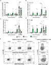
Fig. 5
Fig. 5. Persistent CysVac2-specific IL-17 production by lung-resident CD4+ T cells after pulmonary vaccination with CysVac2/Advax.
C57BL/6 mice (n = 3–5) were vaccinated by the i.t. route with CysVac2/Advax or CysVac2 protein alone (three times, 2 weeks apart). Eight weeks after the last immunization mice were challenged with M. tuberculosis H37Rv by aerosol (~100 CFU). At 2, 4, or 8 weeks after last immunization (solid bars), and at 4 weeks after infection (striped bars), lung cells were restimulated ex vivo with CysVac2, and the production of IFN-γ (a), IL-17 (b), IL-2 (c), or TNF (d) by CD4+ T cells determined by flow cytometry using the gating strategy described in Supplementary Fig. 1. Representative dot plots of co-expression of CD45 IV or RORγT with IL-17 by total CD4+ T cells (e) or Ag85B:I-Ab tetramer-positive cells (f) at 8 weeks after last vaccination. Data are represented as the percentage of cytokine-producing CD4+ T cells ± SEM and are representative of two independent experiments. The significance of differences between the groups was determined by ANOVA (*p < 0.05; **p < 0.01; ***p < 0.001).
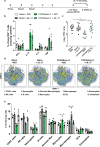
Fig. 6
Fig. 6. Protection afforded by pulmonary CysVac2/Advax against aerosol M. tuberculosis is dependent on IL-17 and correlates with lung phagocytic cell recruitment.
C57BL/6 mice (n = 5–6) were vaccinated by the i.t. route with CysVac2/Advax (three times, 2 weeks apart), and at 8 weeks after the last immunization mice were challenged with M. tuberculosis H37Rv by aerosol (~100 CFU). One day before aerosol, mice were treated i.p. with anti-IL-17 or an isotype control mAb (twice weekly for 3 weeks) (a). Cells from the lungs of infected mice were restimulated ex vivo with CysVac2 and cytokines secretion (IFN-γ, IL-2, IL-17, TNF) determined by flow cytometry using the gating strategy described in Supplementary Fig. 1 (b). Bacterial load was assessed in the lungs and is presented as Log10 of the mean CFU ± SEM (c). Representative tSNE dimensions 1 and 2 plots of the total live cells in the lung (d). Bar graphs show the average of the percentage ± SEM of identified lung cell subsets (e). Data are pooled from two independent experiments. The significance of differences between the groups was determined by ANOVA (*p < 0.05; **p < 0.01; ***p < 0.001).
Fig. 7
Fig. 7. Blocking IL-17 during M. tuberculosis infection impairs the proliferation of pathogen-specific CD4+ T cells in the mediastinal lymph nodes.
C57BL/6 mice (n = 5–6) were vaccinated with CysVac2/Advax and treated i.p. with anti-IL-17 mAb, as described in Fig. 5. Representative dot plot of the expression of Ki67 and RORγT on CD4+ T cells (a). Bar graphs show the average numbers of the total (b) and RORγT+ (c) proliferating CD4+ T cells enumerated in the mLN ± SEM. Representative dot plots show CD44 and either Ag85B:I-Ab or ESAT6:I-Ab staining on CD4+ T cells in the mLN (d), with total number ± SEM of Ag85B:I-Ab+ (e), and ESAT6:I-Ab+ (f) CD4+ T cells in the mLN. Data are pooled from two independent experiments. The significance of differences between the groups was determined by ANOVA (*p < 0.05; **p < 0.01; ***p < 0.001).

Similar articles

Cited by

References

    1. World Health Organisation. Global Tuberculosis Report 2019 (WHO, 2019).
    1. Mangtani P, et al. Protection by BCG vaccine against tuberculosis: a systematic review of randomized controlled trials. Clin. Infect. Dis. 2014;58:470–480. doi: 10.1093/cid/cit790. - DOI - PubMed
    1. Knight GM, et al. Impact and cost-effectiveness of new tuberculosis vaccines in low- and middle-income countries. Proc. Natl Acad. Sci. USA. 2014;111:15520–15525. doi: 10.1073/pnas.1404386111. - DOI - PMC - PubMed
    1. Kaufmann SH, Weiner J, von Reyn CF. Novel approaches to tuberculosis vaccine development. Int J. Infect. Dis. 2017;56:263–267. doi: 10.1016/j.ijid.2016.10.018. - DOI - PubMed
    1. Van Der Meeren O, et al. Phase 2b controlled trial of M72/AS01E vaccine to prevent tuberculosis. N. Engl. J. Med. 2018;379:1621–1634. doi: 10.1056/NEJMoa1803484. - DOI - PMC - PubMed