. 2020 Dec 9;10(1):21555.
doi: 10.1038/s41598-020-78019-5.

Wnt-inducible Lrp6-APEX2 interacting proteins identify ESCRT machinery and Trk-fused gene as components of the Wnt signaling pathway

Affiliations

Wnt-inducible Lrp6-APEX2 interacting proteins identify ESCRT machinery and Trk-fused gene as components of the Wnt signaling pathway

Gabriele Colozza et al. Sci Rep. .

Abstract

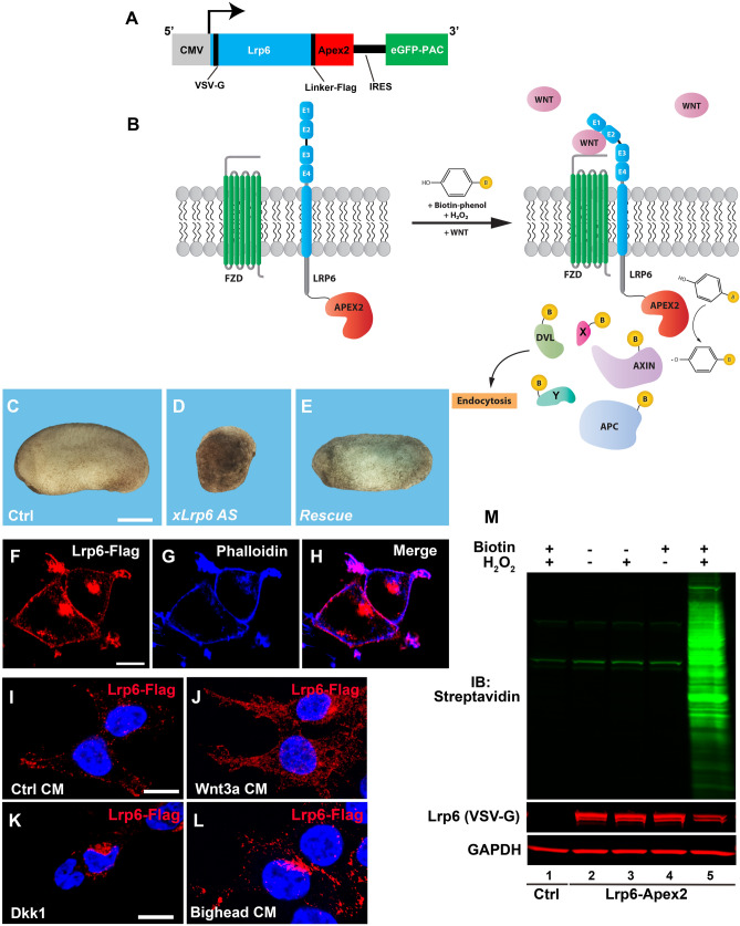
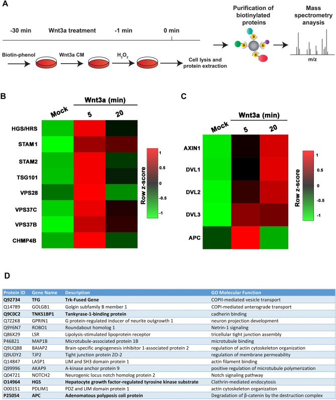
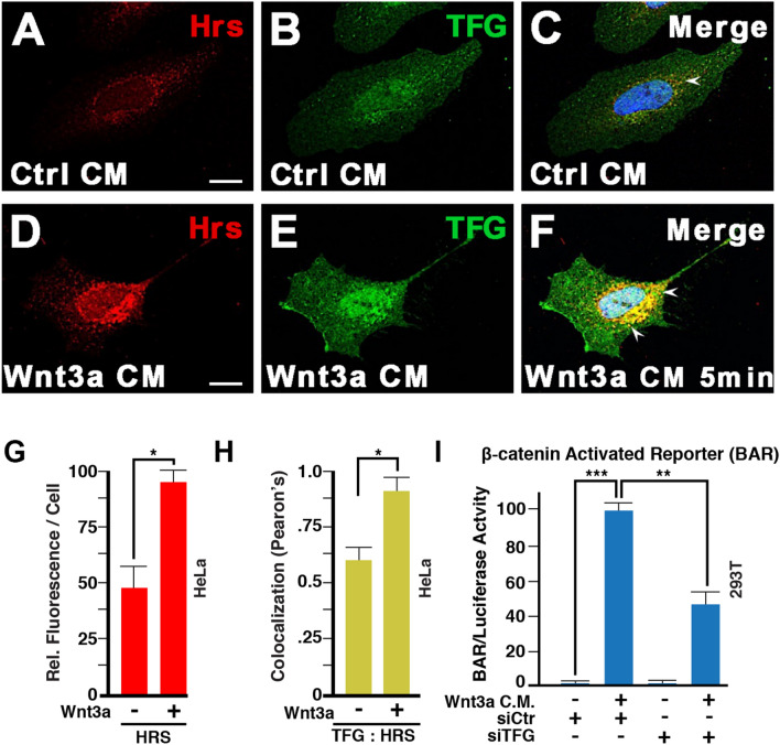
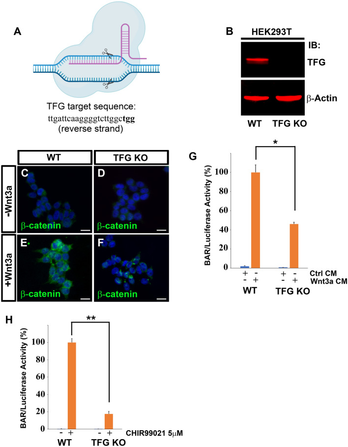
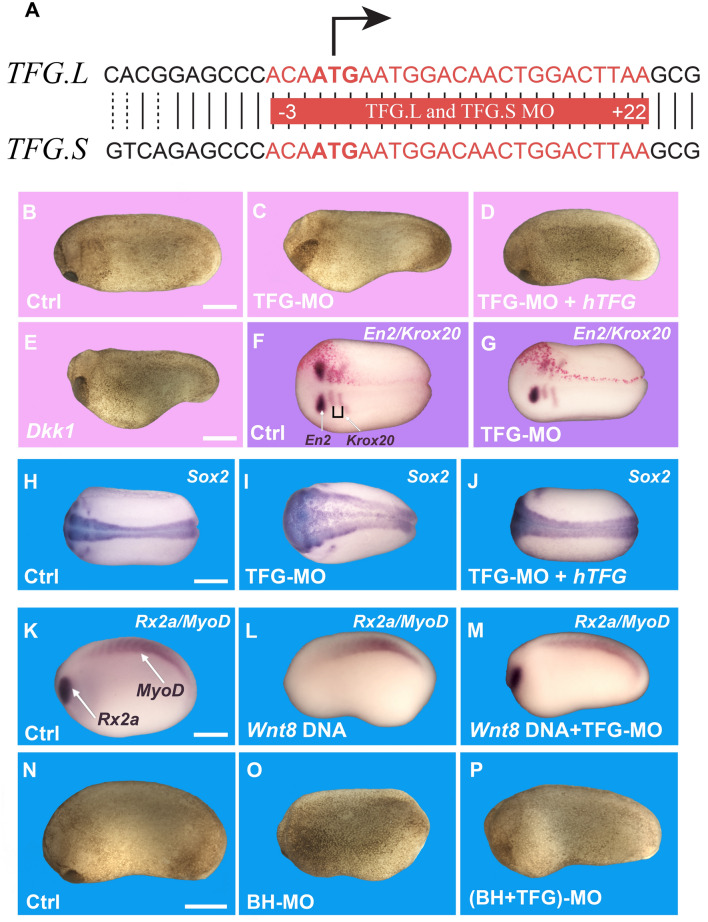
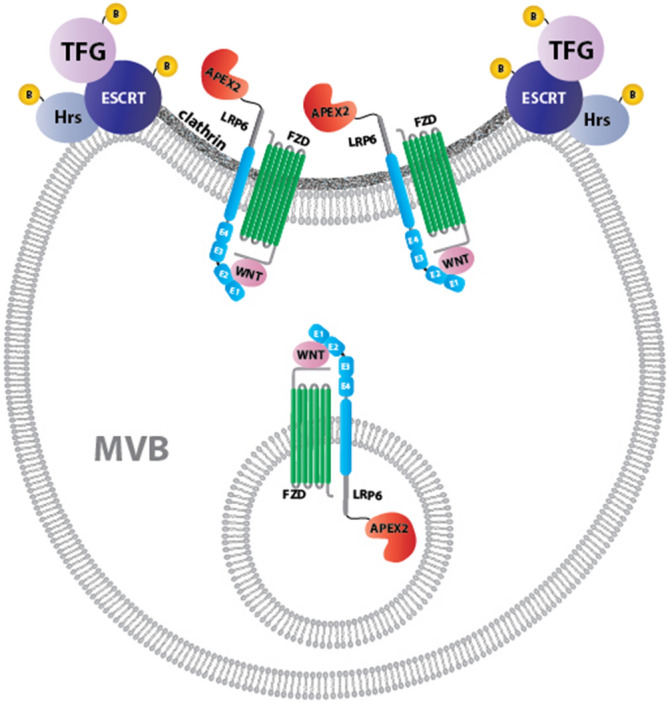
The canonical Wnt pathway serves as a hub connecting diverse cellular processes, including β-catenin signaling, differentiation, growth, protein stability, macropinocytosis, and nutrient acquisition in lysosomes. We have proposed that sequestration of β-catenin destruction complex components in multivesicular bodies (MVBs) is required for sustained canonical Wnt signaling. In this study, we investigated the events that follow activation of the canonical Wnt receptor Lrp6 using an APEX2-mediated proximity labeling approach. The Wnt co-receptor Lrp6 was fused to APEX2 and used to biotinylate targets that are recruited near the receptor during Wnt signaling at different time periods. Lrp6 proximity targets were identified by mass spectrometry, and revealed that many endosomal proteins interacted with Lrp6 within 5 min of Wnt3a treatment. Interestingly, we found that Trk-fused gene (TFG), previously known to regulate the cell secretory pathway and to be rearranged in thyroid and lung cancers, was strongly enriched in the proximity of Lrp6. TFG depletion with siRNA, or knock-out with CRISPR/Cas9, significantly reduced Wnt/β-catenin signaling in cell culture. In vivo, studies in the Xenopus system showed that TFG is required for endogenous Wnt-dependent embryonic patterning. The results suggest that the multivesicular endosomal machinery and the novel player TFG have important roles in Wnt signaling.


Conflict of interest statement

The authors declare no competing interests.

Figures

Figure 1
Figure 1
The Lrp6-APEX2 fusion protein receptor retains signal transducing activity, proper subcellular localization, and biotinylates cellular proteins. (A) Schematic diagram of the Lrp6-APEX2 construct. The human Lrp6 contains an N-terminal VSV-G and a linker Flag-tag followed by the APEX2 enzyme. The construct also contains an internal ribosomal entry site (IRES) followed by a GFP-tagged Puromycin N-acetyltransferase (PAC) used for selection of permanently transfected cells. (B) Diagram depicting the biotinylation of Lrp6-APEX2 proximal targets. During the Wnt signal, in the presence of Biotin-phenol (BP) and H2O2, proteins recruited to the receptor are biotinylated by the APEX2 peroxidase, including known Wnt targets (Dvl, Axin, APC, etc.) and novel targets (X, Y). (C–E) control embryos from oocyte-host transfer experiments show normal development (100%, n = 24), as compared to oocytes depleted with a phosphorothioate antisense DNA oligo targeting Xenopus Lrp6, which develop as ventralized embryos (75%, n = 22). Co-injection of 300 pg of Lrp6-APEX2 mRNA into oocytes completely rescued axis formation (60%, n = 17), indicating that the fusion protein is fully active. Scale bar represents 500 µm. (F–H) Lrp6-APEX2 is trafficked to the plasma membrane in stably transfected HEK293T cells. Lrp6 was detected through its Flag-tag and Phalloidin, which stains cortical F-Actin, confirmed cell surface localization of Lrp6. Scale bar represents 10 µm. (IL) Lrp6-APEX2 changes subcellular localization by Wnt, Dkk1 and Bighead treatments. In presence of control conditioned medium (CM), Lrp6 is located mostly at the plasma membrane (79%). Treatment with Wnt3a CM for 30 min increases the number of intracellular vesicles containing Lrp6 (53%). Treatment with Dkk1 protein (200 ng/ml) or Bighead CM induced relocation of Lrp6 to the juxtanuclear bay area where lysosomes are located (63% and 53%, respectively). Quantification was obtained counting cells showing the observed immunostaining over total nuclei, in 10 different micrographs. Scale bars represent 10 µm. (M) western blot stained with Streptavidin-IRDye 800 (in green) showing controls indicating that Lrp6-APEX2 biotinylates proximal proteins only in the presence of both BP and H2O2 (compare lane 5 with 2–4). Negative control cells not expressing the APEX2 peroxidase had no biotinylation even in presence of BP/H2O2 (lane 1). Note the presence of three Streptavidin-positive bands in all lanes, which correspond to endogenous biotinylated carboxylases. Gapdh served as a loading control.
Figure 2
Figure 2
Analysis of Lrp6-APEX2 proximal targets reveal rapid Wnt3a-induced interaction between Lrp6 and ESCRT proteins. (A) Schematic of the biotinylation proteomic experiments reported in this study. (B) Heatmap of Lrp6-APEX2 proximity labeled ESCRT proteins using normalized intensities at 5 and 20 min time points, showing the horizontal row z-scores of proteins over time. Mock indicates cells treated with control conditioned medium from cells not expressing Wnt3a. Row z-scores were calculated from average intensities of proteins. Average intensity values were obtained from biological duplicates. Highly biotinylated proteins are shown in red and lower ones in green. Note the enrichment in ESCRT proteins particularly after 5 min of Wnt3a treatment. (C) Heatmap of Lrp6-APEX2 proximity labeled known canonical Wnt signaling pathway target proteins using normalized intensities at 5- and 20-min time points, showing the horizontal row z-scores of proteins over time. Average intensity values were obtained from biological duplicates. Highly biotinylated proteins are shown in red and lower ones in green. Note the interaction between Lrp6-APEX2 and Dvl 1–3 and Axin proteins after 20 min of Wnt3a treatment while APC peaks at 5 min; these interactions support the specificity of our Wnt3a signaling experiments. (D) The top 15 biotinylated targets (ranked according to their spectral counts) from Supplementary Table S1 Tab 1. Note the presence of the ESCRT-0 protein Hrs/Hgs, the Wnt inhibitor APC and Actin remodeling proteins such as BAIAP2, LASP1 and PDLIM1. Unexpectedly, the Trk-fused gene (TFG) was the most enriched protein in this list.
Figure 3
Figure 3
TFG co-localizes with the ESCRT-0 protein Hrs/Hgs and is required for Wnt signaling. (AF) Immunostaining on HeLa cells for endogenous Hrs and TFG. Weak co-localization was observed in absence of Wnt signaling (arrow in panel C). However, 5 min of Wnt3a treatment was sufficient to strikingly increase the number of endocytic vesicles containing both Hrs and TFG (arrows). Scale bar 10 µm. (G) Wnt signaling (5 min) increased the number of Hrs positive vesicles quantified as fluorescence per cell; this supports the rapid formation of MVBs in Wnt signaling. (H) Wnt3a increased co-localization of TFG puncta with the Hrs MVB marker by Pearson’s correlation coefficient. (I) TFG knock-down by siRNA reduced Wnt signaling, as assessed by β-catenin Activated Reporter (BAR/Renilla) Luciferase assay in HEK293T cells.
Figure 4
Figure 4
CRISPR-Cas9 mediated TFG knock-out inhibits Wnt-dependent β-catenin stabilization and Wnt-induced reporter activity. (A) Schematic diagram of Cas9 cleaving the TFG genomic DNA target sequence. The target sequence (which is on the reverse strand) is shown in 5′–3′ orientation (left–right); the PAM (protospacer adjacent motif) sequence is in bold. (B) Western blot of HEK293T cells confirming complete elimination of TFG protein in Cas9 knock-out cells. β-actin was used as loading control. (CF) Immunostaining for β-catenin on WT or TFG KO cells. Cells were treated with control conditioned medium or with Wnt3a conditioned medium for 3 h before immunostaining. Note that TFG knock-out decreases Wnt3a-induced β-catenin accumulation; DAPI was used for nuclear counter-staining. Scale bars represent 20 µm. (G) Cas9-mediated TFG knock-out reduces response to Wnt3a by 50% in HEK293T BAR/Renilla β-catenin reporter cells. Cells were treated with Wnt3a or control medium for 16 h before luciferase analysis. (H) Luciferase assay of HEK293T BAR reporter cells treated with the GSK3 inhibitor CHIR99021 at 5 µM concentration for 16 h before being processed for luciferase assay. Note that TFG KO causes an 80% reduction of β-catenin luciferase activity in response to the GSK3 inhibition. Error bars represent standard deviation from triplicate experiments. Statistical significance was calculated with a paired 2-tailed t-Student test. *P < 0.05; **P < 0.01.
Figure 5
Figure 5
TFG regulates antero-posterior (A-P) patterning in the Xenopus embryo and is required for Wnt activity. (A) Diagram showing the TFG morpholino (MO) target sequence (in red), which is the same for both Xenopus L and S homeologs and span the ATG initiation codon. (B) Stage 24 embryos injected with control morpholino (Co-MO), showing normal development (92%, n = 127). (C) Embryos injected with 36 ng of TFG-MO showed severe enlargement of the head region (65%, n = 196). (D) Normal antero-posterior development was rescued by co-injecting 400 pg/embryo of human Flag-tagged TFG mRNA (77%, n = 52). (E) Embryos injected with 50 pg of mRNA encoding the Wnt antagonist Dkk1 showed head enlargement (100%, n = 35). (F,G) Embryos injected unilaterally with 18 ng of Co-MO or TFG-MO, together with 200 pg of nuclear LacZ mRNA, processed for in situ hybridization for Engrailed-2 (En2) and Krox20. LacZ lineage tracing (in red) shows the injected side. TFG-MO reduces En2 expression and shifts Krox20 posteriorly in 65% of the embryos (n = 20), compared to control MO (n = 33). (H,I) In situ hybridization for the pan-neural marker Sox2 at stage 20 showing enlargement of the neural plate following injection 4 times at the 2-cell stage with a total of 72 ng of TFG-MO per embryo (70%, n = 65), compared to the Ctrl-MO embryos (n = 35). (J) Co-injection with 800 pg of hTFG mRNA rescued the neural plate expansion (78%, n = 23). (K,M) Control embryos at stage 22 showed normal eye (Rx2a) and somite (MyoD) development (n = 18), while embryos injected with 32 pg of xWnt8 DNA together with 72 ng of scrambled Ctrl-MO showed reduced or no Rx2a expression, but maintained MyoD expression (66%, n = 18). However, embryos injected with the same amount of Wnt8 DNA together with 72 ng of TFG-MO showed rescue of Rx2a expression (84%, n = 32), indicating that TFG is required for xWnt8 signaling. (NP) Embryos injected with Bighead morpholino (BH-MO) showed reduced head and cement gland development (71%, n = 21), compared to control embryos (n = 37), due to an increase in endogenous Wnt signaling (31). Co-injecting TFG-MO rescued normal head development in Bighead MO embryos (90%). Scale bars represent 500 µm.
Figure 6
Figure 6
Model of how Lrp6-APEX2 may interact with the ESCRT machinery and TFG in multivesicular body formation during Wnt-induced endocytosis. The endocytosed Wnt receptor complex on the surface of multivesicular endosomes becomes clustered in clathrin-containing invaginations that give rise to the intraluminal vesicles (ILVs) of late endosomes. MVBs are the obligatory pathway through which plasma membrane proteins reach the lysosome. We found that most ESCRT GO components are brought into proximity of Lrp6-APEX2 5 min after treatment with Wnt3a conditioned medium. The top interactor with Lrp6-APEX2 after 20 min of Wnt3a was TFG, which we show here is required for Wnt signaling. TFG protein co-localizes with the ESCRT-0 protein Hrs/Hgs after Wnt stimulation. We did not detect interactions with intraluminal vesicle components such as CD63; a possible explanation is that the acidic medium or lysosomal proteases might interfere with APEX2 activity.

References

    1. Nusse R, Clevers H. Wnt/β-Catenin signaling, disease, and emerging therapeutic modalities. Cell. 2017;169:985–999. doi: 10.1016/j.cell.2017.05.016. - DOI - PubMed
    1. Clevers H, Nusse R. Wnt/β-Catenin signaling and disease. Cell. 2012;149:1192–1205. doi: 10.1016/j.cell.2012.05.012. - DOI - PubMed
    1. MacDonald BT, Tamai K, He X. Wnt/beta-catenin signaling: components, mechanisms, and diseases. Dev. Cell. 2009;17:9–26. doi: 10.1016/j.devcel.2009.06.016. - DOI - PMC - PubMed
    1. Del Valle-Pérez B, Arqués O, Vinyoles M, de Herreros AG, Duñach M. Coordinated action of CK1 isoforms in canonical Wnt signaling. Mol. Cell Biol. 2011;14:2877–2888. doi: 10.1128/MCB.01466-10. - DOI - PMC - PubMed
    1. Bilic J, et al. Wnt induces LRP6 signalosomes and promotes dishevelled-dependent LRP6 phosphorylation. Science. 2007;316:1619–1622. doi: 10.1126/science.1137065. - DOI - PubMed

Publication types

MeSH terms

Substances