. 2021 Apr;34(4):748-757.
doi: 10.1038/s41379-020-00719-0. Epub 2020 Dec 9.

Immunohistochemical detection of PAX-FOXO1 fusion proteins in alveolar rhabdomyosarcoma using breakpoint specific monoclonal antibodies

Affiliations

Immunohistochemical detection of PAX-FOXO1 fusion proteins in alveolar rhabdomyosarcoma using breakpoint specific monoclonal antibodies

David O Azorsa et al. Mod Pathol. 2021 Apr.

Abstract

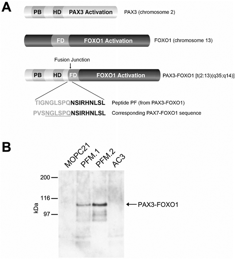
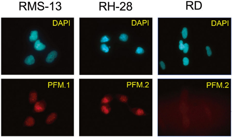
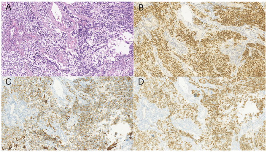
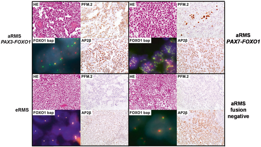
Alveolar Rhabdomyosarcoma (ARMS) is an aggressive pediatric cancer with about 80% of cases characterized by either a t(1;13)(p36;q14) or t(2;13)(q35;q14), which results in the formation of the fusion oncogenes PAX7-FOXO1 and PAX3-FOXO1, respectively. Since patients with fusion-positive ARMS (FP-RMS) have a poor prognosis and are treated with an aggressive therapeutic regimen, correct classification is of clinical importance. Detection of the translocation by different molecular methods is used for diagnostics, including fluorescence in situ hybridization and RT-PCR or NGS based approaches. Since these methods are complex and time consuming, we developed specific monoclonal antibodies (mAbs) directed to the junction region on the PAX3-FOXO1 fusion protein. Two mAbs, PFM.1 and PFM.2, were developed and able to immunoprecipitate in vitro-translated PAX3-FOXO1 and cellular PAX3-FOXO1 from FP-RMS cells. Furthermore, the mAbs recognized a 105 kDa band in PAX3-FOXO1-transfected cells and in FP-RMS cell lines. The mAbs did not recognize proteins in fusion-negative embryonal rhabdomyosarcoma cell lines, nor did they recognize PAX3 or FOXO1 alone when compared to anti-PAX3 and anti-FOXO1 antibodies. We next evaluated the ability of mAb PFM.2 to detect the fusion protein by immunohistochemistry. Both PAX3-FOXO1 and PAX7-FOXO1 were detected in HEK293 cells transfected with the corresponding cDNAs. Subsequently, we stained 26 primary tumor sections and a rhabdomyosarcoma tissue array and detected both fusion proteins with a positive predictive value of 100%, negative predictive value of 98%, specificity of 100% and a sensitivity of 91%. While tumors are stained homogenously in PAX3-FOXO1 cases, the staining pattern is heterogenous with scattered positive cells only in tumors expressing PAX7-FOXO1. No staining was observed in stromal cells, embryonal rhabdomyosarcoma, and fusion-negative rhabdomyosarcoma. These results demonstrate that mAbs specific for the chimeric oncoproteins PAX3-FOXO1 and PAX7-FOXO1 can be used efficiently for simple and fast subclassification of rhabdomyosarcoma in routine diagnostics via immunohistochemical detection.


Conflict of interest statement

Conflict of interest The authors declare that they have no conflict of interest.

Figures

Fig. 1
Fig. 1. Development of monoclonal antibodies to the chimeric protein PAX3-FOXO1.
A The transcription factor PAX3 contains a paired boxed domain (PB), homeodomain-DNA-binding domain (HD) and a PAX3 transactivation domain. FOXO1 contains a forkhead DNA-binding domain (FD) and a transactivation domain. The sequence of peptide PF1 spanning the fusion junction site is shown with the PAX3 amino acids in gray and the FOXO1 amino acids in black. The corresponding PAX7-FOXO1 sequence is shown with identical amino acids underlined. Adapted from [24, 25]. B Recognition of PAX3-FOXO1 by anti-PAX3-FOXO1 peptide mAbs. PAX3-FOXO1 was immunoprecipitated specifically from cellular lysate from the FP-RMS cell line RH-30 by anti-PAX3-FOXO1 mAbs PFM.1 (lane 2) and PFM.2 (lane 3), but not by IgG1 control mAb MOPC21 (lane 1) or by anti-NCOA3 peptide mAb AC3 (lane4). Immunoprecipitated proteins were separated by SDS-PAGE, transferred to nylon membranes and detected using anti-FOXO1 antibodies.
Fig. 2
Fig. 2. Specificity of anti-PAX3-FOXO1 peptide mAbs.
A Antibody reactivity of mAb PFM.2 was compared to anti-PAX3 and anti-FOXO1 Abs by western blot analysis of NIH-3T3 cells stably-transfected with vector alone (NIL.C), PAX3 (PAX3.1), or PAX3-FOXO1 (PF.1). MAb PFM.2 recognizes only PAX3-FOXO1 and not PAX3 in NIH-3T3. Immunoblots were re-probed with an anti-tubulin antibody as a loading control. B Expression of PAX3-FOXO1 in rhabdomyosarcoma cell lines. MAbs PFM.1 and PFM.2 recognize PAX3-FOXO1 on FP-RMS cell lines RH-4, RH-28, RH-30, and RMS-13 but not on FN-RMS cell lines RD and CTR. Immunoblots were re-probed with an anti-tubulin antibody as a loading control.
Fig. 3
Fig. 3. Nuclear localization of PAX3-FOXO1 in FP-RMS cells.
FP-RMS cell lines RMS-13 and RH-28, and the FN-RMS cell line RD were incubated with mAbs PFM.1 or PFM.2. Antibody binding was visualized using a Cy3-goat anti-mouse antibody and DAPI was used to visualize the nucleus.
Fig. 4
Fig. 4. Immunofluorescence detection of PAX-FOXO1 fusion proteins.
293T cells transfected with indicated wild-type or fusion proteins were stained with mAb PFM.2 or antibodies directed against PAX3 or FOXO1. Scale bar, 100 μm.
Fig. 5
Fig. 5. Detection of PAX3-FOXO1-positive tumors by immunohistochemistry.
PAX3-FOXO1 was detected in a tumor sample from a patient with alveolar rhabdomyosarcoma containing the t(2;13) using mAb PFM.2. Sections were stained with A Hematoxylin and Eosin, B PFM.2, C desmin, and D myogenin.
Fig. 6
Fig. 6. Immunohistochemical staining and FOXO1-FISH of rhabdomyosarcoma tumor sections.
Tissue sections from the four depicted tumors were stained with Hematoxylin and Eosin (upper left picture), with the mAb PFM.2 (upper right picture), an antibody directed against AP2β (lower right picture) and by FISH using a FOXO1 break-apart probe (bap) (lower left picture). The depicted tumors are positive for PAX3-FOXO1 (upper left case), PAX7-FOXO1 (upper right case), or negative for known fusion proteins in poorly differentiated ERMS (lower left case) and translocation negative ARMS (lower right case).

References

    1. Pappo AS. Rhabdomyosarcoma and other soft tissue sarcomas in children. Curr Opin Oncol. 1996;8:311–6. - PubMed
    1. WHO Classification of Tumours Editorial Board. Soft tissue and bone tumours: WHO classification of tumours. 5th ed. Lyon: IARC Press; 2020.
    1. Shern JF, Chen L, Chmielecki J, Wei JS, Patidar R, Rosenberg M, et al. Comprehensive genomic analysis of rhabdomyosarcoma reveals a landscape of alterations affecting a common genetic axis in fusion-positive and fusion-negative tumors. Cancer Discov. 2014;4:216–31. - PMC - PubMed
    1. Barr FG, Galili N, Holick J, Biegel JA, Rovera G, Emanuel BS. Rearrangement of the PAX3 paired box gene in the paediatric solid tumour alveolar rhabdomyosarcoma. Nat Genet. 1993;3:113–7. - PubMed
    1. Chen X, Stewart E, Shelat AA, Qu C, Bahrami A, Hatley M, et al. Targeting oxidative stress in embryonal rhabdomyosarcoma. Cancer Cell. 2013;24:710–24. - PMC - PubMed

Publication types

MeSH terms

Substances