. 2021 Jan;589(7842):437-441.
doi: 10.1038/s41586-020-3018-x. Epub 2020 Dec 9.

Arterialization requires the timely suppression of cell growth

Affiliations

Arterialization requires the timely suppression of cell growth

Wen Luo et al. Nature. 2021 Jan.

Abstract

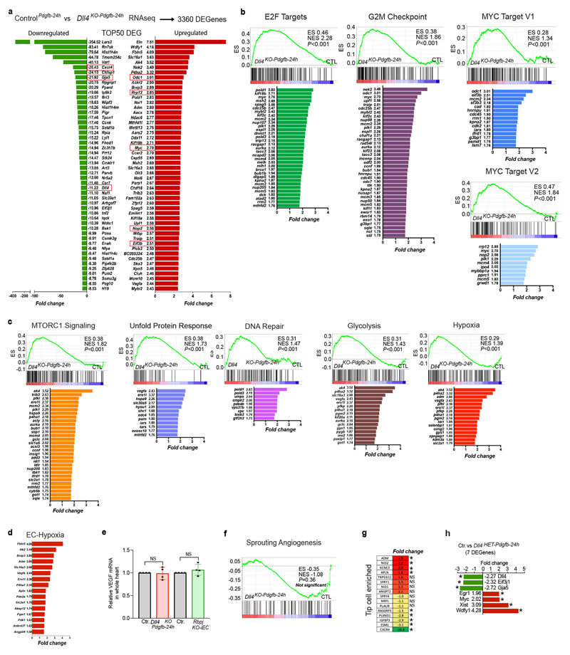
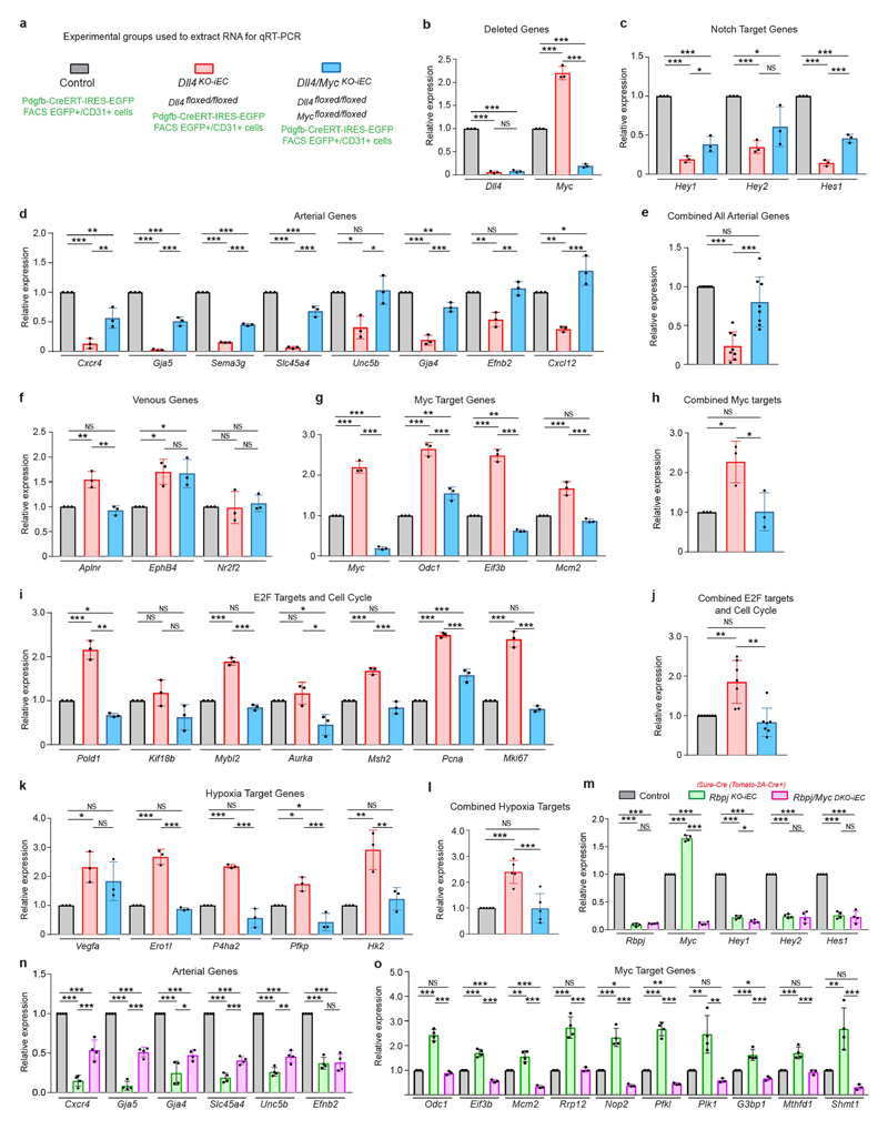
The formation of arteries is thought to occur by the induction of a highly conserved arterial genetic programme in a subset of vessels that will later experience an increase in oxygenated blood flow1,2. The initial steps of arterial specification require both the VEGF and Notch signalling pathways3-5. Here, we combine inducible genetic mosaics and transcriptomics to modulate and define the function of these signalling pathways in cell proliferation, arteriovenous differentiation and mobilization. We show that endothelial cells with high levels of VEGF or Notch signalling are intrinsically biased to mobilize and form arteries; however, they are not genetically pre-determined, and can also form veins. Mechanistically, we found that increased levels of VEGF and Notch signalling in pre-arterial capillaries suppresses MYC-dependent metabolic and cell-cycle activities, and promotes the incorporation of endothelial cells into arteries. Mosaic lineage-tracing studies showed that endothelial cells that lack the Notch-RBPJ transcriptional activator complex rarely form arteries; however, these cells regained the ability to form arteries when the function of MYC was suppressed. Thus, the development of arteries does not require the direct induction of a Notch-dependent arterial differentiation programme, but instead depends on the timely suppression of endothelial cell-cycle progression and metabolism, a process that precedes arterial mobilization and complete differentiation.


Conflict of interest statement

Competing interests The authors declare no competing interests.

Figures

Extended Data Fig. 1
Extended Data Fig. 1. Multispectral genetic mosaics to analyse the proliferation and arteriovenous fate of heart and retina ECs.
a, Schematic of Dual ifgMosaic technology to multispectrally barcode cells with different combinations of chromatin (iChr2-Mosaic) or membrane (iMb2-Mosaic) tagged fluorescent proteins. b, Combined or individual channels of multispectral confocal imaging of the heart-surface coronary vasculature and sections. c, iChr-Notch-Mosaic allele targeted to the Rosa26 locus. In cells expressing Cre, the allele undergoes stochastic recombination, generating a mosaic of cells expressing either H2B-Cherry (control wild-type cells with Cherry FP bound to chromatin/nuclei), H2B-GFP-2A-DN-MAML1 (GFP labels the nuclei of cells with induced downregulation of Notch/Rbpj signalling) or HA-H2B-Cerulean-2A-N1ICDP (Cerulean containing the HA epitope labels the nuclei of cells with induced upregulation of NOTCH1 signalling). d, qRT–PCR analysis of Notch target-genes in H2B-GFP+ and H2B-Cherry+ ECs obtained from iChr-NotchTie2-Mosaic E15.5 hearts by FACS. e, Survival rate of adult iChr-NotchTie2-Mosaic mice follows expected Mendelian frequencies indicating no lethality. f, g, Confocal images of iChr-NotchNfatc1-Mosaic embryonic hearts sections showing the localization and relative frequency of ECs with distinct Notch signalling levels. h, Quantification of experiments in f and g showing how the relative percentage of each type of cell changes throughout development according to their location. i, Proliferation (EdU+) of coronary ECs (ICAM2+/ERG+ in myocardium) and endocardium (EMCN+/ERG+) at E12.5 and E15.5. j, k, Analysis of cell proliferation in iChr-NotchTie2-Mosaic ECs. l, Illustration showing the iFlpMTomato-cre/MYfp mosaic allele and expected outcomes in cells expressing Flp/FlpO recombinase. m, qRT–PCR analysis of Rbpj mRNA levels in CD31+/MbYFP+ and CD31+/MbTomato+ ECs collected by FACS from Apln-FlpO;iFlpMTomato-cre/MYfp; Rbpjf/f E15.5 hearts. Residual Rbpj expression is due to the FACS and qRT–PCR assay as previously described (see Methods). n, Confocal images and quantification of postnatal day 6 (P6) retinas showing the localization and frequency of MTomato+ (RbpjKO) and MYFP+ (wild-type) cells in retina arteries (A), veins (V) and intermediate capillaries. Data shown as mean ± s.d. *P < 0.05, **P < 0.01, ***P < 0.001. Scale bars, 100 μm. For statistics, see Supplementary Data 1.
Extended Data Fig. 2
Extended Data Fig. 2. Ectopic Notch activation is compatible with vein development and impairs coronary artery development.
a, Genetic construct used to generate mice with Cre-dependent conditional expression of MbTomato, H2B-GFP and N1ICDP in Tie2-Cre + coronary vessels which results in increased expression of canonical Notch target genes. b, Bright-field images of E12.5 and E15.5 embryos and hearts showing the appearance of microcirculatory defects at E15.5. c, Statistical analysis of mouse survival rate per stage indicates that most mutant mice do not pass embryonic stage E16.5. d, Cardiovascular development defects in N1ICDPTie2-cre hearts may result in tissue hypoxia and the observed upregulation of Vegf mRNA. e, f, Myocardial EC proliferation analysis. g, h, CD31 staining of control and N1ICDPTie2-cre heart sections showing vascular defects and thinner myocardial wall (inset) in mutant hearts. i–m, Analysis of the heart surface coronary vein diameter and plexus development and density. n, Confocal images showing immunostaining of the heart surface venous plexus for the arterial marker CX40. ECs with high Notch activity (Tomato+) do not express CX40 and are able to form veins. o, Confocal images of E14.5 heart veins showing that Tomato/N1ICDP+ cells (yellow arrowheads) express EPHB4 like wild-type cells (white arrowheads). p, Confocal images of E14.5 heart veins showing that GFP+/N1ICDP+ cells (yellow arrowheads) express COUP-TFII like GFP-/wild-type cells (white arrowheads). q, Chart showing the quantification of COUP-TFII immunosignals intensity in 280GFP+and 280GFP- cells present in the veins of 3 independent N1ICDPTie2-cre hearts (2 veins per heart were quantified). r, Schematic of the mouse postnatal retina vascular development, in which tamoxifen was induced at P1, when the vessels start to grow, and analysis was carried at P6. sv, Cells with induced high Notch activity (MbTomato+or H2B-GFP+, yellow arrowheads) can differentiate and form veins like wild-type cells (white arrowheads). The Tomato+/N1ICDP+ cells found in veins do not express the arterial marker CX40, and instead express the venous markers VEGFR3, COUP-TFII and EPHB4. w, Quantification of signals (in pictures like shown in sv) indicate no change in the expression of arteriovenous genes. x, Analysis of coronary artery development. y, z, Confocal images of heart sections and quantifications of immunosignals in the myocardium showing an increase in CX40, DLL4 and SM22α in N1ICDPTie2-cre myocardium vessels. Data shown as mean ± s.d. *P < 0.05, **P < 0.01, ***P < 0.001. Scale bars, 100 μm. For statistics, see Supplementary Data 1.
Extended Data Fig. 3
Extended Data Fig. 3. Overactivation of Notch in PDGFB+ coronary ECs, but not in NFATC1+ endocardium, compromises coronary vessel and heart development.
a, Schematic of the inducible Notch gain-of-function allele, that once crossed with the Pdgfb-icreERT2-ires-egfp allele enables the co-expression of Mbtomato, H2B-GFP and N1ICDP in PDGFB+ coronary ECs. b, Confocal picture showing the strong expression of the Pdgfb-icreERT2-ires-egfp allele in coronary vessels of the myocardium (m), but not in the EMCN+ endocardium (e). c, Stereomicroscope analysis of mutant embryos and control littermates showing growth retardation at E16.5. d, Immunostaining for the reporter MbTomato shows the induction of the allele in Pdgfb-icreERT2-ires-egfp+ myocardium vessels (ICAM2+) but not endocardium. e, g, Thinner myocardial wall and vascular defects in N1ICDPPdgfb-creERT hearts. f, h, Malformation and reduced coronary arterial diameter in N1ICDPPdgfb-creERT hearts. ik, Reduced coronary vessel density and vein diameter in N1ICDPPdgfb-creERT hearts. l, m, Expression of the arterial proteins DLL4 and CX40 in control and N1ICDPPdgfb-creERT coronary vessels. n, o, Reduced proliferation (ERG+/EdU+) of N1ICDPPdgfb-creERT subepicardium venous vessels (whole-mount and sections). p, Induction of N1ICDP expression (MbTomato+) in the NFATC1+ endocardium and its lineages does not cause major cardiovascular development defects at E15.5. qt, Comparative analysis of coronary artery and vein development in control and N1ICDPNfatc1-cre hearts. Note the absence of GFP+/N1ICDP+ cells in veins derived from Nfatc1-cre lineage in N1ICDPNfatc1-cre mice. u, Haematoxylin and eosin staining of 7-month-old hearts of control and N1ICDPNfatc1-cre mice. Mutant hearts are larger. Quantifications of heart weight per body weight and left ventricle (LV) fraction shortening are shown. Data shown as mean ± s.d. *P < 0.05, **P < 0.01, ***P < 0.001. Scale bars, 100 μm. For statistics, see Supplementary Data 1.
Extended Data Fig. 4
Extended Data Fig. 4. Induction of cell-cycle and metabolic pathways in coronary ECs 24 h after Dll4 deletion.
a, List of the 50 most up- and downregulated genes (from a total of 3,360) after Dll4 deletion in coronary ECs for 24 h. b, c, List of dysregulated genes within a selected enriched gene set (gene set enrichment analysis, GSEA). d, Fold changes for a selected list of genes previously shown to be upregulated when HUVECs are exposed to hypoxia,. e, Vegf mRNA levels do not change in hearts from Dll4KO-Pdgfb-24h (E14.5) and RbpjKO-iEC (E15.5) embryos, suggesting the absence of cardiac hypoxia, despite the existence of endothelial hypoxia. f, The group of genes belonging to the sprouting angiogenesis GSEA pathway are not differentially regulated. g, The expression of most genes previously found to be upregulated in tip cells is not increased after the loss of DLL4–NOTCH signalling. h, Only 7 genes are significantly dysregulated in Dll4Het-Pdgfb-24h samples. Data shown as mean ± s.d. *P < 0.05, Benjamini and Hochberg adjusted.
Extended Data Fig. 5
Extended Data Fig. 5. DLL4 signalling dose is essential for coronary proliferation and arterialization but not sprouting or connection to the aorta.
a, Dll4 deletion in Pdgfb-icreERT2-ires-egfp + coronary ECs for 24 h (from E11.5–E12.5 or E13.5–E14.5) does not induce endothelial sprouting, but slightly reduces the mean density of heart subepicardial vessels at E14.5. be, The coronary plexus forming the coronary artery connects (yellow arrows) to the main aorta of control, Dll4Het-iEC and Dll4KO-iEC hearts. However, despite no defects in the connection, the calibre of the coronary artery stem is reduced and most Dll4Het-iEC hearts and all Dll4KO-iEC hearts do not develop proper coronary arteries with CX40+signals (pink arrows) at E14.5. f, At E17.5, the surviving Dll4Het-iEC embryos also show a major developmental defect or delay in coronary artery development. Of note, the DLL4 heterozygous haploinsufficiency is variable and more pronounced on the C57BL/6J background used in this study, as previously reported,. g, h, Dll4 and Rbpj deletion reduces the frequency of EdU+ ECs in subepicardium vessels. i, Dll4 deletion increases p-ERK activity in subepicardium vessels. j, k, Dll4 and Rbpj deletion increases the frequency of p21+/ERG+ ECs in subepicardium vessels. l, m, Dll4 and Rbpj deletion increases the frequency of proliferation (EdU+/ERG+) in arterial zone coronary vessels. Data shown as mean ± s.d. *P < 0.05, **P < 0.01, ***P < 0.001. Scale bars, 100 μm. For statistics, see Supplementary Data 1.
Extended Data Fig. 6
Extended Data Fig. 6. Regulation of MYC by DLL4–NOTCH signalling in vivo and in vitro.
a, MYC–GFP fusion protein expression is upregulated in coronary vessels (ICAM2+) after inducing heterozygous Dll4 deletion. b, Comparison of MYC protein expression in wild-type and Rbpj mutant ERG+ ECs. c, Western blot analysis showing no major changes in MYC protein levels after stimulation of HUVECs with DLL4 ligands for 6 or 24 h, in the presence or absence of the Notch inhibitor DBZ. d, qRT–PCR analysis showing significant changes of Notch target genes (Dll4, Hey1, Hey2, Hes1), but not Myc and Odc1, after stimulation of HUVECs with DLL4 ligands for 8 h in the presence or absence of the Notch inhibitor DBZ. eh, Western blot and qRT–PCR analysis showing that an even stronger activation of Notch signalling (for 24 h, e and f; and 48 h g and h) and its downstream target genes (HEY1,HEY2,NRARP and HES1) in HUVECs overexpressing Dox-inducible N1ICD-V5 has relatively minor or non-significant effects on MYC expression. ODC1 expression is 40% decreased. i, Whole-mount analysis of control and MYC mutant hearts showing that global MYC deletion compromises the development of coronary vessels and consequently arteries (CX40+).j, Sectional analysis of control and MYC mutant hearts showing a severe depletion of coronary vessels in the myocardium which causes a reduction in its thickness. k, Mosaic induction of Mycflox/flox Cdh5-creERT2 iSuRe-cre mice shows that MYC-null ECs (MbTomato-2A-Cre+) form well coronary arteries, in contrast to hearts with full induction of Myc deletion. Data shown as mean ± s.d. *P < 0.05, **P < 0.01, ***P < 0.001. Scale bars, 100 μm. For statistics, see Supplementary Data 1. For western blot gel source data, see Supplementary Fig. 1. Western blot controls (tubulin) were run on separate gels, owing to overlapping size with MYC, as sample processing controls.
Extended Data Fig. 7
Extended Data Fig. 7. Loss of Myc rescues the proliferation and arterialization defects caused by Rbpj or Dll4 deletion.
a, Sectional analysis of control and Rbpj/MycDKO-iEC iSuRe-cre mutant hearts showing that MbTomato-2A-Cre-expressing cells (from the induced iSuRe-cre allele) form CX40+ coronary arteries. b, qRT–PCR analysis showing the efficient deletion of Rbpj and Myc in Rbpj/MycDKO-iEC mutant cells. Note residual expression detected is due to the FACS and qRT–PCR assay as previously described (see Methods). c, Confocal pictures of the heart deeper arterial zone and quantifications showing that the additional loss of Myc rescues the proliferation (ERG+/EdU+) defects of RbpjKO ECs, rescuing also arterial development. df, Confocal pictures and quantifications showing that loss of Myc rescues the proliferation and arterial development defects induced by hemizygous loss of Dll4. g, h, The additional use of the iSuRe-cre allele, ensures that Tomato-2A-Cre expressing cells have deletion of all floxed alleles (in this case Dll4 and Myc). These cells do not form arteries on a Dll4floxed background but yes on a Dll4/Mycfloxed background. Note that Tomato-cells also express Cdh5-creERT2, which also efficiently deletes Dll4 and Myc, independently of recombination of the iSuRe-cre transgene. i, Arterial development in control, RbpjKO-iEC and Rbpj/MycDKO-iEC P6 retinas. Tamoxifen was injected at P2 and P3. Data shown as mean ± s.d. ***P < 0.001. Scale bars, 100 μm. For statistics, see Supplementary Data 1.
Extended Data Fig. 8
Extended Data Fig. 8. Gene expression signatures of Dll4/Myc and Rbpj/Myc mutant coronary vessels.
a–l, qRT–PCR analysis of the indicated genes in the samples mentioned in a. Each dot in a chart column bar represents the relative measure in a sample of cells obtained by FACS from an entire litter containing several Pdgfb-icreERT2-ires-egfp+ hearts. Charts in e, h, j and l represent the fold change of all genes belonging to the indicated group and shows that the additional deletion of Myc rescues the expression of all sets of genes to control sample levels. mo, qRT–PCR analysis of the indicated genes in the samples mentioned in m. Each dot in a chart column bar represents the relative measure in a sample of cells obtained by FACS from an entire litter containing several Tomato- and Tomato+ hearts. Data shown as mean ± s.d. *P < 0.05, **P < 0.01, ***P < 0.001. For statistics, see Supplementary Data 1.
Extended Data Fig. 9
Extended Data Fig. 9. iFlpMTomato-cre/MYfp genetic mosaics reveal the long term fate of RbpjKO and Rbpj/MycDKO ECs in different organs.
a, Schematic of the Apln-FlpO and iFlpMTomato-cre/MYfp alleles used to induce full loss-of-function genetic mosaics in the mouse heart, retina, liver and aorta. b, Lineage qRT–PCR analysis of Tomato+ and YFP+ cells isolated from Apln-FlpO;iFlpMTomato-cre/MYfp;Rbpjf/f and Apln-FlpO;iFlpMTomato-cre/MYfp;Rbpjf/f;Mycf/f E15.5 hearts. Note that Apln-FlpO recombines the alleles at an early stage and that mutant cells differentially segregate throughout development, leading to gene expression changes at E15.5 that may reflect not only the regulation by Rbpj and Myc (shown in Extended Data Fig. 8) but also their segregational arteriovenous fate. c, Apln-FlpO;iFlpMTomato-cre/MYfp;Rbpjf/f;Mycf/f mouse survival rate. d, Expression of Apln-HA-FlpO in retina vessels at P6 shows the localization of HA-FlpO in the nuclei of angiogenic front ECs, and not in arteries. e, Schematic of recombination of the Apln-FlpO and iFlpMTomato-cre/MYfp alleles in the retina angiogenic front at P2 and distribution of MbTomato+ and MbYFP+ cells at P6. fh, Representative confocal images of P6 retinas from mice with the indicated genotypes, and immunostained for isolectinB4, Tomato/Dsred and YFP. Arteries are easily distinguishable from veins by their relatively stronger IsolectinB4 signal and morphology/diameter. i, Quantifications shown are complementary to those in Fig. 4g and allow the direct comparison of significance in the differential distribution of Tomato+ cells only within arteries, or only within capillaries, or only within veins, without taking into account their distinct capillary frequencies and developmental proliferation bias. The results indicate that Rbpj/MycDKO ECs are less frequently found in capillaries, and therefore proliferate less, but have a normal ability of forming arteries, unlike RbpjKO ECs, that occur at a relatively high frequency in capillaries and at very low frequency in arteries (see also log2-transformed fold change indicated in Fig. 4g). j, k, Sectional analysis of control and mutant genetic mosaics of embryonic, newborn and adult hearts showing the very low frequency or absence of Tomato+/mutant cells in coronary arteries of Rbpjflox/flox hearts, and their presence in the coronary arteries of control and Rbpjflox/flox/Mycflox/flox hearts. For quantifications of adult hearts data, see Fig. 4e. l, Sectional analysis of control and mosaic mutant adult livers showing portal arteries (PA) and portal veins (PV) and respective quantifications in these oxygen-rich (DLL4+ and EFNB2+) liver portal vessels. m, Confocal imaging of the inner surface of aortas, stained for CD144 (VE-cadherin), showing the relative occurrence of control and mutant cells and chart with the respective quantifications. Data shown as mean ± s.d. *P < 0.05, **P < 0.01, ***P < 0.001. Scale bars, 100 μm. For statistics, see Supplementary Data 1.
Extended Data Fig. 10
Extended Data Fig. 10. Models for the regulation of arterialization by Notch.
The initial model used to describe arterialization was based on the assumption that an increase in pulsatile and oxygenated blood flow induces the direct differentiation and development of arterial vessels. The current model is based on the evidence that pre-arterial capillary ECs have higher Notch–RBPJ activity and higher expression of arterial genes. This model assumes that arterial fate is determined before the increase in arterial blood flow, through the direct induction of arterial gene expression via the Notch–RBPJ transcriptional complex. Here, we propose a model in which Notch–RBPJ suppresses MYC-dependent cell cycle or biosynthetic activity, rendering ECs more permissive to the adoption of an arterial phenotype without the need for Notch-dependent genetic pre-determination or differentiation. This model is supported by the observed phenotypic and transcriptomic outcomes in several mouse mutant models.
Fig. 1
Fig. 1. Fate mapping of heart ECs with distinct Notch signalling levels.
a, Mice containing the iChr-Notch-Mosaic and Tie2-cre alleles undergo induction of a multispectral mosaic of ECs expressing distinct Notch signalling modulators. DN-MAML1, dominant-negative MAML1. N1ICDP, NOTCH1 intracellular domain including its native PEST sequence. b, Schematic of heart coronary development. A, artery; SV, sinus venosus; V, vein. c, Localization of ECs with distinct Notch signalling levels throughout development. Edc., endocardium; EGFP, enhanced green fluorescent protein; EMCN, endomucin. d, Contribution of ECs with distinct Notch signalling levels to the development of coronary veins or arteries. PECAM1 is used as a vascular cell marker. WT, wild type. e, Arterial fate analysis of Apln-FlpO × iFlpMTomato-cre/MYfp genetic mosaics. See also Extended Data Fig. 1. Scale bars, 100 μm. Error bars indicate s.d. NS, not significant. *P < 0.05, ***P < 0.001. For statistics, see Supplementary Data 1.
Fig. 2
Fig. 2. Transcriptional and phenotypic changes after loss of DLL4 or Notch signalling in coronary ECs.
a, The Pdgfb-icreERT2-ires-egfp allele is expressed only in myocardium vessels. b, Validation of Dll4 deletion. c, Fold change in expression of arterial and venous genes. d, Top upregulated gene sets in Dll4KO-Pdgfb-24h samples. Adj. FDR, adjusted false discovery rate; NES, normalized enrichment score. e, Fold change of MYC target genes in Dll4KO-Pdgfb-24h samples. f, Differentially expressed genes (DEG) after heterozygous and full loss of DLL4. Ctrl, control. g, ICAM2 immunostaining showing lack of coronary artery (CA) in Dll4 mutants (Dll4Het and Dll4KO). h, Arterial gene expression in Dll4 mutants. i, Endothelial MYC pathway gene expression in Dll4 mutants. Scale bars, 100 μm. *P < 0.05, Benjamin–Hochberg adjusted FDR. ‘#’ denotes not significant.
Fig. 3
Fig. 3. High VEGF-ERK signalling induces arterialization by suppressing EC proliferation.
a, Mice containing an iMb-Vegfr2-Mosaic allele were crossed with Tie2-cre mice to generate a mosaic of coronary ECs with distinct levels of VEGFR2 signalling (expressing constitutively active VEGFR2 (VEGFR2Ac) or a tyrosine kinase mutant (VEGFR2TK-). b–e, Relative p-ERK levels and localization of MbTomato+ (VEGFR2high) and MbYFP+ (VEGFR2low) in coronary vessels and endocardium. f, Analysis of proliferation (EdU+) of ECs with distinct VEGFR2–ERK signalling levels. g, Comparison of MYC expression in wild-type (Tomato-) and VEGFR2high (Tomato+) ERG+ ECs. Scale bars, 100 μm. Data are mean and s.d. *P < 0.05, ***P < 0.001. For statistics, see Supplementary Data 1.
Fig. 4
Fig. 4. Arterial development occurs in the absence of NOTCH–RBPJ signalling when MYC function is suppressed.
a, b, Reduced EC proliferation in MycKO-iEC coronary vessels. PA, periarterial; PV, perivenous. c, Arterial fate analysis of Myc loss-of-function genetic mosaics. d, Loss of Myc rescues the defects in arterial differentiation (CX40) and proliferation (EdU+) induced by full loss of Rbpj (see also Extended Data Figs. 7a–c, 8, 9). e, Arterial fate analysis of indicated genetic mosaics in adult heart coronary arteries. f, g, Arterial, capillary and venous frequency analysis of the indicated genetic mosaics in P6 retinas. The log2-transformed fold change indicates comparisons between arteries and capillaries of the same genotype. Only Rbpj-knockout (RbpjKO) cells are underrepresented in arteries. Scale bars, 100 μm. Data are mean and s.d. *P < 0.05, **P < 0.01, ***P < 0.001. For statistics, see Supplementary Data 1.

Comment in

References

    1. Red-Horse K, Siekmann AF. Veins and arteries build hierarchical branching patterns differently: bottom-up versus top-down. BioEssays. 2019;41:e1800198. - PMC - PubMed
    1. Simons M, Eichmann A. Molecular controls of arterial morphogenesis. Circ Res. 2015;116:1712–1724. - PMC - PubMed
    1. Phng LK, Gerhardt H. Angiogenesis: a team effort coordinated by notch. Dev Cell. 2009;16:196–208. - PubMed
    1. Benedito R, Hellström M. Notch as a hub for signaling in angiogenesis. Exp Cell Res. 2013;319:1281–1288. - PubMed
    1. Lawson ND, Vogel AM, Weinstein BM. Sonic hedgehog and vascular endothelial growth factor act upstream of the Notch pathway during arterial endothelial differentiation. Dev Cell. 2002;3:127–136. - PubMed

Publication types

MeSH terms

Substances