

Pheochromocytoma of the organ of Zuckerkandl
- PMID: 33299507
- PMCID: PMC7708655
- DOI: 10.1016/j.radcr.2020.11.024
Pheochromocytoma of the organ of Zuckerkandl
Erratum in
-
Erratum regarding missing declaration of competing interest and patient consent statements in previously published articles.Radiol Case Rep. 2023 Jan 25;18(4):1643-1644. doi: 10.1016/j.radcr.2023.01.017. eCollection 2023 Apr. Radiol Case Rep. 2023. PMID: 36895588 Free PMC article.
Abstract
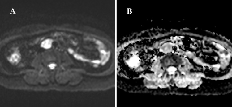
Paragangliomas are uncommon neuroendocrine neoplasms that occur in characteristic locations. While parasympathetic paragangliomas are mainly located at the head and neck, sympathetic paragangliomas are mostly located below the neck. Among parasympathetic paragangliomas, pheochromocytomas are the most common. Ninety percent of cases of pheochromocytomas arise within the adrenal gland. We report a case of a 63-year-old woman with an extra-adrenal pheochromocytoma of the organ of Zuckerkandl detected by CT and MRI and subsequently confirmed by postoperative histology and immunohistochemistry.
Keywords: Histopathology; Immunohistochemical; MRI; Paraganglioma; Pheochromocytoma; Zuckerkandl.
© 2020 The Authors. Published by Elsevier Inc. on behalf of University of Washington.
Figures


References
-
- Lee K.Y., Oh Y.-W., Noh H.J. Extraadrenal paragangliomas of the body: imaging features. Am. J. Roentgenol. 2006;187(2):492–504. - PubMed
-
- Cheung V.K.Y., Gill A.J., Chou A. Old, new, and emerging immunohistochemical markers in pheochromocytoma and paraganglioma. Endocr Pathol. 2018;29(2):169–175. - PubMed
Publication types
LinkOut - more resources
Full Text Sources
Other Literature Sources
