

Meningeal tuberculoma mimicking a brain tumor
- PMID: 33299510
- PMCID: PMC7708657
- DOI: 10.1016/j.radcr.2020.11.028
Meningeal tuberculoma mimicking a brain tumor
Erratum in
-
Erratum regarding missing declaration of competing interest and patient consent statements in previously published articles.Radiol Case Rep. 2023 Jan 25;18(4):1643-1644. doi: 10.1016/j.radcr.2023.01.017. eCollection 2023 Apr. Radiol Case Rep. 2023. PMID: 36895588 Free PMC article.
Abstract
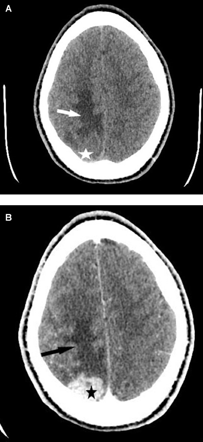
Meningeal tuberculoma is one of the most serious sites of tuberculosis. Its incidence varies depending on the geographical area, rare in Western countries and frequent in developing countries where it represents 5% to 10% of intracranial masses. We report the case of a 21-year-old male patient with no particular medical history from Africa and living in Europe for more than a year, is hospitalized for an isolated inaugural, generalized, afebrile seizure in whom the scanner and cerebral magnetic resonance imaging (MRI) revealed a meningeal mass with significant glove finger edema suggesting a primary brain tumor. Surgical excision and anatomopathological analysis of the excisional piece allowed the diagnosis of tuberculoma. Meningeal tuberculoma is a source of diagnostic error because its clinical and radiological expression can mimic a brain tumor. This is an etiology that should not be ignored in the face of a meningeal mass in any subject coming from or living in a region with a high endemic tuberculosis.
Keywords: Brain tumor; MRI; Meningeal tuberculoma; Scanner.
© 2020 The Authors. Published by Elsevier Inc. on behalf of University of Washington.
Figures


References
-
- Muralidhar K., Katti MK. Pathogenesis, diagnosis, treatment, and outcome aspects of cerebral tuberculosis. Med Sci Monit. 2004;10:215–229. - PubMed
-
- Semlali S., El Kharras A., Mahi M., Hsaini Y., Benameur M., Aziz N. The imaging aspects of tuberculosis of the central nervous system. J Radiol. 2008;89:209–220. - PubMed
-
- Nouira K., Allani R., Abdelmalek R., Azaiez O., Laamari L., Benmessaoud M contribution of MRI in the diagnosis of tuberculosis of the central nervous system. La Presse Médicale 2008; 37: 634–642. - PubMed
-
- Suslu H.T., Bozbuga M., Bayindir C. Cerebral tuberculoma mimicking high grade glial tumor. Turkish Neurosurg. 2011;21:427–429. - PubMed
-
- Batra A., Tripathi R.P. Diffusion-weighted magnetic resonance imaging and magnetic resonance spectroscopy in the evaluation of focal cerebral tubercular lesions. Acta Radiol. 2004;45:679–688. - PubMed
Publication types
LinkOut - more resources
Full Text Sources
Other Literature Sources
