. 2020 Nov 23:2020:7627934.
doi: 10.1155/2020/7627934. eCollection 2020.

Paricalcitol Attenuates Contrast-Induced Acute Kidney Injury by Regulating Mitophagy and Senescence

Affiliations

Paricalcitol Attenuates Contrast-Induced Acute Kidney Injury by Regulating Mitophagy and Senescence

Eunjin Bae et al. Oxid Med Cell Longev. .

Abstract

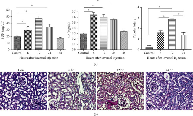
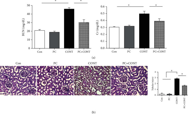
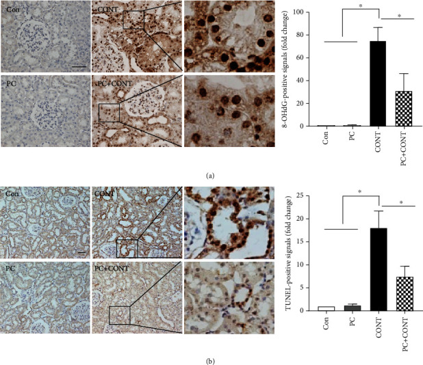
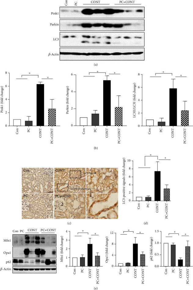
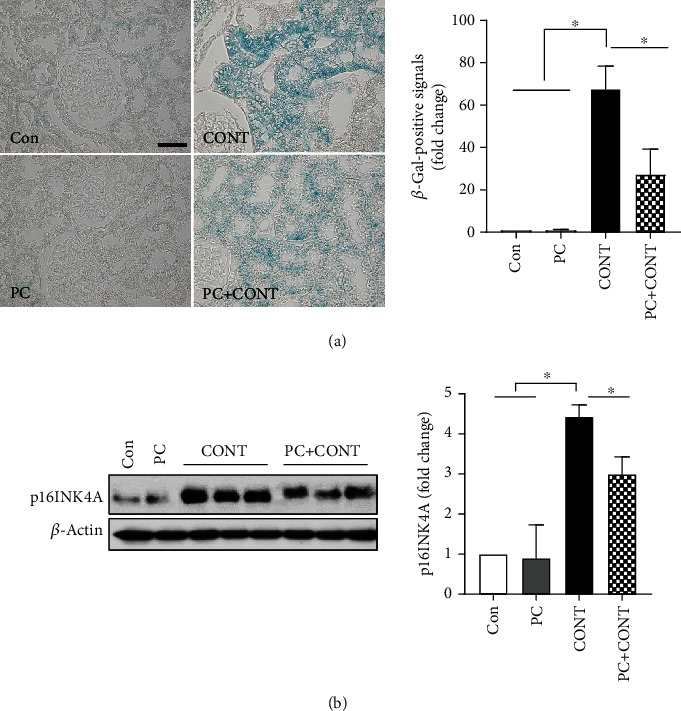
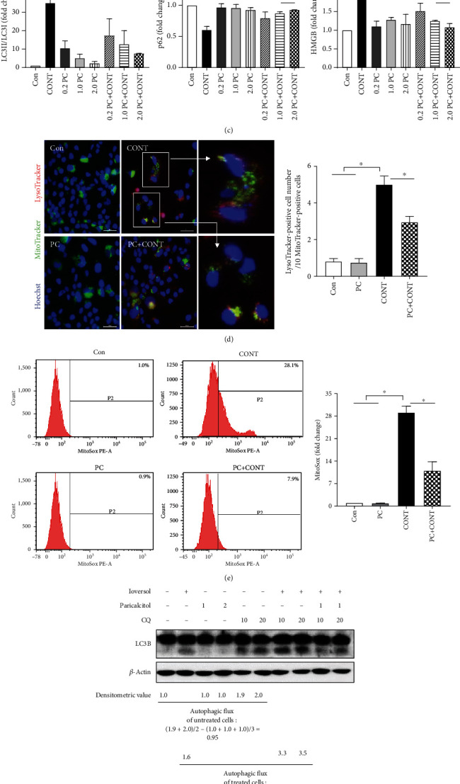
Contrast-induced acute kidney injury (CI-AKI) is the third most common cause of hospital-acquired renal failure, with an incidence of 11%. However, the disease mechanism remains unclear, and no effective treatment is available. Paricalcitol has been reported to be effective in animal models of kidney injury. We hypothesized that paricalcitol could play a renoprotective role against CI-AKI. Rats were divided into control, paricalcitol, contrast, and paricalcitol-plus-contrast groups. We used a previously published protocol to produce CI-AKI. Paricalcitol (0.3 μg/kg) was administered intraperitoneally before 24 h and 30 min before indomethacin. We used HK-2 cells to evaluate the effects of paricalcitol on mitophagy and senescence. Ioversol triggered renal dysfunction, increasing blood urea nitrogen and serum creatinine. Significant tubular damage, increased 8-OHdG expression, and apoptosis were apparent. Ioversol injection induced high expression levels of the mitophagy markers Pink1, Parkin, and LC3 and the senescence markers β-galactosidase and p16INK4A. Paricalcitol pretreatment prevented renal dysfunction and reduced tissue damage by reducing both mitophagy and senescence. Cellular morphological changes were found, and expression of LC3B and HMGB1 was increased by ioversol in HK-2 cells. Paricalcitol countered these effects. This study showed that mitochondria might drive injury phenotypes in CI-AKI, and that paricalcitol protects against CI-AKI by decreasing mitochondrial damage.


Conflict of interest statement

The authors declare no competing interests.

Figures

Figure 1
Figure 1
The contrast-induced renal dysfunction and histological injury in rats. (a) BUN and serum creatinine levels at 6, 12, 24, and 48 hours after contrast infusion. (b) Representative images of PAS staining in the tubular injury at 6, 12, and 24 hours after contrast infusion. Data were presented as mean ± SEM (P < 0.05).
Figure 2
Figure 2
Effect of paricalcitol on biochemical tests and histological injuries in the kidney after ioversol administration. (a) Serum BUN and creatinine levels at 12 hours and (b) PAS staining in the kidney after ioversol administration. The injury score was determined as described in Materials and Methods. Images are representative of each group. Con: PBS-treated group; PC: paricalcitol-treated group; CONT: ioversol-treated group; PC+CONT: paricalcitol- and ioversol-treated group. Data were presented as mean ± SEM (P < 0.05).
Figure 3
Figure 3
Effects of paricalcitol on oxidative stress and apoptosis in the kidney after ioversol administration. Immunohistochemical staining was performed with a specific antibody against 8-OHdG. Densitometric quantification for 8-OHdG was applied to each group. Ioversol-induced apoptosis was detected using the TUNEL assay. TUNEL-positive cells were stained with dense brown spots and counted as described in Materials and Methods. Apoptotic signals were found by TUNEL staining. Images are representative of each group. Con: PBS-treated group; PC: paricalcitol-treated group; CONT: ioversol-treated group; PC+CONT: paricalcitol- and ioversol-treated group. Data were presented as mean ± SEM (P < 0.05).
Figure 4
Figure 4
Effects of paricalcitol on mitophagy in the kidney after ioversol administration. (a, b) Immunoblot analysis was performed with a specific antibody against PINK1, Parkin, and LC3. β-Actin was used as loading control, and data were normalized against the density of β-actin. Blots are representative of each group. Cropped blots are displayed here, and full-length blots are included in the section of Supplementary Information. (c, d) Immunohistochemical staining was performed with a specific antibody against LC3. Densitometric quantification for LC3 was applied to each group. Images are representative of each group. (e) Immunoblot analysis for Mfn1 and Opa1 was performed to confirm the effect of paricalcitol on mitophagy, and p62 is for the effect of paricalcitol on the autophagic flux. Con: PBS-treated group; PC: paricalcitol-treated group; CONT: ioversol-treated group; PC+CONT: paricalcitol- and ioversol-treated group. Data were presented as mean ± SEM (P < 0.05).
Figure 5
Figure 5
Effects of paricalcitol on senescence in the kidney after ioversol administration. (a) β-Galactosidase staining was performed with a specific assay kit. Densitometric quantification for β-galactosidase was applied to each group. Positive signals for β-galactosidase were observed as blue in the ioversol-treated kidney. Images are representative of each group. (b) Immunoblot analysis was performed with a specific antibody against p16INK4A. β-Actin was used as loading control, and data were normalized against the density of β-actin. Blots are representative of each group. Cropped blots are displayed here, and full-length blots are included in the section of Supplementary Information. Con: PBS-treated group; PC: paricalcitol-treated group; CONT: ioversol-treated group; PC+CONT: paricalcitol- and ioversol-treated group. Data were presented as mean ± SEM (P < 0.05).
Figure 6
Figure 6
Effects of paricalcitol on contrast-induced cellular senescence in HK-2 cells. (a) Cell morphology was observed in each group at 60 min using phase-contrast microscopy under 200x magnification. HK-2 cells were treated with 100 mg/mL ioversol and 0.2, 1.0, and 2.0 ng/mL paricalcitol for 60 min. (b, c) Immunoblot analysis was performed with a specific antibody against LC3 and HMGB. β-Actin was used as loading control, and the ratio of LC3II/LC3I expression and the level of HMGB expression were analyzed by a densitometer. Blots are representative of each group. Cropped blots are displayed here, and full-length blots are included in the section of Supplementary Information. (d) Representative images of colocalization of lysosome (red—LysoTracker) and mitochondria (green—MitoTracker) in HK-2 cells. Mitophagy was detected by dual-positive staining. Hoechst (blue) was used for nuclear staining. (e) The mitochondria-associated ROS levels were measured by staining the cells with MitoSox, using flow cytometry. (f) Paricalcitol inhibited ioversol from inducing an autophagic flux. CQ is used as an inhibitor of an autophagic flux, and LC3B-II (lower band) expression is analyzed by Western blot. Con: no treatment; CONT: ioversol-treated group; PC+CONT: paricalcitol and ioversol-treated group. Data were presented as mean ± SEM (P < 0.05).

References

    1. Andreucci M., Solomon R., Tasanarong A. Side effects of radiographic contrast media: pathogenesis, risk factors, and prevention. BioMed Research International. 2014;2014:20. doi: 10.1155/2014/741018.741018 - DOI - PMC - PubMed
    1. Giacoppo D., Madhavan M. V., Baber U., et al. Impact of contrast-induced acute kidney injury after percutaneous coronary intervention on short- and long-term outcomes: pooled analysis from the HORIZONS-AMI and ACUITY trials. Circulation: Cardiovascular Interventions. 2015;8(8) doi: 10.1161/CIRCINTERVENTIONS.114.002475. - DOI - PubMed
    1. McCullough P. A., Choi J. P., Feghali G. A., et al. Contrast-induced acute kidney injury. Journal of the American College of Cardiology. 2016;68(13):1465–1473. doi: 10.1016/j.jacc.2016.05.099. - DOI - PubMed
    1. Wong P. C., Li Z., Guo J., Zhang A. Pathophysiology of contrast-induced nephropathy. International Journal of Cardiology. 2012;158(2):186–192. doi: 10.1016/j.ijcard.2011.06.115. - DOI - PubMed
    1. Lei R., Zhao F., Tang C. Y., et al. Mitophagy plays a protective role in iodinated contrast-induced acute renal tubular epithelial cells injury. Cellular Physiology and Biochemistry. 2018;46(3):975–985. doi: 10.1159/000488827. - DOI - PubMed

LinkOut - more resources