

Clinical Assessment of Breast Volume: Can 3D Imaging Be the Gold Standard?
- PMID: 33299702
- PMCID: PMC7722547
- DOI: 10.1097/GOX.0000000000003236
Clinical Assessment of Breast Volume: Can 3D Imaging Be the Gold Standard?
Abstract
Three-dimensional (3D) camera systems are increasingly used for computerized volume calculations. In this study we investigate whether the Vectra XT 3D imaging system is a reliable tool for determination of breast volume in clinical practice. It is compared with the current gold standard in literature, magnetic resonance imaging (MRI), and current clinical practice (plastic surgeon's clinical estimation).
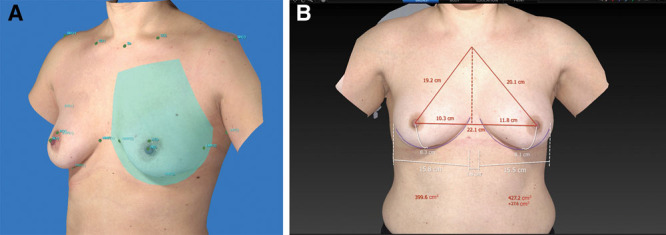
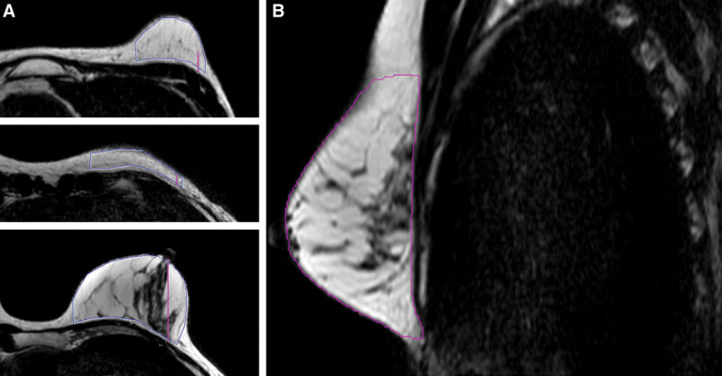
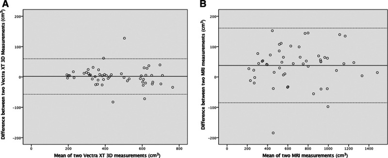
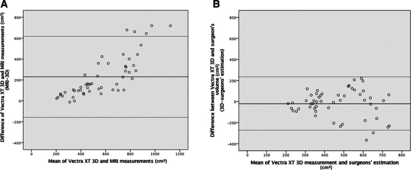
Methods: Breast volumes of 29 patients (53 breasts) were evaluated. 3D images were acquired by Vectra XT 3D imaging system. Pre-existing breast MRI images were collected. Both imaging techniques were used for volume analyses, calculated by two independent investigators. Breast volume estimations were done by plastic surgeons during outpatient consultations. All volume measurements were compared using paired samples t-test, intra-class correlation coefficient, Pearson's correlation, and Bland-Altman analysis.
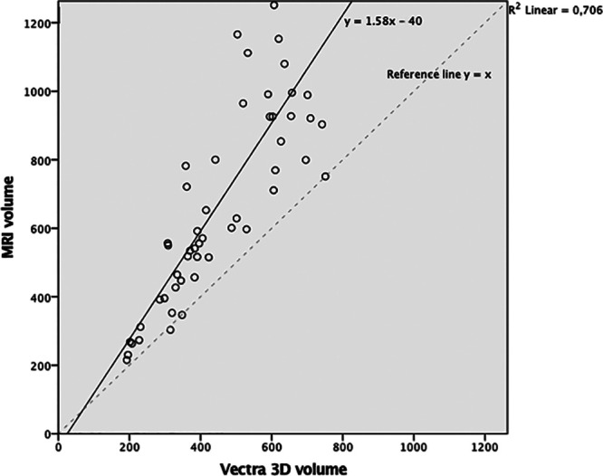
Results: Two 3D breast volume measurements showed an excellent reliability (intra-class correlation coefficient: 0.991), which was comparable to the reliability of MRI measurements (intra-class correlation coefficient: 0.990). Mean (SD) breast volume measured with 3D breast volume was 454 cm3 (157) and with MRI was 687 cm3 (312). These volumes were significantly different, but a linear association could be found: y(MRI) = 1.58 × (3D) - 40. Three-dimensional breast volume was not significantly different from volume estimation made by plastic surgeons (472 cm3 (69), P = 0.323).
Conclusions: The 3D imaging system measures lower volumes for breasts than MRI. However, 3D measurements show a linear association with MRI and have excellent reliability, making them an objective and reproducible measuring method suitable for clinical practice.
Copyright © 2020 The Authors. Published by Wolters Kluwer Health, Inc. on behalf of The American Society of Plastic Surgeons.
Conflict of interest statement
Figures
References
-
- Herold C, Ueberreiter K, Busche MN, et al. Autologous fat transplantation: Volumetric tools for estimation of volume survival. A systematic review. Aesthetic Plast Surg. 2013;37:380–387. - PubMed
-
- Yip JM, Mouratova N, Jeffery RM, et al. Accurate assessment of breast volume: A study comparing the volumetric gold standard (direct water displacement measurement of mastectomy specimen) with a 3D laser scanning technique. Ann Plast Surg. 2012;68:135–141. - PubMed
-
- Choppin SB, Wheat JS, Gee M, et al. The accuracy of breast volume measurement methods: A systematic review. Breast. 2016;28:121–129. - PubMed
LinkOut - more resources
Full Text Sources