. 2020 Nov 13;23(12):101799.
doi: 10.1016/j.isci.2020.101799. eCollection 2020 Dec 18.

Neutrophil Elastase Facilitates Tumor Cell Intravasation and Early Metastatic Events

Affiliations

Neutrophil Elastase Facilitates Tumor Cell Intravasation and Early Metastatic Events

Elena Deryugina et al. iScience. .

Abstract

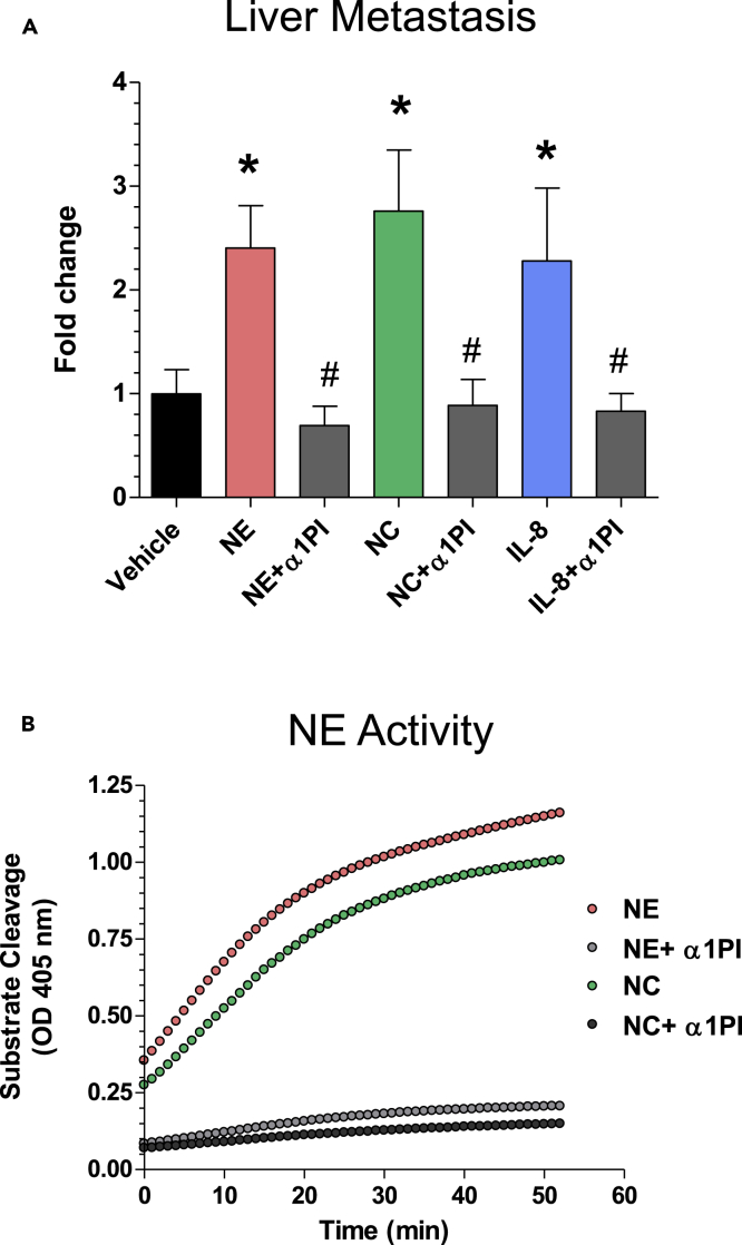
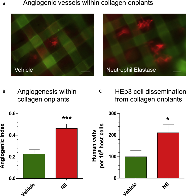
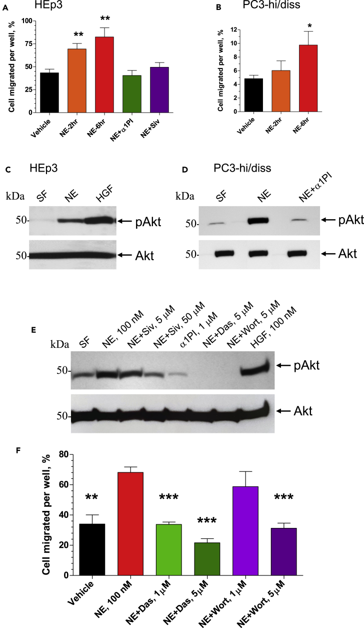
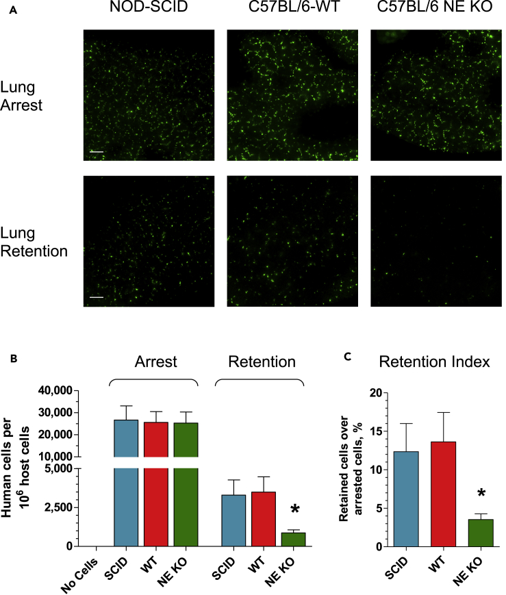
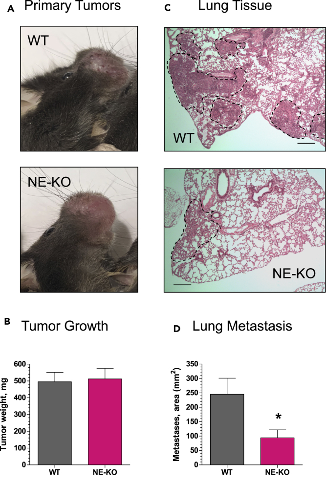
Functional roles of neutrophil elastase (NE) have not been examined in distinct steps of the metastatic cascade. NE, delivered to primary tumors as a purified enzyme or within intact neutrophils or neutrophil granule content, enhanced human tumor cell intravasation and subsequent dissemination via NE-mediated formation of dilated intratumoral vasculature. These effects depended on picomole range of NE activity, sensitive to its natural inhibitor, α1PI. In Elane-negative mice, the lack of NE decreased lung retention of human tumor cells in experimental metastasis. Furthermore, NE was essential for spontaneous metastasis of murine carcinoma cells in a syngeneic orthotopic model of oral cancer. NE also induced tumor cell survival and migration via Src/PI3K-dependent activation of Akt signaling, vital for tumor cell dissemination in vivo. Together, our findings implicate NE, a potent host enzyme specific for first-responding innate immune cells, as directly involved in early metastatic events and a potential target for therapeutic intervention.

Keywords: Cancer; Cell Biology.


Conflict of interest statement

The authors declare no competing interests.

Figures

None
Graphical abstract
Figure 1
Figure 1
NE Is Involved in Tumor Cell Metastasis (A) Human HEp3 carcinoma cells were grafted onto the CAM of live, day 10 chick embryos (6 sites each containing 1 × 105 cells in 10 μL of native type I collagen at 2.3 mg/mL). Developing tumors were treated daily with topical applications of 10 μL solutions of PBS/1% DMSO, containing purified neutrophil elastase (NE) (100 nM), NE mixed with α1PI (0.3 μM), neutrophil cavitated (NC) preparations (100 nM of NE specific activity), NC mixed with α1PI (0.3 μM), IL-8 (100 ng), and IL-8 mixed with α1PI (0.3 μM). Control embryos were treated with vehicle solution (PBS/1% DMSO). On day 5, liver tissue was harvested, total DNA prepared and processed for human-specific Alu-qPCR to quantify the number of HEp3 cells within the chicken embryo background (106 cells). The data from individual experiments (5–7 embryos per variable) were normalized to the number of disseminated cells in the vehicle control group (fold differences), and the normalized data (up to 5 independent experiments) were combined for statistical assessment. ∗, p < 0.05 in Student's t test or Mann-Whitney U test compared with control. #, p < 0.05 in Student's t test or Mann-Whitney U-test compared with the corresponding group containing no α1PI. Data are presented as mean ± SEM. (B) Inhibition of NE activity by α1PI. Enzymatic activity of NE and NC preparations was analyzed for the sensitivity to α1PI in a substrate cleavage assay. Purified human NE (100 nM) and NC (diluted 1:3 to achieve ~100 nM of NE activity), used alone or pre-mixed with 0.3 μM α1PI, were added to a 350 nM elastase-specific colorimetric substrate. Generation of the cleaved substrate was monitored for 60 min at 405 nM.
Figure 2
Figure 2
NE Affects Tumor Angiogenesis and Angiogenesis-Dependent Intravasation (A) Grid rafts, each containing 30 μL of type I collagen (2.3 mg/mL) populated with GFP-tagged HEp3 cells (1 × 10⁶/mL reconstituted collagen), were planted on the CAM of chick embryos. The “onplants” were treated on days 1 and 2 with purified neutrophil elastase (NE) (10 μL of 100 nM solution) or vehicle (PBS/1% DMSO). On day 3, the embryos were inoculated i.v. with red-fluorescent lectin, Rhodamine-conjugated LCA (100 μg in 0.1 mL PBS), to contrast blood vessels (red fluorescence) against the grids of onplant-supporting meshes. Scale bars, 100 μm. (B) Angiogenic vessels containing visible circulating erythrocytes were counted between mesh grids of the collagen raft, and the angiogenic index was calculated as the ratio of grids containing newly formed functional blood vessels versus total number of grids. ∗∗∗, p < 0.05 in Student's t test compared with the vehicle control. Data are presented as mean ± SEM. (C) On day 5, the levels of HEp3 cell dissemination from the 3D collagen rafts were determined with human-specific Alu-qPCR in portions of CAM harvested far distal (1–5 cm) to the sites of onplant grafting. ∗, p < 0.05 in Student's t test compared with the vehicle control. Data are presented as mean ± SEM.
Figure 3
Figure 3
NE and Neutrophil Granule Content Facilitate the Development of an Intravasation-Sustaining Intratumoral Vasculature (A) GFP-tagged HEp3 cells were grafted on the CAM and treated daily (10 μL per tumor, topical application) with vehicle (PBS/1% DMSO), purified neutrophil elastase (NE), alone (100 nM) or mixed with α1PI (0.3 μM), and neutrophil cavitated preparations (NC), alone (at 100 nM NE activity) or mixed with α1PI (0.3 μM). On day 5, the embryos were inoculated with Rhodamine-conjugated LCA (0.1 mL of 1 mg/mL solution in PBS) to contrast the intratumoral angiogenic vasculature against green fluorescence of human tumor cells. (Top) Tumor images with the signals of HEp3-GFP cells (green) and LCA-stained vessels (red) merged. Scale bars, 100 μm. (Bottom) Monochromatic images depicting intratumoral blood vessels only. (B) The microtumors were analyzed for vessel diameter distribution on day 5 after cell grafting. Data are presented as mean ± SEM of at least 10 microtumors analyzed for each of control group and groups treated with NE (red bars) or NC (blue bars) with (hatched bars) or without (non-hatched, open bars) α1PI (from a total of 3 independent experiments). ∗, p < 0.05 in Student's t test compared with the vehicle control. (C) Levels of intravasation were quantified by human-specific Alu-qPCR in the portions of the CAM harvested distal to primary tumors and presented as fold differences relative to vehicle control. Data are presented as mean ± SEM determined for three independent experiments involving from 7 to 38 embryos per variable. ∗, p < 0.05 in Student's t test compared with the vehicle control. #, p < 0.05 in Student's t test compared with the corresponding group containing no α1PI.
Figure 4
Figure 4
Intact NE-Competent Neutrophils Facilitate the Development of Intravasation-Supporting Intratumoral Vasculature (A) GFP-tagged HEp3 cells were grafted on the CAM within six 10-μL-collagen droplets, each containing 1 × 10⁵ cells. On day 3, the embryos were inoculated with 2 × 10⁶ neutrophils freshly isolated from human peripheral blood. In a set of embryos, the developing tumors were pre-treated with α1PI (0.3 μM) on day 1 and 2 and additionally treated after neutrophil inoculations on day 4. On day 5, the embryos were inoculated with Rhodamine-conjugated LCA (0.1 mL of 1 mg/mL solution in PBS) to contrast the intratumoral angiogenic vasculature against green fluorescence of human tumor cells. (Top) Representative IF images of tumors with signals for HEp3-GFP cells (green) and LCA-stained vessels (red) merged. Scale bars, 100 μm. (Bottom) Monochromatic images depicting intratumoral blood vessels only. (B) The microtumors were analyzed for vessel diameter distribution on day 5 after cell grafting. Data are presented as mean ± SEM of at least 10 microtumors analyzed for each of control group and groups treated with neutrophils with or without α1PI (4 independent experiments). ∗, p < 0.05 in Student's t test compared with the vehicle control. (C) On day 5 of tumor development, the numbers of human tumor cells within portions of the CAM distal to primary tumors were quantified by human-specific Alu-qPCR using a standard curve. The levels of intravasation are presented as fold differences relative to the buffer control. The data are mean ± SEM of four independent experiments involving a total of 24 embryos in the control group, 21 embryos in the group inoculated with neutrophils, and 6 embryos in the group treated with both neutrophils and α1PI. ∗, p < 0.05 in Student's t test compared with the vehicle control. #, p < 0.05 in Student's t test compared with the corresponding group containing no α1PI.
Figure 5
Figure 5
NE Induces Tumor Cell Migration via Src/PI3K-Dependent Akt Signaling (A and B) Subconfluent cultures of HEp3 cells (A) and PC3-hi/diss cells (B) were pre-treated with 100 nM purified NE for 2 or 6 h and then washed twice in serum-free (SF) medium. NE inhibitors α1PI (1 μM) and Sivelestat (Siv; 50 μM) were added along with 100 nM NE to HEp3 cells for the 6-h incubation (A). Vehicle-treated cells (negative control) and NE-treated cells were placed into Transwell inserts (1 × 105) and allowed to migrate toward chemoattractants in lower chamber (5% FCS). After 18- to 24-h incubation, the percentage of cells transmigrated into the lower chamber was quantified. From 2 to 3 experiments were performed for both cell types, each experiment in triplicate. ∗ and ∗∗, p < 0.05 and p < 0.01, respectively, compared with vehicle control; two-tailed Student's t test. Data are presented as mean ± SEM. (C and D) Serum-starved HEp3 (C) and PC3-hi/diss (D) cells were pre-treated for 20 min with purified NE (100 nM), alone or along with a1PI (1 mM), or hepatocyte growth factor (HGF; 50 nM), washed and lysed. Western blot analysis was performed on equal protein content (20 μg) under reducing conditions using antibodies against phosphorylated Akt (pAkt; upper panels) and total Akt (lower panels). (E) Serum-starved HEp3 cells were pre-treated for 20 min with the NE inhibitor Sivelestat (5 μM or 50 μM) and α1PI (1 μM) or signaling inhibitors Dasatinib (Das, 5 μM) and Wortmannin (5 μM). Then, NE or HGF were added, both at 100 nM. After 20 min incubation, the cells were washed and lysed. Western blot analysis was performed on equal protein content (40 μg) under reducing conditions using antibodies against phosphorylated Akt (pAkt). Equal protein loading is indicated by the blot re-probed with antibodies against total Akt protein (Akt). (F) HEp3 cells were pretreated for 6 h with vehicle or NE (100 nM), alone or mixed with Src inhibitor Dasatinib (Das; 1 μM or 5 μM) or PI3K inhibitor Wortmannin (Wort, 1 μM or 5 μM). Vehicle-treated cells (negative control) and NE-treated cells were placed into Transwell inserts (1 × 105) and allowed to migrate toward 5% FCS in the lower chamber. After 18- to 24-h incubation, the percentage of cells transmigrated into the lower chamber was quantified. ∗∗ and ∗∗∗, p < 0.01 and p < 0.001, respectively, in comparison to the treatment with NE alone; two-tailed Student's t test. Data are presented as mean ± SEM. See also Figures S1–S3.
Figure 6
Figure 6
NE Contributes to Vascular Arrest and Tissue Retention of Human Carcinoma Cells (A) GFP-tagged HEp3 cells were inoculated into the tail vein of immunodeficient SCID mice or C57BL/6 mice, WT type or NE-KO (5 × 10⁵ cells per mouse). The lungs were excised 20 min or 14 h later to determine the levels of tumor cell arrest in the lung vasculature or tissue retention of vascular arrested cells, respectively. Green fluorescent HEp3 cells were imaged in the lungs at original 200× magnification in an immunofluorescence microscope equipped with a digital video camera. Scale bars, 100 μm. (B) The number of inoculated HEp3 cells arrested and retained in the lung vasculature was measured in the lung tissue with human-specific Alu-qPCR. The lung tissue of mice that received no human tumor cells was used as negative control. Three independent experiments were performed involving a total of 14–17 mice per variable. ∗, p < 0.05, unpaired two-tailed Student's t test. Data are presented as mean ± SEM. (C) Retention indices were quantified for each mouse as the percentage of HEp3 cells retained in the lung compared with the number of cells arrested in the lung vasculature, based on human-specific Alu-qPCR analyses. ∗, p < 0.01, unpaired two-tailed Student's t test. Data are presented as mean ± SEM. See also Figures S4 and S5.
Figure 7
Figure 7
NE Is Critical for Spontaneous Metastasis in a Syngeneic Mouse Model of Head and Neck Cancer Murine head and neck cancer carcinoma cells, MOC2, were inoculated into the buccal mucosa of syngeneic WT or NE-KO C57BL/6 mice at 1 × 10⁶ cells per site. Twelve to 14 days later (A), the mice were sacrificed and their primary buccal tumors weighed (B). The lungs were excised, fixed, and processed for staining with H&E (C). Scale bars, 1 mm. The areas of lung parenchyma filled with tumor cells (C, areas indicated by dashed lines) were quantified for each mouse against total area of lung tissue and expressed as “Metastases” area (D). Two independent experiments were performed, involving a total of 16 WT and 12 NE-KO mice. ∗, p < 0.05; unpaired two-tailed Student's t test. The numerical data are presented as mean ± SEM.

References

    1. Aikawa N., Kawasaki Y. Clinical utility of the neutrophil elastase inhibitor sivelestat for the treatment of acute respiratory distress syndrome. Ther. Clin. Risk Manag. 2014;10:621–629. - PMC - PubMed
    1. Aikawaa N., Ishizaka A., Hirasawa H., Shimazaki S., Yamamoto Y., Sugimoto H., Shinozaki M., Taenaka N., Endo S., Ikeda T. Reevaluation of the efficacy and safety of the neutrophil elastase inhibitor, Sivelestat, for the treatment of acute lung injury associated with systemic inflammatory response syndrome; a phase IV study. Pulm. Pharmacol. Ther. 2011;24:549–554. - PubMed
    1. Akizuki M., Fukutomi T., Takasugi M., Takahashi S., Sato T., Harao M., Mizumoto T., Yamashita J. Prognostic significance of immunoreactive neutrophil elastase in human breast cancer: long-term follow-up results in 313 patients. Neoplasia. 2007;9:260–264. - PMC - PubMed
    1. Albrengues J., Shields M.A., Ng D., Park C.G., Ambrico A., Poindexter M.E., Upadhyay P., Uyeminami D.L., Pommier A., Kuttner V. Neutrophil extracellular traps produced during inflammation awaken dormant cancer cells in mice. Science. 2018;361:eaao4227. - PMC - PubMed
    1. Altomare D.A., Testa J.R. Perturbations of the AKT signaling pathway in human cancer. Oncogene. 2005;24:7455–7464. - PubMed

LinkOut - more resources