[Preprint]. 2020 Dec 1:2020.12.01.406611.
doi: 10.1101/2020.12.01.406611.

Designed proteins assemble antibodies into modular nanocages

Affiliations

Designed proteins assemble antibodies into modular nanocages

Robby Divine et al. bioRxiv. .

Update in

  • Designed proteins assemble antibodies into modular nanocages.
    Divine R, Dang HV, Ueda G, Fallas JA, Vulovic I, Sheffler W, Saini S, Zhao YT, Raj IX, Morawski PA, Jennewein MF, Homad LJ, Wan YH, Tooley MR, Seeger F, Etemadi A, Fahning ML, Lazarovits J, Roederer A, Walls AC, Stewart L, Mazloomi M, King NP, Campbell DJ, McGuire AT, Stamatatos L, Ruohola-Baker H, Mathieu J, Veesler D, Baker D. Divine R, et al. Science. 2021 Apr 2;372(6537):eabd9994. doi: 10.1126/science.abd9994. Science. 2021. PMID: 33795432 Free PMC article.

Abstract

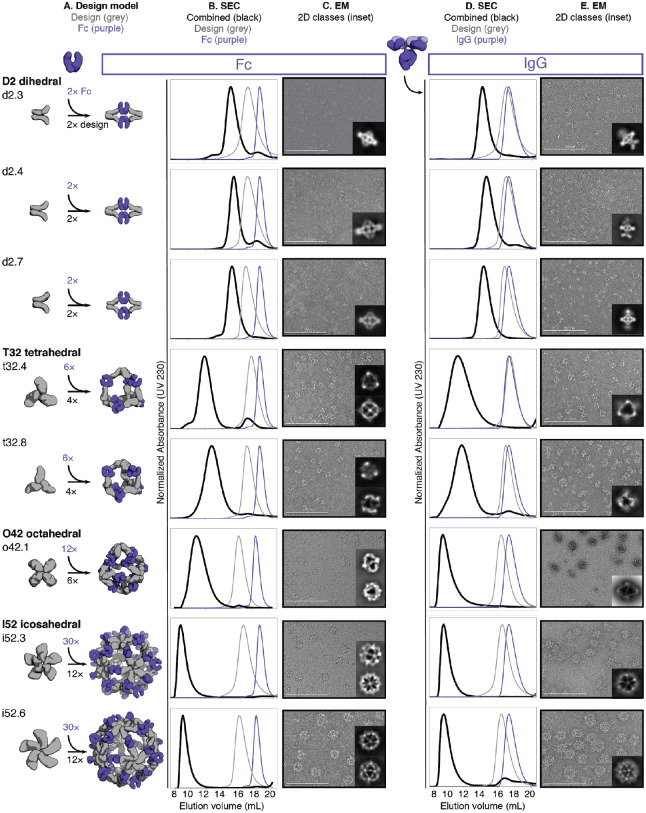
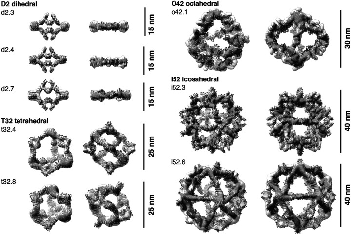
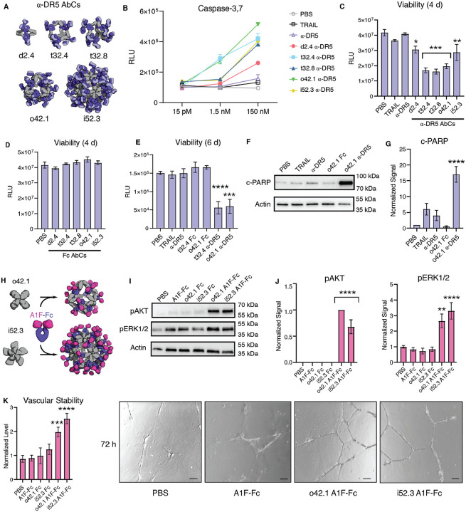
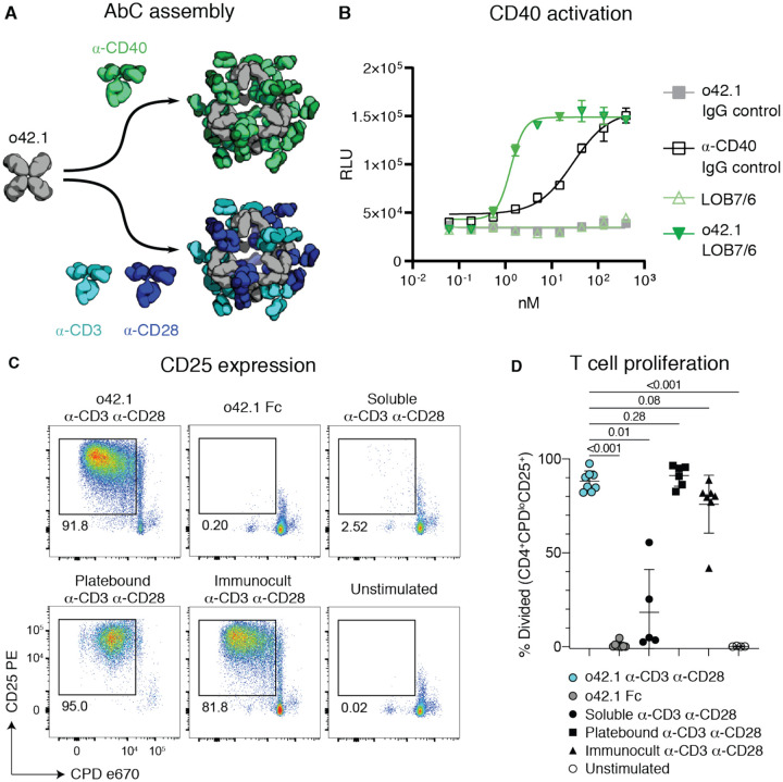
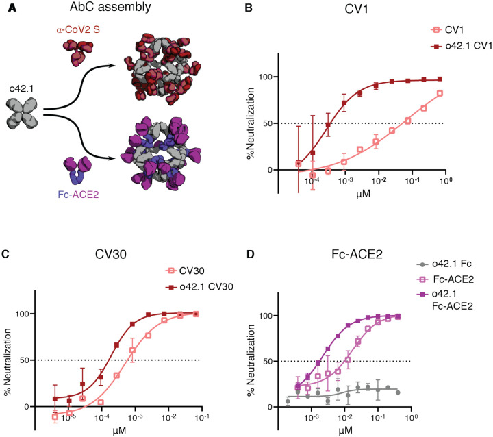
Antibodies are widely used in biology and medicine, and there has been considerable interest in multivalent antibody formats to increase binding avidity and enhance signaling pathway agonism. However, there are currently no general approaches for forming precisely oriented antibody assemblies with controlled valency. We describe the computational design of two-component nanocages that overcome this limitation by uniting form and function. One structural component is any antibody or Fc fusion and the second is a designed Fc-binding homo-oligomer that drives nanocage assembly. Structures of 8 antibody nanocages determined by electron microscopy spanning dihedral, tetrahedral, octahedral, and icosahedral architectures with 2, 6, 12, and 30 antibodies per nanocage match the corresponding computational models. Antibody nanocages targeting cell-surface receptors enhance signaling compared to free antibodies or Fc-fusions in DR5-mediated apoptosis, Tie2-mediated angiogenesis, CD40 activation, and T cell proliferation; nanocage assembly also increases SARS-CoV-2 pseudovirus neutralization by α-SARS-CoV-2 monoclonal antibodies and Fc-ACE2 fusion proteins. We anticipate that the ability to assemble arbitrary antibodies without need for covalent modification into highly ordered assemblies with different geometries and valencies will have broad impact in biology and medicine.


Conflict of interest statement

Competing Interests Provisional patents have been filed on the AbC-forming designs, α-DR5 AbCs, A1F-Fc AbCs, α-CD40 AbCs, and α-CoV-2 S AbCs. A provisional patent application (U.S. Provisional Application No. 63/016268) has been filed on the SARS-CoV-2 specific monoclonal antibodies discussed here. D.V. is a consultant for Vir Biotechnology Inc. The Veesler laboratory has received an unrelated sponsored research agreement from Vir Biotechnology Inc. The other authors declare no competing interests.

Figures

Figure 1.
Figure 1.. Antibody nanocage (AbC) design.
A, Polyhedral geometry is specified. Clockwise from top left: icosahedral, dihedral, octahedral, and tetrahedral geometries are shown. B, An antibody Fc model from hIgG1 is aligned to one of the C2 axes (in this case, a D2 dihedron is shown). C, Antibody Fc-binders are fused to helical repeat proteins that are then fused to the monomeric subunit of helical cyclic oligomers. All combinations of building blocks and building block junctions are sampled (below inset, grey). D, Tripartite fusions are checked to ensure successful alignment of the C2 Fc symmetry axes with that of the polyhedral architecture (in the case of the D2 symmetry shown here, the C2 axes must intersect at a 90° angle). E, Fusions that pass the geometric criteria move forward with sidechain redesign, where e.g. amino acids are optimized to ensure that core-packing residues are nonpolar and solvent-exposed residues are polar. F, Designed AbC-forming oligomers are bacterially expressed, purified, and assembled with antibody Fc or IgG.
Figure 2.
Figure 2.. Structural characterization of AbCs.
A, Design models, with antibody Fc (purple) and designed AbC-forming oligomers (grey). B, Overlay of representative SEC traces of assembly formed by mixing design and Fc (black) with those of the single components in grey (design) or purple (Fc). C, EM images with reference-free 2D class averages in inset; all data is from negative-stain EM with the exception of designs o42.1 and i52.3 (cryo-EM). D-E, SEC (D) and NS-EM representative micrographs with reference-free 2D class averages (E) of the same designed antibody cages assembled with full human IgG1 (with the 2 Fab regions intact). In all EM cases shown in C and E, assemblies were first purified via SEC, and the fractions corresponding to the left-most peak were pooled and used for imaging; this was mainly done to remove any excess of either design or Ig component.
Figure 3.
Figure 3.. 3D reconstructions of AbCs formed with Fc.
Computational design models (cartoon representation) of each AbC are fit into the experimentally-determined 3D density from EM. Each nanocage is viewed along an unoccupied symmetry axis (left), and after rotation to look down one of the C2 axes of symmetry occupied by the Fc (right). 3D reconstructions from o42.1 and i52.3 are from cryo-EM analysis; all others, from NS-EM.
Figure 4.
Figure 4.. AbCs activate apoptosis and angiogenesis signaling pathways.
Caspase-3,7 is activated by AbCs formed with α-DR5 antibody (A), but not the free antibody, in RCC4 renal cancer cells (B). C-D, controls (α-DR5 AbCs (C), but not Fc AbC D) reduce cell viability 4 days after treatment. E, α-DR5 AbCs reduce viability 6 days after treatment. F-G, o42.1 α-DR5 AbCs enhance PARP cleavage, a marker of apoptotic signaling; G, quantification of F relative to PBS control. H, The F-domain from Angiopoietin-1 was fused to Fc (A1F-Fc) and assembled into octahedral (o42.1) and icosahedral (i52.3) AbCs. I, Representative Western blots show that A1F-Fc AbCs, but not controls, increase pAKT and pERK1/2 signals. J, quantification of I: pAKT quantification is normalized to o42.1 A1F-Fc signaling (no pAKT signal in the PBS control); pERK1/2 is normalized to PBS. K, A1F-Fc AbCs increase vascular stability after 72 hours. Left: quantification of vascular stability compared to PBS. Right: representative images. All error bars represent means ± SEM; means were compared using ANOVA and Dunnett post-hoc tests (Tables S8, S9).
Figure 5.
Figure 5.. Activation of immune cells by α-CD40 and α-CD3/28 AbCs.
A, Octahedral AbCs are produced with either α-CD40 or pre-mixed α-CD3 and α-CD28 antibodies. B, CD40 pathways are activated by α-CD40 LOB7/6 octahedral nanocages but not by free α-CD40 LOB7/6 or controls. Scale bars represent means ± SD, n=3; EC50s reported in Table S10. C-D, T cell proliferation and activation is strongly induced by α-CD3 α-CD28 mosaic AbCs compared to unassembled (soluble) α-CD3 α-CD28 antibodies. Representative plots (C) show the frequency of dividing, activated cells (CPDloCD25+). Mosaic AbC-induced proliferation is comparable to traditional positive controls, platebound or tetrameric α-CD3 α-CD28 antibody bead complex (Immunocult). Gated on live CD4+ T cells. Summary graph (D) shows mean ± SD. Significance was determined by Kruskal-Wallis tests correcting for multiple comparisons using FDR two-stage method (n=4–8 per condition). Adjusted p values are reported. CPD, Cell Proliferation Dye.
Figure 6.
Figure 6.. Nanocage assembly enhances SARS-CoV-2 pseudovirus neutralization.
A, Octahedral AbCs are produced with either α-CoV-2 S IgGs or Fc-ACE2 fusion. B-C, SARS-CoV-2 S pseudovirus neutralization by octahedral AbC formed with α-CoV-2 S IgGs CV1 (B) or CV30 (C) compared to un-caged IgG. D, SARS-CoV-2 S pseudovirus neutralization by Fc-ACE2 octahedral AbC compared to un-caged Fc-ACE2. Error bars represent means ± SD, n=2; IC50s reported in Table S11.

References

    1. Lu R.-M., Hwang Y.-C., Liu I.-J., Lee C.-C., Tsai H.-Z., Li H.-J., Wu H.-C., Development of therapeutic antibodies for the treatment of diseases. J. Biomed. Sci. 27, 1–30 (2020). - PMC - PubMed
    1. Cuesta A. M., Sainz-Pastor N., Bonet J., Oliva B., Alvarez-Vallina L., Multivalent antibodies: when design surpasses evolution. Trends Biotechnol. 28, 355–362 (2010). - PubMed
    1. Nuñez-Prado N., Compte M., Harwood S., Álvarez-Méndez A., Lykkemark S., Sanz L., Álvarez-Vallina L., The coming of age of engineered multivalent antibodies. Drug Discov. Today. 20, 588–594 (2015). - PubMed
    1. Laursen N. S., Friesen R. H. E., Zhu X., Jongeneelen M., Blokland S., Vermond J., van Eijgen A., Tang C., van Diepen H., Obmolova G., van der Neut Kolfschoten M., Zuijdgeest D., Straetemans R., Hoffman R. M. B., Nieusma T., Pallesen J., Turner H. L., Bernard S. M., Ward A. B., Luo J., Poon L. L. M., Tretiakova A. P., Wilson J. M., Limberis M. P., Vogels R., Brandenburg B., Kolkman J. A., Wilson I. A., Universal protection against influenza infection by a multidomain antibody to influenza hemagglutinin. Science. 362, 598–602 (2018). - PMC - PubMed
    1. Seifert O., Plappert A., Fellermeier S., Siegemund M., Pfizenmaier K., Kontermann R. E., Tetravalent antibody-scTRAIL fusion proteins with improved properties. Mol. Cancer Ther. 13, 101–111 (2014). - PubMed

Publication types