. 2021 Dec 1;43(6):607-613.
doi: 10.1093/ejo/cjaa065.

The effect of differential force system and minimal surgical intervention on orthodontic tooth movement and root resorption

Affiliations

The effect of differential force system and minimal surgical intervention on orthodontic tooth movement and root resorption

Anjali Nanda et al. Eur J Orthod. .

Abstract

Objective: The primary objective of this study was to quantify the orthodontic tooth movement (OTM) and orthodontically induced root resorption (OIRR) with differential force system in conjunction with minimal surgical insult.

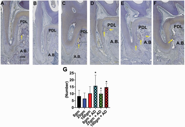
Material and methods: 15-week-old, 48 male Wistar rats were used in the research and were randomly divided into six groups: 1. Group 1 (8 Wistar rats): OTM for 14 days with 8-g force; 2. Group 2 (8 Wistar rats): OTM for 14 days with 25-g force; 3. Group 3 (8 Wistar rats): OTM for 14 days with 100-g force; 4. Group 4 (8 Wistar rats): OTM for 14 days with 8-g force and alveolar decortications (ADs); 5. Group 5 (8 Wistar rats): OTM for 14 days with 25-g force and ADs; 6. Group 6 (8 Wistar rats): OTM for 14 days with 100-g force and ADs. A nickel-titanium spring was used to protract the molar mesially using maxillary incisors as an anchorage. ADs (minimal surgical insult) were done using a hand piece and a round bur, adjacent to the left first maxillary molar on the palatal alveolar bone. After 14 days of OTM, Wistar rats were killed and microfocus computed tomography and histological analysis were performed.

Results: The 100-g group showed significant increase (P < 0.05) in OTM. However, with ADs, the OTM was significantly higher (P < 0.05) in 8 and 100 g. In addition, with ADs, there is significant increase (P < 0.05) in OIRR and significant decrease (P < 0.05) in bone volume fraction. Histological quantification of tartrate-resistant acid phosphatase indicated a significant increase (P < 0.05) in the number of osteoclasts with ADs when compared without ADs.

Conclusions: Light force in conjunction with ADs are optimal to accelerate the OTM. Additionally, ADs increases the OIRR.


Figures

Figure 1.
Figure 1.
(A) Schematic depicting Ni–Ti coil spring for orthodontic force application and minimal surgical intervention through alveolar decortication (AD). (B–G) Intermolar distance (M1–M2) as a measure of orthodontic tooth movement in different groups with and without AD: (B) intermolar distance in 8-g group at Day 14; (C) intermolar distance in 25-g group at Day 14; (D) intermolar distance in 100-g group at Day 14; (E) intermolar distance in 8 g + AD group at Day 14; (F) intermolar distance in 25 g + AD group at Day 14; (G) intermolar distance in 100 g + AD group at Day 14. (H) histogram showing the significant increase (P < 0.05) in the intermolar distance in 100-g (*) group when compared to 8 and 25 g and significant increase (P < 0.05) in the intermolar distance in 8 g + AD (#) and 100 g + AD (#) group when compared to 25 g. Yellow line depicts the intermolar distance.
Figure 2.
Figure 2.
Micro-CT data showing bone volume fraction (BVF) and tissue density in different groups with and without alveolar decortication (AD). (A) Coronally reconstructed micro-CT image of 8-g group at Day 14. (B) Coronally reconstructed micro-CT image of 25-g group at Day 14. (C) Coronally reconstructed micro-CT image of 100-g group at Day 14. (D) Coronally reconstructed micro-CT image of 8 g + AD group at Day 14. (E) Coronally reconstructed micro-CT image of 25 g + AD group at Day 14. (F) Coronally reconstructed micro-CT image of 100 g + AD group at Day 14. (G) Histogram showing the significant decrease (P < 0.05) in the BVF with AD in 8-g (*), 25-g (*), and 100-g (*) groups when compared to same groups without AD. (H) Histogram showing the tissue density with and without AD in different groups at Day 14. The yellow rectangle in Figure 3A is the region of interest where the bone parameters (BVF and tissue density were measured).
Figure 3.
Figure 3.
Micro-CT data showing root volume in different groups with and without alveolar decortication (AD). (A) Reconstructed micro-CT image of mesio-buccal root in 8-g group at Day 14. (B) Reconstructed micro-CT image of mesio-buccal root in 25-g group at Day 14. (C) Reconstructed micro-CT image of mesio-buccal root in 100-g group at Day 14. (D) Reconstructed micro-CT image of mesio-buccal root in 8 g + AD group at Day 14. (E) Reconstructed micro-CT image of mesio-buccal root in 25 g + AD group at Day 14. (F) Reconstructed micro-CT image of mesio-buccal root in 100 g + AD group at Day 14. (G) Histogram showing the significant decrease (P < 0.05) in the root volume with AD in 8-g (*), 25-g (*), and 100-g (*) groups when compared to same groups without AD and 8 g had significantly (#) more root volume when compared to 25 and 100 g.
Figure 4.
Figure 4.
Histologic examination and quantification of osteoclast number with and without alveolar decortication (AD) with differential force system. (A) TRAP-positive cells in 8-g group at Day 14. (B) TRAP-positive cells in 25-g group at Day 14. (C) TRAP-positive cells in 100-g group at Day 14. (D) TRAP-positive cells in 8 g + AD group at Day 14. (E) TRAP-positive cells in 25 g + AD group at Day 14. (F) TRAP-positive cells in 100 g + AD group at Day 14. (G) Histogram showing the significant increase (P < 0.05) in the osteoclast number with AD. Yellow arrows depict the number of TRAP-positive cells.
Figure 5.
Figure 5.
Immunohistochemical staining of sclerostin (sost) showed increased expression with alveolar decortications (AD) when compared to groups without AD. (A–F) Compression side. (G–L) Tension side. (A) Immunohistochemical staining of sost in 8-g group at Day 14. (B) Immunohistochemical staining of sost in 25-g group at Day 14. (C) Immunohistochemical staining of sost in 100-g group at Day 14. (D) Immunohistochemical staining of sost in 8-g + AD group at Day 14. (E) Immunohistochemical staining of sost in 25-g + AD group at Day 14. (F) Immunohistochemical staining of sost in 100-g + AD group at Day 14. (G) Immunohistochemical staining of sost in 8-g group at Day 14. (H) Immunohistochemical staining of sost in 25-g group at Day 14. (I) Immunohistochemical staining of sost in 100-g group at Day 14. (J) Immunohistochemical staining of sost in 8 g + AD group at Day 14. (K) Immunohistochemical staining of sost in 25 g + AD group at Day 14. (L) Immunohistochemical staining of sost in 100 g + AD group at Day 14. Six animals were used in each group for sost immunostaining.
Figure 6.
Figure 6.
Immunohistochemical staining of OPG showed increased expression with alveolar decortications (AD) when compared to groups without AD. (A–F) Compression side. (G–L) Tension side. (A) Immunohistochemical staining of OPG in 8-g group at Day 14. (B) Immunohistochemical staining of OPG in 25-g group at Day 14. (C) Immunohistochemical staining of OPG in 100-g group at Day 14. (D) Immunohistochemical staining of OPG in 8 g + AD group at Day 14. (E) Immunohistochemical staining of OPG in 25 g + AD group at Day 14. (F) Immunohistochemical staining of OPG in 100 g + AD group at Day 14. (G) Immunohistochemical staining of OPG in 8-g group at Day 14. (H) Immunohistochemical staining of OPG in 25-g group at Day 14. (I) Immunohistochemical staining of OPG in 100-g group at Day 14. (J) Immunohistochemical staining of OPG in 8 g + AD group at Day 14. (K) Immunohistochemical staining of OPG in 25 g + AD group at Day 14. (L) Immunohistochemical staining of OPG in 100 g + AD group at Day 14. Six animals were used in each group for OPG immunostaining.
Figure 7.
Figure 7.
Immunohistochemical staining of TUNEL showed increased expression with alveolar decortications (AD) when compared to groups without AD. (A) Immunohistochemical staining of TUNEL in 8-g group at Day 14. (B) Immunohistochemical staining of TUNEL in 25-g group at Day 14. (C) Immunohistochemical staining of TUNEL in 100-g group at Day 14. (D) Immunohistochemical staining of TUNEL in 8 g + AD group at Day 14. (E) Immunohistochemical staining of TUNEL in 25 g + AD group at Day 14. (F) Immunohistochemical staining of TUNEL in 100 g + AD group at Day 14. Six animals were used in each group for TUNEL immunostaining.
Figure 8.
Figure 8.
Immunohistochemical staining of RANKL showed no difference in the expression with alveolar decortications (AD) when compared to groups without AD. (A) Immunohistochemical staining of RANKL in 8-g group at Day 14. (B) Immunohistochemical staining of RANKL in 25-g group at Day 14. (C) Immunohistochemical staining of RANKL in 100-g group at Day 14. (D) Immunohistochemical staining of RANKL in 8 g + AD group at Day 14. (E) Immunohistochemical staining of RANKL in 25 g + AD group at Day 14. (F) Immunohistochemical staining of RANKL in 100 g + AD group at Day 14. Six animals were used in each group for RANKL immunostaining.

References

    1. Shoji-Matsunaga, A., Ono, T., Hayashi, M., Takayanagi, H., Moriyama, K. and Nakashima, T. (2017) Osteocyte regulation of orthodontic force-mediated tooth movement via RANKL expression. Scientific Reports, 7, 8753. - PMC - PubMed
    1. Masella, R.S. and Meister, M. (2006) Current concepts in the biology of orthodontic tooth movement. American Journal of Orthodontics and Dentofacial Orthopedics, 129, 458–468. - PubMed
    1. Jiang, N., Guo, W., Chen, M., Zheng, Y., Zhou, J., Kim, S.G., Embree, M.C., Songhee Song, K., Marao, H.F. and Mao, J.J. (2016) periodontal ligament and alveolar bone in health and adaptation: tooth movement. Frontiers of Oral Biology, 18, 1–8. - PMC - PubMed
    1. Huang, H., Williams, R.C., Kyrkanides, S.(2014) Accelerated orthodontic tooth movement: molecular mechanisms. American Journal of Orthodontics and Dentofacial Orthopedics, 146, 620–632. - PubMed
    1. von Böhl, M. and Kuijpers-Jagtman, A.M. (2009) Hyalinization during orthodontic tooth movement: a systematic review on tissue reactions. European Journal of Orthodontics, 31, 30–36. - PubMed

Publication types