. 2021 Jan 25;6(2):e142189.
doi: 10.1172/jci.insight.142189.

Downregulation of epithelial DUOX1 in chronic obstructive pulmonary disease

Affiliations

Downregulation of epithelial DUOX1 in chronic obstructive pulmonary disease

Caspar Schiffers et al. JCI Insight. .

Abstract

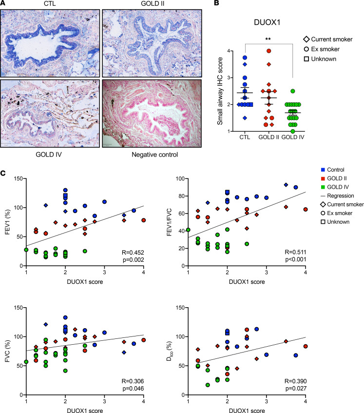
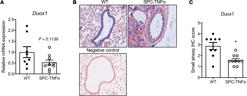
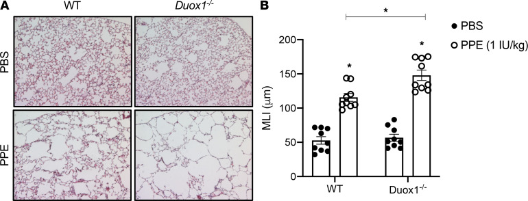
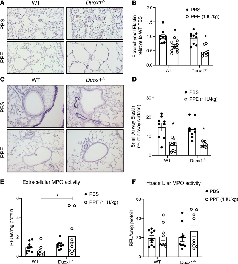
Chronic obstructive pulmonary disease (COPD) is a chronic respiratory disease characterized by small airway remodeling and alveolar emphysema due to environmental stresses such as cigarette smoking (CS). Oxidative stress is commonly implicated in COPD pathology, but recent findings suggest that one oxidant-producing NADPH oxidase homolog, dual oxidase 1 (DUOX1), is downregulated in the airways of patients with COPD. We evaluated lung tissue sections from patients with COPD for small airway epithelial DUOX1 protein expression, in association with measures of lung function and small airway and alveolar remodeling. We also addressed the impact of DUOX1 for lung tissue remodeling in mouse models of COPD. Small airway DUOX1 levels were decreased in advanced COPD and correlated with loss of lung function and markers of emphysema and remodeling. Similarly, DUOX1 downregulation in correlation with extracellular matrix remodeling was observed in a genetic model of COPD, transgenic SPC-TNF-α mice. Finally, development of subepithelial airway fibrosis in mice due to exposure to the CS-component acrolein, or alveolar emphysema induced by administration of elastase, were in both cases exacerbated in Duox1-deficient mice. Collectively, our studies highlight that downregulation of DUOX1 may be a contributing feature of COPD pathogenesis, likely related to impaired DUOX1-mediated innate injury responses involved in epithelial homeostasis.

Keywords: COPD; Cell Biology; Extracellular matrix; Pulmonology.


Conflict of interest statement

Conflict of interest: The authors have declared that no conflict of interest exists.

Figures

Figure 1
Figure 1. DUOX1 loss in small airways of patients with COPD is associated with loss of lung function, emphysema, and airway remodeling.
(A) IHC for DUOX1 (blue) with Nuclear Fast Red counterstaining (red) in lung tissue of a representative control, GOLD II, and GOLD IV patient (including a negative control; original magnification, ×100). (B) Small airway score of DUOX1 in control, GOLD II, and GOLD IV patients. Data are shown as mean ± SEM. **P < 0.01, by 1-way ANOVA. (C) Pearson’s correlations (2-tailed significance) between DUOX1 scores and percentages of FEV1, FVC, and DLCO and FEV1/FVC ratio (determined in ref. 32) in all subjects studied. Colored labels highlight controls (blue), GOLD II (red), and GOLD IV (green) patients, and symbol shapes represent smoking status (current or former smokers). DUOX1, dual oxidase 1; COPD, chronic obstructive pulmonary disease; GOLD II, moderate COPD; GOLD IV, very severe COPD.
Figure 2
Figure 2. Airway epithelial DUOX1 is decreased in SPC-TNF-α mice.
(A) Relative Duox1 mRNA expression in lungs of 6-month-old SPC-TNF-α mice and littermate controls (n = 8–9 from 2 separate experiments; 2-tailed unpaired nonparametric t test). (B and C) Representative staining of DUOX1 protein (in blue; original magnification, ×400) with corresponding small airway IHC score of airway epithelial DUOX1 in SPC-TNF-α mice compared with WT littermate controls (n = 9–10). Data are shown as mean ± SEM. *P < 0.05, by 2-tailed unpaired t test. DUOX1, dual oxidase 1.
Figure 3
Figure 3. Airway epithelial DUOX1 loss in SPC-TNF-α mice is associated with elastin remodeling.
(A–L) IHC analysis (original magnification, ×200) and quantification (percentage of surface area) of elastin and collagen in parenchyma (A–F) and small airways (G–L) in 6-month-old WT and SPC-TNF-α mice. Quantified stainings (n = 9–10) were correlated to small airway DUOX1 scores. Data are shown as mean ± SEM. *P < 0.05, by 2-tailed unpaired t test. DUOX1, dual oxidase 1.
Figure 4
Figure 4. Duox1 deficiency sensitizes airways to acrolein-induced peribronchiolar fibrosis.
C57BL/6J mice were exposed to acrolein (ACR) and analyzed (original magnification, ×200) for collagen by Masson’s trichrome (A and B) or α-smooth muscle actin (C and D). Quantification of staining was based on 6–8 mice per group from 2 separate experiments. Data are shown as mean ± SEM. *P < 0.05, by 2-way ANOVA.
Figure 5
Figure 5. Duox1 deficiency enhances development of elastase-induced emphysema.
WT and Duox1–/– mice were exposed to 50 μL porcine pancreatic elastase (PPE) or PBS control, and lung tissues were analyzed by H&E staining (original magnification, ×100) for alveolar enlargement (A), with corresponding (B) calculation of alveolar mean linear intercept (MLI, μm) as a measure of emphysema. Data are shown as mean ± SEM; n = 9 per group, from 2 separate experiments. *P < 0.05 by 2-way ANOVA.
Figure 6
Figure 6. Development of elastase-induced emphysema is associated with decreased alveolar and small airway elastin levels, and Duox1 deficiency enhances elastase-induced extracellular MPO activity.
Analysis of parenchymal (A and B) and small airway (C and D) elastin levels (original magnification, ×200) in both WT and Duox1–/– mice in response to porcine pancreatic elastase (PPE) or PBS control with Weigert’s Resorcin Fuchsin staining. Analysis of extracellular (E) and intracellular (F) MPO activity in lung tissue homogenates. Data are shown as mean ± SEM; n = 9 per group, from 2 separate experiments. *P < 0.05 by 2-way ANOVA. MPO, myeloperoxidase.

Similar articles

Cited by

References

    1. Hogg JC, et al. The nature of small-airway obstruction in chronic obstructive pulmonary disease. N Engl J Med. 2004;350(26):2645–2653. doi: 10.1056/NEJMoa032158. - DOI - PubMed
    1. McDonough JE, et al. Small-airway obstruction and emphysema in chronic obstructive pulmonary disease. N Engl J Med. 2011;365(17):1567–1575. doi: 10.1056/NEJMoa1106955. - DOI - PMC - PubMed
    1. World Health Organization. Chronic obstructive pulmonary disease (COPD) (Fact Sheet March 2015). http://www.who.int/respiratory/copd/en/ Accessed December 4, 2020.
    1. Rahman I, Adcock IM. Oxidative stress and redox regulation of lung inflammation in COPD. Eur Respir J. 2006;28(1):219–242. doi: 10.1183/09031936.06.00053805. - DOI - PubMed
    1. Janssen R, Wouters EFM. Loss of alveolar attachments as a pathomechanistic link between small airway disease and emphysema. Am J Respir Crit Care Med. 2020;201(7):878–879. doi: 10.1164/rccm.201910-1981LE. - DOI - PMC - PubMed

Publication types

MeSH terms