. 2020 Dec 10;15(12):e0242445.
doi: 10.1371/journal.pone.0242445. eCollection 2020.

ACAD10 protein expression and Neurobehavioral assessment of Acad10-deficient mice

Affiliations

ACAD10 protein expression and Neurobehavioral assessment of Acad10-deficient mice

Kaitlyn Bloom et al. PLoS One. .

Abstract

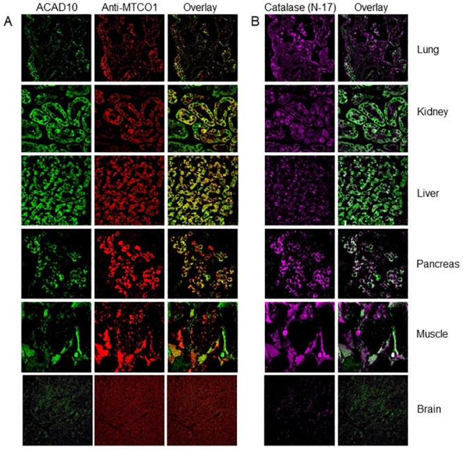
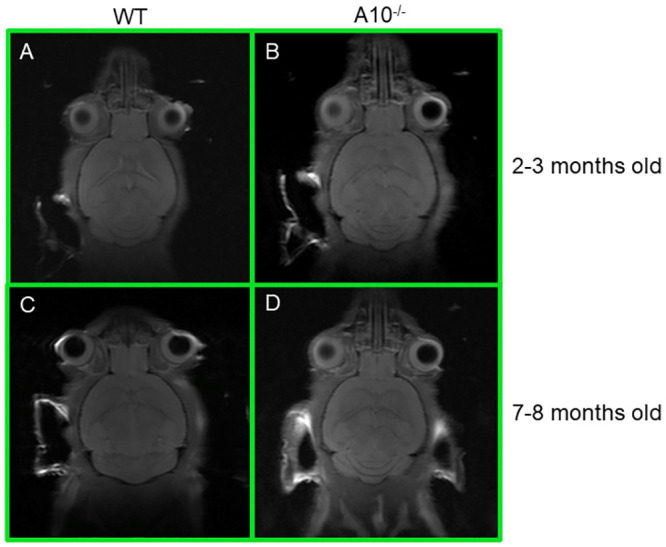
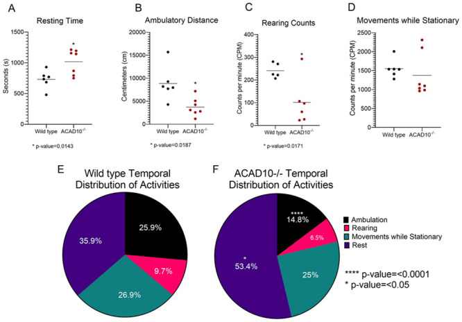
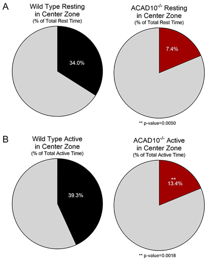
Acyl-CoA dehydrogenase 10 (Acad10)-deficient mice develop impaired glucose tolerance, peripheral insulin resistance, and abnormal weight gain. In addition, they exhibit biochemical features of deficiencies of fatty acid oxidation, such as accumulation of metabolites consistent with abnormal mitochondrial energy metabolism and fasting induced rhabdomyolysis. ACAD10 has significant expression in mouse brain, unlike other acyl-CoA dehydrogenases (ACADs) involved in fatty acid oxidation. The presence of ACAD10 in human tissues was determined using immunohistochemical staining. To characterize the effect of ACAD10 deficiency on the brain, micro-MRI and neurobehavioral evaluations were performed. Acad10-deficient mouse behavior was examined using open field testing and DigiGait analysis for changes in general activity as well as indices of gait, respectively. ACAD10 protein was shown to colocalize to mitochondria and peroxisomes in lung, muscle, kidney, and pancreas human tissue. Acad10-deficient mice demonstrated subtle behavioral abnormalities, which included reduced activity and increased time in the arena perimeter in the open field test. Mutant animals exhibited brake and propulsion metrics similar to those of control animals, which indicates normal balance, stability of gait, and the absence of significant motor impairment. The lack of evidence for motor impairment combined with avoidance of the center of an open field arena and reduced vertical and horizontal exploration are consistent with a phenotype characterized by elevated anxiety. These results implicate ACAD10 function in normal mouse behavior, which suggests a novel role for ACAD10 in brain metabolism.


Conflict of interest statement

The authors have declared that no competing interests exist.

Figures

Fig 1
Fig 1. Western blot survey of ETC subunits and ACADs in mouse heart, liver, muscle, and brain.
A 4–15% SDS-PAGE gel with 50 μg of tissue extract applied per lane was visualized by western blotting with antiserum to the indicated protein. (A) Mitochondrial protein subunits complex III (55 kDa), complex V (48 kDa), complex IV (40 kDa), complex II (30 kDa), and complex I (20 kDa) were visualized in heart, liver, muscle, and brain using a mitochondrial cocktail antibody. (B) Immunostaining for citrate synthase revealed a 37 kDa band. Western blots using anti VLCAD (C) and MCAD (D) antisera showed the appropriate protein sizes of 62 kDa and 42 kDa, respectively.
Fig 2
Fig 2. Immunofluorescent staining of lung, kidney, liver, pancreas, muscle, and brain from human tissues.
(A) Immunofluorescent staining of lung, kidney, liver, pancreas, muscle, and brain from human tissues with antiserum to ACAD10 (left column), the mitochondrial marker MTCO1 (middle column), and the merged image (right column). Lung, pancreas, muscle, and kidney showed co-localization of ACAD10 with the mitochondrial marker. (B) In addition, human tissues were stained with antibodies to the peroxisomal marker catalase (left column). The right column shows the merged images and identifies colocalization of ACAD10 to peroxisomes as well as mitochondria in lung and pancreas. Slides were analyzed using an Olympus FluoView FV1000 confocal microscope with a magnification of 60X.
Fig 3
Fig 3. Micro-MRI of mouse brain.
Mice were visualized using Horizontal bore 7-T MRI system, Bruker Biospin 70/30 with full vital monitoring system. (A) and (B) is the same wild type mouse at 2–3 months old and 7–8 months old, respectively, using RARE-T1 Imaging. The coronal image is whole, live, mouse brain section 11 of 15 sections. (C) and (D) is the same Acad10-deficient mouse at 2–3 months old and 7–8 months old, respectively, using RARE-T1 Imaging. The coronal image is a representative whole, live, mouse brain section 11 of 15 sections. The Acad10-deficient mice do not show any significant changes in brain structure and morphology over time as compared to wild type control mice of the same background.
Fig 4
Fig 4. Open field measures for wild-type and Acad10-deficient mice.
(A-D) Group averages (horizontal lines) of rest time, distance travelled, counts per minute (CPM) of rearing, and CPM of movements while stationary for wild-type (black dots) and Acad10-deficient mice (red dots). During the 30-minute test, Acad10-deficient animals spent statistically significantly more time resting (A), travelled a statistically significantly shorter distance (B), and reared statistically significantly less frequently (C) compared to wild-type mice of the same background (unpaired Student’s T-test with Welch’s correction, p-value <0.05 to < 0.0005). The frequency of movements while stationary (D) did not differ significantly between Acad10-deficient mice and wild-type mice of the same background (unpaired Student’s T-test with Welch’s correction). (E-F) Group averages of the percent of time of the total 30-minute test wild-type mice (E) and Acad10-deficient mice (F) engaged in resting, ambulating, rearing, and movements while stationary (e.g., grooming). Acad10-deficient mice spent a statistically significantly greater percent of time resting and, conversely, statistically significantly smaller percentage of time ambulating, rearing, or engaged in movements while stationary, compared to wild-type mice of the same background (unpaired Student’s T-test with Welch’s correction, p-value <0.05).
Fig 5
Fig 5. Temporal distribution of activities in the open field test by wildtype and Acad10-deficient mice.
(A-B) Group averages of the percent of total rest time or percent of total active time spent in the center (colored segments) versus the surround (grey segments) of the the open field arena for wild-type mice (black) and Acad10-deficient (red) mice. Of the total amount of time mice rested (A) or were active (i.e., engaged in locomotion, rearing, or movements while stationary; B), the percent of time mice did so in the center of the test arena was statistically significantly lower for Acad10-deficient mice compared to wild-type mice of the same background (unpaired Student’s T-test with Welch’s correction, p-value <0.05).

References

    1. Wang Y., et al., Evidence for physical association of mitochondrial fatty acid oxidation and oxidative phosphorylation complexes. J Biol Chem, 2010. 285(39): p. 29834–41. 10.1074/jbc.M110.139493 - DOI - PMC - PubMed
    1. Lehninger, N., and Cox, Principles of Biochemistry, 2nd edition. 1982.
    1. Vockley, J., Bennett MJ, Gillingham MB Mitochondrial Fatty Acid Oxidation Disorders. in The Online Metabolic and Molecular Bases of Inherited Disease 2016.
    1. He M.e.a., Identification and characterization of new long chain Acyl-CoA dehydrogenases. Molecular Genetics and Metabolism, 2011. 102(4): p. 418–429. - PMC - PubMed
    1. Hong M.G., et al., A genome-wide assessment of variability in human serum metabolism. Hum Mutat, 2013. 34(3): p. 515–24. 10.1002/humu.22267 - DOI - PubMed

Publication types

MeSH terms