. 2021 Mar 3;144(2):473-486.
doi: 10.1093/brain/awaa394.

Modulation of beta bursts in subthalamic sensorimotor circuits predicts improvement in bradykinesia

Affiliations

Modulation of beta bursts in subthalamic sensorimotor circuits predicts improvement in bradykinesia

Yasmine M Kehnemouyi et al. Brain. .

Abstract

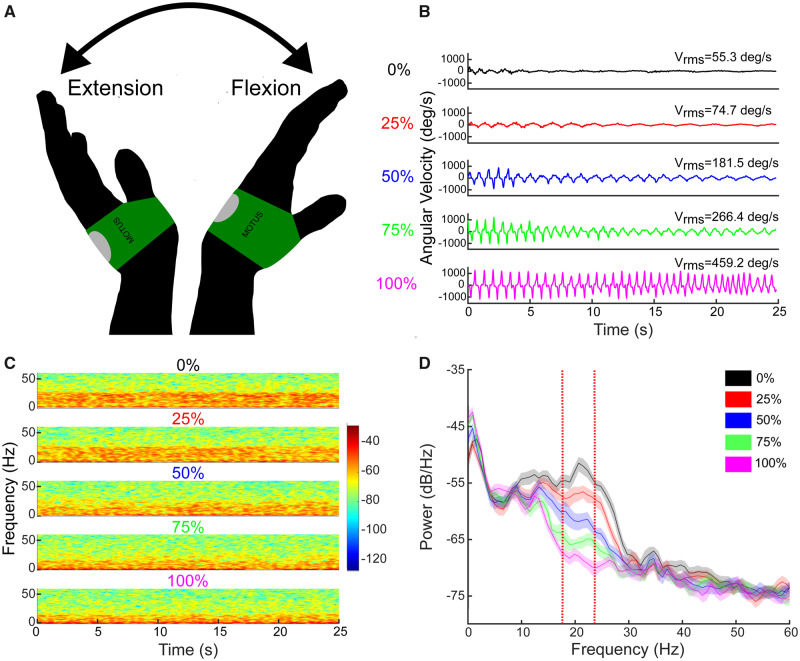
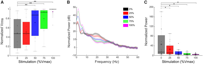
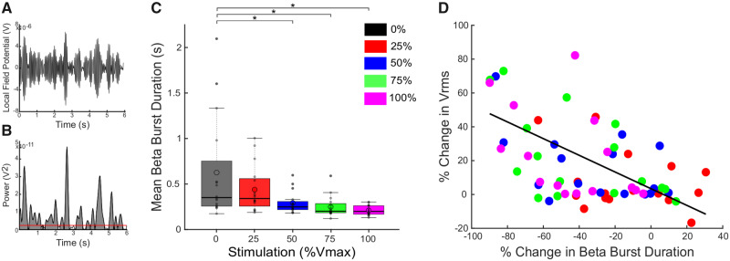
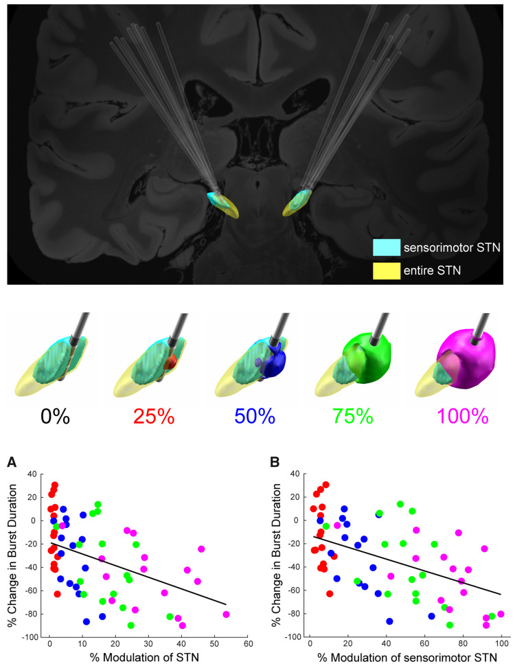
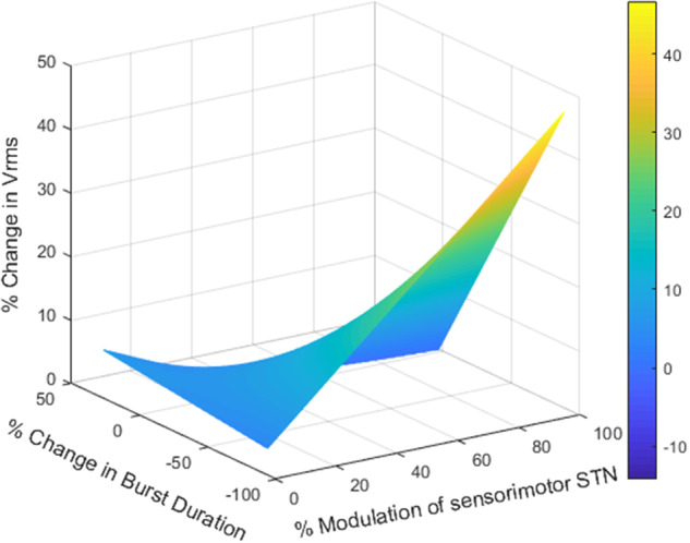
No biomarker of Parkinson's disease exists that allows clinicians to adjust chronic therapy, either medication or deep brain stimulation, with real-time feedback. Consequently, clinicians rely on time-intensive, empirical, and subjective clinical assessments of motor behaviour and adverse events to adjust therapies. Accumulating evidence suggests that hypokinetic aspects of Parkinson's disease and their improvement with therapy are related to pathological neural activity in the beta band (beta oscillopathy) in the subthalamic nucleus. Additionally, effectiveness of deep brain stimulation may depend on modulation of the dorsolateral sensorimotor region of the subthalamic nucleus, which is the primary site of this beta oscillopathy. Despite the feasibility of utilizing this information to provide integrated, biomarker-driven precise deep brain stimulation, these measures have not been brought together in awake freely moving individuals. We sought to directly test whether stimulation-related improvements in bradykinesia were contingent on reduction of beta power and burst durations, and/or the volume of the sensorimotor subthalamic nucleus that was modulated. We recorded synchronized local field potentials and kinematic data in 16 subthalamic nuclei of individuals with Parkinson's disease chronically implanted with neurostimulators during a repetitive wrist-flexion extension task, while administering randomized different intensities of high frequency stimulation. Increased intensities of deep brain stimulation improved movement velocity and were associated with an intensity-dependent reduction in beta power and mean burst duration, measured during movement. The degree of reduction in this beta oscillopathy was associated with the improvement in movement velocity. Moreover, the reduction in beta power and beta burst durations was dependent on the theoretical degree of tissue modulated in the sensorimotor region of the subthalamic nucleus. Finally, the degree of attenuation of both beta power and beta burst durations, together with the degree of overlap of stimulation with the sensorimotor subthalamic nucleus significantly explained the stimulation-related improvement in movement velocity. The above results provide direct evidence that subthalamic nucleus deep brain stimulation-related improvements in bradykinesia are related to the reduction in beta oscillopathy within the sensorimotor region. With the advent of sensing neurostimulators, this beta oscillopathy combined with lead location could be used as a marker for real-time feedback to adjust clinical settings or to drive closed-loop deep brain stimulation in freely moving individuals with Parkinson's disease.

Keywords: Parkinson’s disease; beta oscillations; bradykinesia; deep brain stimulation; local field potentials.


Figures

Figure 1
Figure 1
Example of the effect of stimulation on behaviour and beta power. (A) Illustration of the repetitive wrist flexion-extension (rWFE) task with the Motus gyroscope attached to the dorsum of the hand. (B) Example of angular velocity traces during the rWFE task at each stimulation condition from a representative individual. (C) Spectrogram of STN LFPs during the rWFE task at each stimulation condition. The colour bar represents the power in dB/Hz. (D) PSD diagrams averaged across the rWFE for each stimulation condition. Vertical red lines identify the 6 Hz beta band of interest. Shading represents 95% confidence intervals.
Figure 2
Figure 2
DBS improved behaviour and attenuated beta power. (A) Box plots with individual data overlaid depict the Vrms during rWFE normalized to the maximum Vrms observed for each STN. DBS improved Vrms, with significant differences from no stimulation occurring at 50%, 75%, and 100% Vmax. (B) Grand average PSDs for all STNs during rWFE at each DBS intensity. (C) Grand average box plots with individual data overlaid depicting averaged normalized power in the movement band of each STN during rWFE at each DBS intensity. Stimulation progressively reduced movement band power, with significant differences from no stimulation occurring at all intensities of DBS. Thick horizontal line represents the median and the open circle represents the mean. *P <0.05, **P <0.01.
Figure 3
Figure 3
DBS reduced movement band burst durations during rWFE and shorter burst durations were associated with improvement in bradykinesia. (A) Bandpass filtered LFP in the 6 Hz movement band during a portion of the rWFE task. (B) Envelope of the squared movement band filtered signal in A. The red horizontal line denotes the baseline used for burst identification. Shaded grey regions represent identified bursts. (C) Box plots with individual data overlaid depicting the movement band burst durations for each stimulation intensity. DBS progressively reduced movement band mean burst duration, with significant differences occurring at 50%, 75%, and 100% Vmax. (D) There was a significant correlation between the % change in movement band burst duration and % change in Vrms as calculated by the linear mixed model (P <0.001). Thick horizontal line represents the median and the open circle represents the mean. *P <0.05.
Figure 4
Figure 4
Change in beta power and burst duration was dependent on theoretical amount of tissue modulated in entire STN and sensorimotor STN.Top: DBS lead placement targeting sensorimotor STN (light blue) for all STN leads. Middle: Example of the VTM in one STN at 0%, 25% (red), 50% (blue), 75% (green), and 100% (magenta) Vmax overlaid on the STN. The whole STN is depicted in yellow and the sensorimotor region is highlighted in light blue. Bottom: (A) Relationship between % change in movement band power and % modulation of the STN. (B) Relationship between the % change in movement band burst duration and % modulation of the sensorimotor portion of the STN. The % modulation of both the STN as a whole and of the sensorimotor region of the STN significantly predicted the change in burst duration, P <0.001, with the stronger relationship observed for the sensorimotor portion of the STN.
Figure 5
Figure 5
Change in movement band burst duration interacted with the amount of overlap of the VTM with the sensorimotor STN to predict the increase in Vrms. The heat map represents the 3D interaction among changes in movement band burst duration, % overlap of the VTM with the sensorimotor STN, and % increase in Vrms. Greater reduction in movement band burst duration and greater overlap of the VTM with the sensorimotor STN were associated with larger increases in the % change in Vrms, P <0.05. Colour bar represents the % change in Vrms.

Comment in

References

    1. Afzal MF, Velisar A, Anidi C, Neuville R, Prabhakar V, Bronte-Stewart HM.. Abstract #96: subthalamic neural closed-loop deep brain stimulation for bradykinesia in Parkinson’s disease. Brain Stimul 2019; 12: E33.
    1. Akram H, Georgiev D, Mahlknecht P, Hyam J, Foltynie T, Limousin P, et al.Subthalamic deep brain stimulation sweet spots and hyperdirect cortical connectivity in Parkinson’s disease. Neuroimage 2017; 158: 332–45. - PMC - PubMed
    1. Anderson RWKehnemouyi YMNeuville RSWilkins KBAnidi CMPetrucci MN, . et al. A novel method for calculating beta band burst durations in Parkinson’s disease using a physiological baseline. J Neurosci Methods 2020; 343: 108811.doi: 10.1016/j.jneumeth.2020.108811. - DOI - PMC - PubMed
    1. Anidi C, O'Day JJ, Anderson RW, Afzal MF, Syrkin-Nikolau J, Velisar A, et al.Neuromodulation targets pathological not physiological beta bursts during gait in Parkinson’s disease. Neurobiol Dis 2018; 120: 107–17. - PMC - PubMed
    1. Arlotti M, Marceglia S, Foffani G, Volkmann J, Lozano AM, Moro E, et al.Eight-hours adaptive deep brain stimulation in patients with Parkinson disease. Neurology 2018; 90: e971–6. - PMC - PubMed

Publication types