Review
. 2021 Jun:100-101:207-220.
doi: 10.1016/j.matbio.2020.12.002. Epub 2020 Dec 7.

Role of autophagy in intervertebral disc and cartilage function: implications in health and disease

Affiliations
Review

Role of autophagy in intervertebral disc and cartilage function: implications in health and disease

Vedavathi Madhu et al. Matrix Biol. 2021 Jun.

Abstract

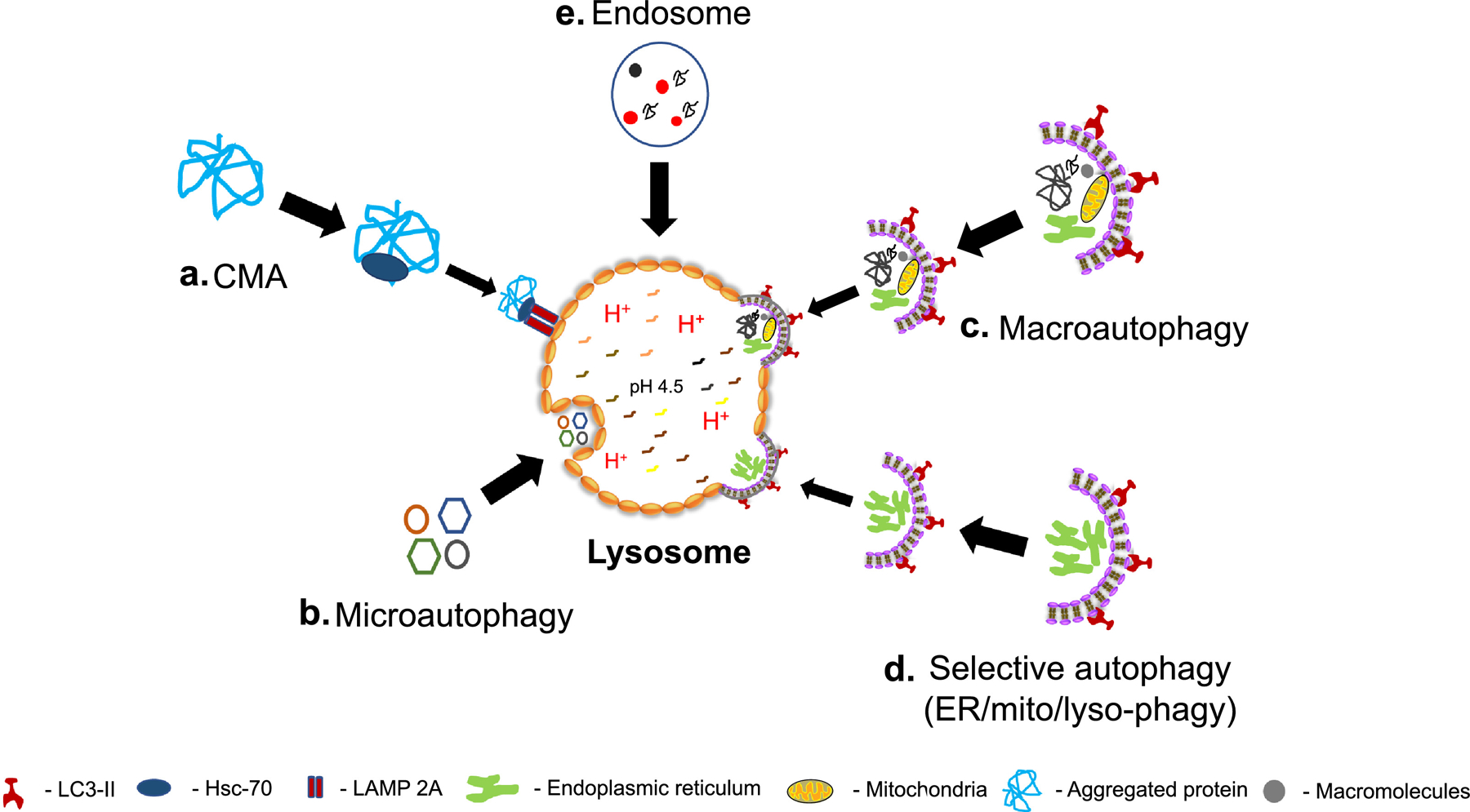
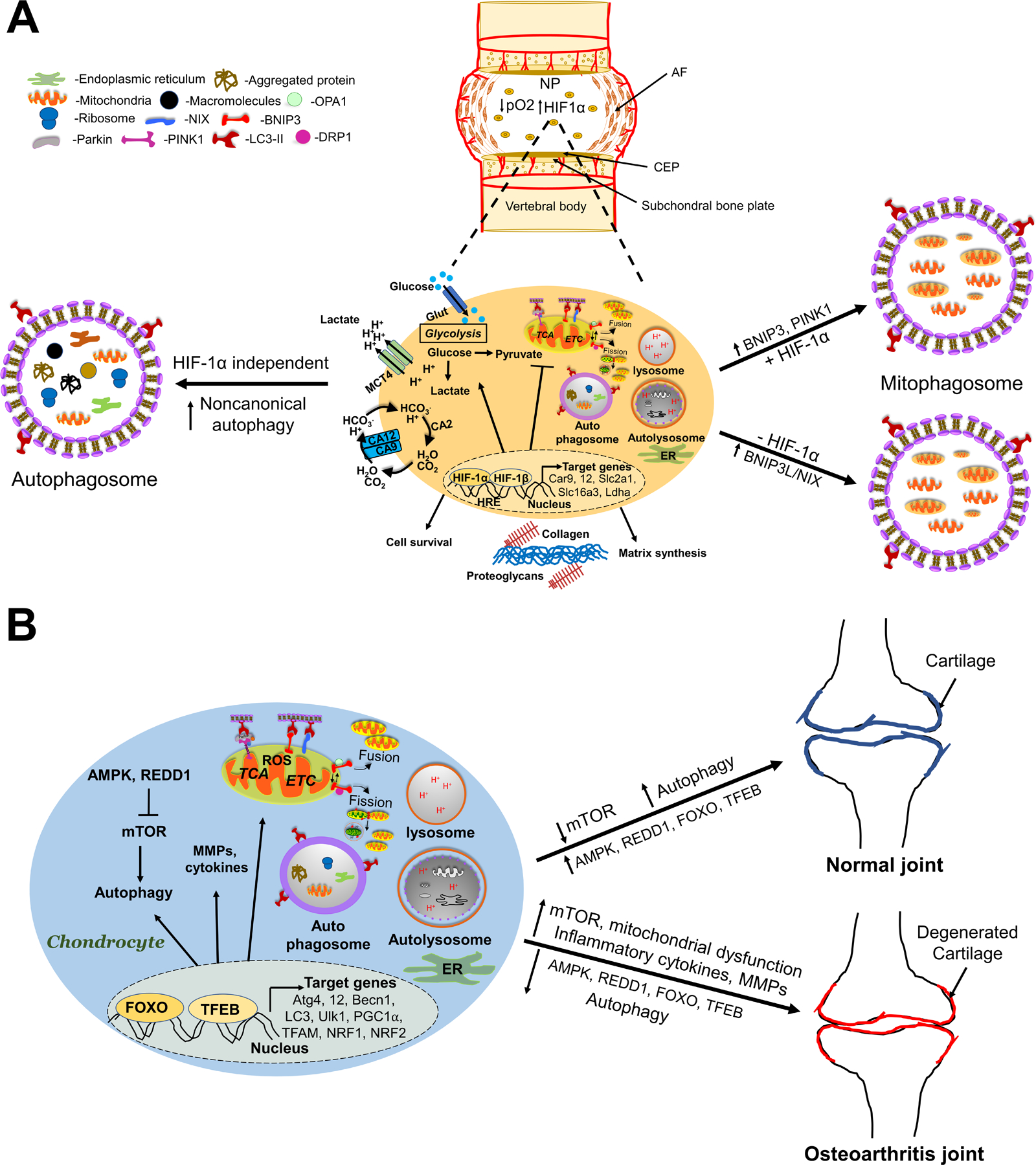
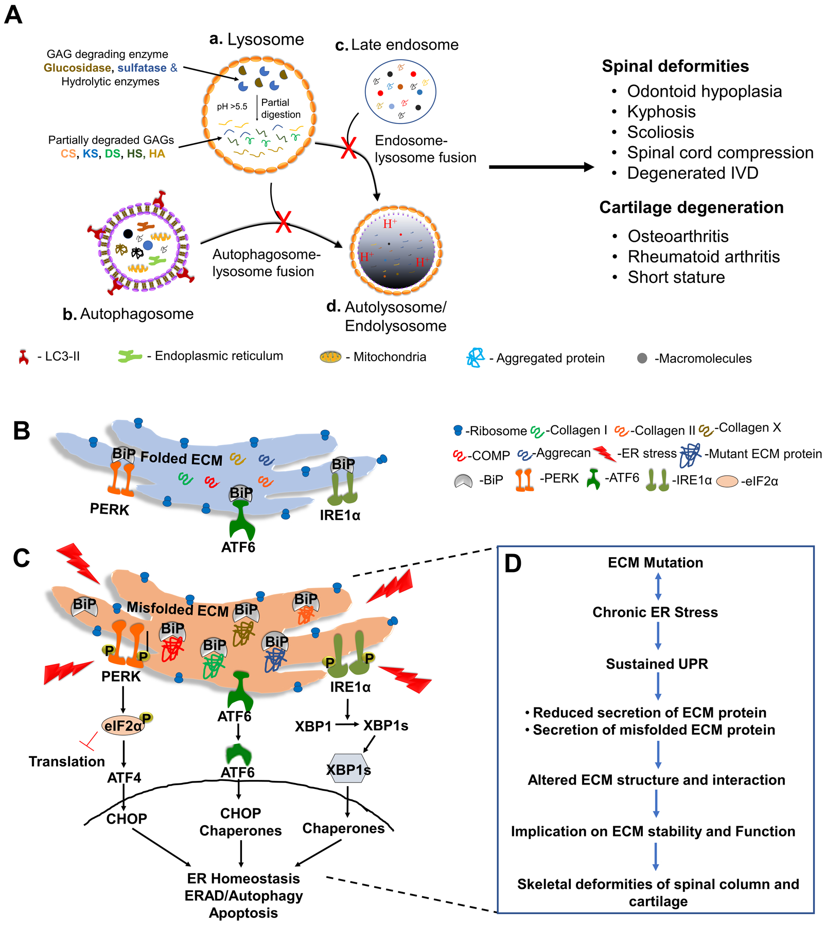
The intervertebral disc and cartilage are specialized, extracellular matrix-rich tissues critical for absorbing mechanical loads, providing flexibility to the joints, and longitudinal growth in the case of growth plate cartilage. Specialized niche conditions in these tissues, such as hypoxia, are critical in regulating cellular activities including autophagy, a lysosomal degradation pathway that promotes cell survival. Mounting evidence suggests that dysregulation of autophagic pathways underscores many skeletal pathologies affecting the spinal column, articular and growth plate cartilages. Many lysosomal storage disorders characterized by the accumulation of partially degraded glycosaminoglycans (GAGs) due to the lysosomal dysfunction thus affect skeletal tissues and result in altered ECM structure. Likewise, pathologies that arise from mutations in genes encoding ECM proteins and ECM processing, folding, and post-translational modifications, result in accumulation of misfolded proteins in the ER, ER stress and autophagy dysregulation. These conditions evidence reduced secretion of ECM proteins and/or increased secretion of mutant proteins, thereby impairing matrix quality and the integrity of affected skeletal tissues and causing a lack of growth and degeneration. In this review, we discuss the role of autophagy and mechanisms of its regulation in the intervertebral disc and cartilages, as well as how dysregulation of autophagic pathways affects these skeletal tissues.

Keywords: Articular cartilage; Autophagy; Chondrodysplasia; Disc degeneration; ECM mutations; ER stress; GAG storage disorders; Growth plate; Intervertebral disc; Lysosomal storage disorders; Osteoarthritis; Unfolded protein response.


Conflict of interest statement

Conflict of Interest

The authors declare no conflicts of interest.

Figures

Figure 1.
Figure 1.. Overview of autophagic pathways.
Lysosomes as catabolic centers of the cell. Lysosomes utilize five distinct pathways for the degradation of cellular material. (a) Chaperone-mediated autophagy (CMA) selectively targets proteins with a KFERQ motif for delivery to lysosomes using Hsc-70 as its chaperone and LAMP-2A as its receptor. (b) Microautophagy involves the pinocytosis of cytosolic regions surrounding lysosomes. (c) Macroautophagy begins with the formation of isolation membranes that sequester regions of the cytosol that include denatured biomolecules and old/damaged organelles into autophagosomes. (d) Selective autophagy results in degradation of organelles by the autolysosome. Cargo is often tagged with ligands such as ubiquitin, enabling it to interact with receptor proteins that can then interact with the autophagosomal membrane. After the cargo is brought to the phagophore, formation of the autophagosome occurs, and this structure can then fuse with the lysosome to form the autolysosome, degrading the cargo. (e) Endosomal degradation by lysosomes predominantly targets late endosomes/multivesicular bodies. Fusion between late endosomes and lysosomes can occur by full fusion/degradation or content mixing, where transient endosomal docking occurs.
Figure 2.
Figure 2.. Schematic showing the role of hypoxia and HIF-1 in regulating autophagic pathways in intervertebral disc.
A. The nucleus pulposus, which is completely devoid of any blood supply and receives its nutrition and O2 from capillaries in the subchondral bone plate by diffusion through the cartilagenous endplates. HIF-1 α, a key transcription factor is robustly expressed in NP cells and is critical for energy metabolism, cell survival, and autophagy. Hypoxia induces a noncanonical autophagy in NP cells, where formation of LC3-positive autophagosomes is independent of HIF-1α. Hypoxia also governs mitochondrial function and morphology through induction of mitochondrial fragmentation. Under hypoxic conditions, expression of PINK1 and translocation of BNIP3 to mitochondria is enhanced, thereby inducing mitophagy. In the absence of HIF-1α, mitophagy in NP cells is maintained through compensatory increases in NIX/BNIP3L levels. B. Autophagic pathways in healthy and OA joints. In healthy joints, as a normal cellular response to stress, AMPK, REDD1, FOXO, and TFEB inhibit mTOR activity and increase autophagy as a cell survival mechanism. In OA joints, autophagy is diminished due to decreased mTOR inhibiting factors, resulting in mitochondrial dysfunction, oxidative stress, inflammatory cytokines, and MMP production.
Figure 3.
Figure 3.. Overview of lysosomal storage disorders and ER-stress-UPR signaling in skeletal pathologies.
A. (a) deficiencies in GAG degradative and proteolytic enzymes prevent complete degradation of GAGs, which leads to accumulation of partially degraded substrates, such as keratin sulfate, dermatan sulfate, heparan sulfate, chondroitin sulfate, and hyaluronic acid, within the lysosomes. Impaired fusion of autophagic and endocytic organelles with lysosomes results in the accumulation of (b) autophagosomes and (c) late endosomes; or the lack of clearance of autophagic and endocytic substrates results in the accumulation of (d) autolysosomes and endolysosomes. These disorders affect the spinal column, cartilage, and other skeletal tissues. B–D. ER-stress-UPR signaling in disc and cartilage. B. Under physiological conditions, Grp78/BiP and other chaperones present in the lumen of ER are responsible for protein folding. Grp78 binds three ER stress receptors – PERK, ATF6, and IRE1 – inhibiting their downstream signaling. C. UPR sensors- IRE1α, PERK, and ATF6α are dissociated from Grp78/BiP and activated when mutated ECM proteins accumulate in the ER. Dissociated Grp78/BiP is sequestered by misfolded protein aggregates. Activated PERK phosphorylates eIF2α, suppressing protein synthesis, decreasing their entry into the ER, and enabling translation of ATF4. ATF4 induces the transcription of genes required to restore ER homeostasis, including that for CHOP. Activated ATF6α is transported to the Golgi apparatus, and its cytosolic domain is cleaved by S1P and S2P proteases, triggering the transcription of the ER chaperones. IRE1 activation leads to splicing of XBP1 RNA to its variant XBP1s, a major transcriptional regulator of UPR genes including BiP, foldases, and protein degradative factors. The initial UPR response is geared towards restoring ER homeostasis. In the case of sustained ER stress, PERK and IRE1 activation mediate ERAD and autophagy. Chronic ER stress ultimately results in cell death/apoptosis. D. The pathophysiology of ECM mutations – accumulation of misfolded protein in the ER lumen induces ER stress, which in turn activates UPR. Consequently, reduced secretion of normal ECM protein, as a result of premature termination or increased secretion of misfolded proteins, compromises ECM structure and function of skeletal tissues, spine/disc, and cartilage.

Similar articles

Cited by

References

    1. Feng Y, He D, Yao Z, Klionsky DJ (2014). The machinery of macroautophagy. Cell Res 24 (1), 24. DOI: 10.1038/cr.2013.168 - DOI - PMC - PubMed
    1. Mizushima N, Levine B, Cuervo AM, Klionsky DJ (2008). Autophagy fights disease through cellular self-digestion. Nature 451, 1069–1075. DOI: 10.1038/nature06639 - DOI - PMC - PubMed
    1. Settembre C, Di Malta C, Polito VA et al. (2011). TFEB links autophagy to lysosomal biogenesis. Science 332, 1429–33. DOI: 10.1126/science.1204592 - DOI - PMC - PubMed
    1. Jin M and Klionsky DJ (2015). The amino acid transporter SLC38A9 regulates MTORC1 and autophagy. Autophagy 11(10), 1709–1710. DOI: 10.1080/15548627.2015.1084461. - DOI - PMC - PubMed
    1. Anding AL and Baehrecke EH (2017). Cleaning house: selective autophagy of organelles. Dev Cell 41(1), 10–22. DOI: 10.1016/j.devcel.2017.02.016 - DOI - PMC - PubMed

Publication types

Substances

LinkOut - more resources