. 2021 Feb 15:227:117617.
doi: 10.1016/j.neuroimage.2020.117617. Epub 2020 Dec 7.

Resolving bundle-specific intra-axonal T2 values within a voxel using diffusion-relaxation tract-based estimation

Affiliations

Resolving bundle-specific intra-axonal T2 values within a voxel using diffusion-relaxation tract-based estimation

Muhamed Barakovic et al. Neuroimage. .

Abstract

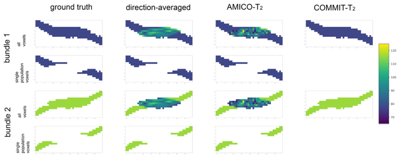
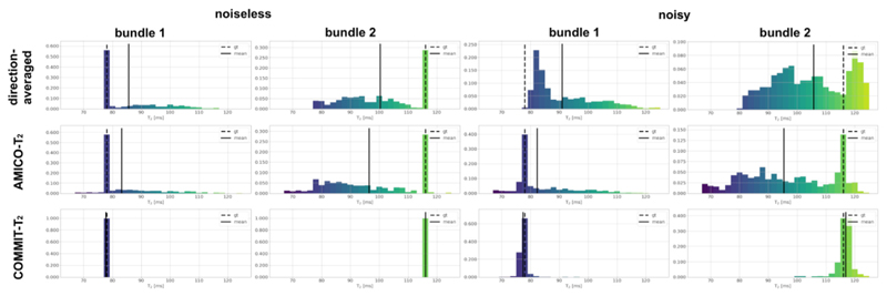
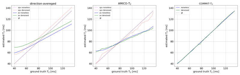
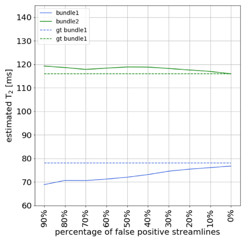
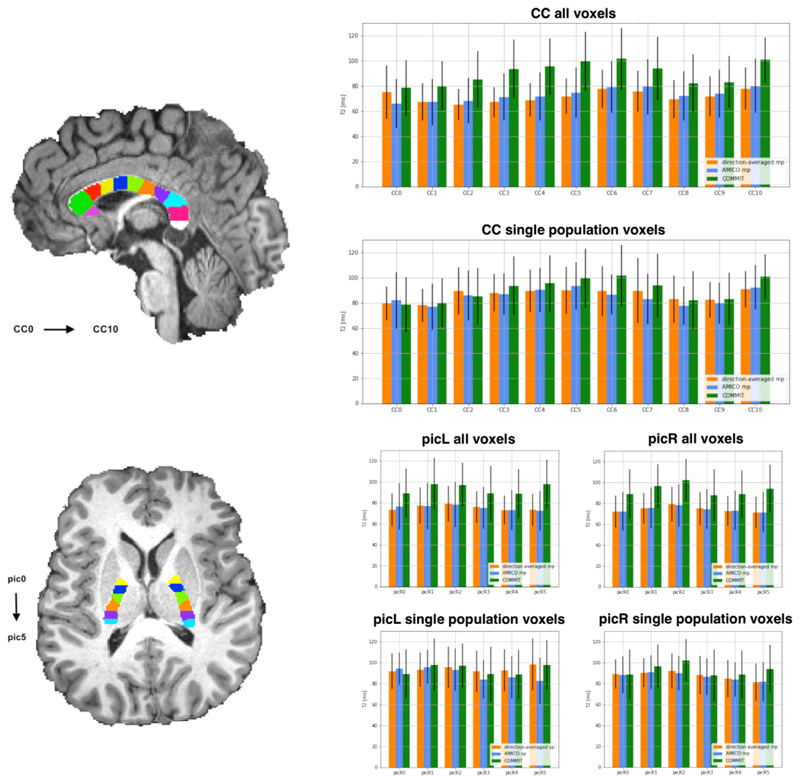
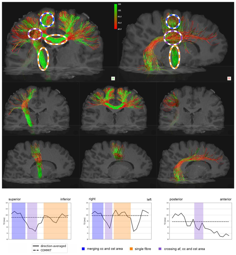
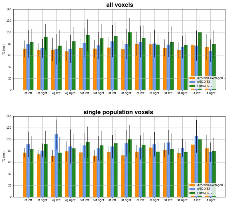
At the typical spatial resolution of MRI in the human brain, approximately 60-90% of voxels contain multiple fiber populations. Quantifying microstructural properties of distinct fiber populations within a voxel is therefore challenging but necessary. While progress has been made for diffusion and T1-relaxation properties, how to resolve intra-voxel T2 heterogeneity remains an open question. Here a novel framework, named COMMIT-T2, is proposed that uses tractography-based spatial regularization with diffusion-relaxometry data to estimate multiple intra-axonal T2 values within a voxel. Unlike previously-proposed voxel-based T2 estimation methods, which (when applied in white matter) implicitly assume just one fiber bundle in the voxel or the same T2 for all bundles in the voxel, COMMIT-T2 can recover specific T2 values for each unique fiber population passing through the voxel. In this approach, the number of recovered unique T2 values is not determined by a number of model parameters set a priori, but rather by the number of tractography-reconstructed streamlines passing through the voxel. Proof-of-concept is provided in silico and in vivo, including a demonstration that distinct tract-specific T2 profiles can be recovered even in the three-way crossing of the corpus callosum, arcuate fasciculus, and corticospinal tract. We demonstrate the favourable performance of COMMIT-T2 compared to that of voxelwise approaches for mapping intra-axonal T2 exploiting diffusion, including a direction-averaged method and AMICO-T2, a new extension to the previously-proposed Accelerated Microstructure Imaging via Convex Optimization (AMICO) framework.

Keywords: COMMIT; Diffusion MRI; Human brain; T(2) relaxometry; Tractography; White matter.


Figures

Fig. 1
Fig. 1. A cross-section of the synthetic phantom.
The phantom simulates a crossing of two fiber bundles with different T2 values: T2,B1 = 78 ms (in blue color) and T2,B2 = 116 ms (in green color). Voxels with a single fiber were differentiated to test the performance of the three methods: the direction-averaged technique, AMICO-T2, and COMMIT-T2.
Fig. 2
Fig. 2
Histograms of the T2i values estimated in the phantom using the three evaluated methods: the direction-averaged technique, AMICO-T2, and COMMIT-T2. The solid black line indicates the mean value of the histogram. The ground-truth (dashed line) values are T2,B1 = 78 ms and T2,B2 116 ms respectively. For more details, see Fig. 1. Results from both the noise-free and noisy datasets are reported. Bundle 1 represents the analysis performed on all the voxels passed by the streamlines defined in bundle 1 (see ground truth Fig. 1). Similarly, Bundle 2 represents the analysis performed on all the voxels passed by the streamlines defined in bundle 2.
Fig. 3
Fig. 3
Comparison of the T2i estimation per bundle in the numerical phantom which varies T2,B2 and keeps a constant T2,B2 =78 ms as described in Section 2.6, against ground-truth for the direction-averaged, AMICO-T2, and COMMIT-T2 methods. The comparison for the direction-averaged and AMICO-T2 methods is performed in a single population voxels (spv) and all voxels (av), which includes a single and multiple populations. The comparison was done on the noiseless data and on noisy data with the same amount of noise estimated after denoising the in-vivo data.
Fig. 4
Fig. 4
Effect of introducing false positive streamlines in the simulated phantom with two fiber bundles with different T2 values: T2,B1 = 78 ms (in red color) and T2,B2 = 116 ms (in green color), respectively. The figure shows the effect of the COMMIT-T2 estimation when a different percent of false positives is introduced and the same numbers of true-positives are removed, while keeping constant the number of streamlines in the dictionary. The ground-truth T2 values are displayed in dashed lines as a reference.
Fig. 5
Fig. 5
T2 estimations using the direction-averaged, AMICO-T2, and COMMIT-T2. The analysis is performed on two commonly-studied white matter fasciculus: the Corpus Callosum (CC) and the Posterior limb Internal Capsule (PIC). The CC was subdivided into 11 ROIs while the PIC was subdivided into 6 ROIs. The ROIs shown in the figure are used to separate the bundles passing through them, i.e. they are not the ROIs from which the measurements are extracted. A comparison is performed for the three methods considering the mean and standard deviation of the voxels appertaining to a bundle. Furthermore, we compared all the voxels of the bundles, multiple populations, and voxels in which only one fiber population is present (defined as having a voxel-based fractional anisotropy (FA) greater than 0.7). Bar height corresponds to mean value and whiskers represent the standard deviation across voxels.
Fig. 6
Fig. 6
Profiles of T2 estimates by using the direction-averaged approach for three well-known bundles: the central parts of the Corticospinal Tract (CST) and the Corpus Callosum (CC), and Arcuate Fasciculus (AF). In the top row of the figure, the three bundles are merged. The orange ovals highlight regions containing a single bundle population, the blue ovals highlight two-bundle populations, and the purple ovals highlight the crossing of all three bundles. The bottom part of the figure shows the profile of the estimated T2s along the CST, CC, and AF bundles, respectively. The profile has been calculated based on the centroid of the bundle; we numbered each voxel of the centroid, then we calculate the distance from each voxel of the bundle mask to the centroid; finally, we calculate the mean of all the voxels associated with each voxel of the centroid. The estimates of intra-axonal T2 from COMMIT-T2 are shown by the dashed line as a reference.
Fig. 7
Fig. 7
T2 values estimated by using the direction-averaged method, AMICO-T2 and COMMIT-T2. The analysis is performed on the arcuate fasciculus (AF), cingulum (CG), inferior fronto-occipital fasciculus (IFOF), inferior longitudinal fasciculus (SLF), optic radiation (OR), superior longitudinal fasciculus (SLF), and uncinate fasciculus (UF). Comparison is performed considering the average along all voxels where the bundle is defined, where multiple populations occurred, and in voxels where only one population is present (by defining a fractional anisotropy (FA) threshold = 0.7, computed by fitting a diffusion tensor model). Bar height indicates the mean value and whiskers represent the standard deviation across voxels.

References

    1. Aboitiz F, Scheibel AB, Fisher RS, Zaidel E. Fiber composition of the human corpus callosum. Brain Res. 1992;598:143–153. - PubMed
    1. Andersson JLR, Skare S, Ashburner J. How to correct susceptibility distortions in spin-echo echo-planar images: application to diffusion tensor imaging. Neuroimage. 2003;20:870–888. doi: 10.1016/S1053-8119(03)00336-7. - DOI - PubMed
    1. Andersson JLR, Sotiropoulos SN. An integrated approach to correction for off-resonance effects and subject movement in diffusion MR imaging. Neuroimage. 2016;125:1063–1078. doi: 10.1016/j.neuroimage.2015.10.019. - DOI - PMC - PubMed
    1. Assaf Y, Basser PJ. Composite hindered and restricted model of diffusion (CHARMED) MR imaging of the human brain. Neuroimage. 2005;27:48–58. doi: 10.1016/j.neuroimage.2005.03.042. - DOI - PubMed
    1. Barakovic M, Girard G, Romascano D, Rafael-Patino J, Descoteaux M, Innocenti GM, Jones DK, Thiran J-P, Daducci A. Assessing feasibility and reproducibility of a bundle-specific framework on in vivo axon diameter estimates at 300mT/m; Proceedings of the ISMRM; 2018.

Publication types