. 2020 Dec 8;6(4):345.
doi: 10.3390/jof6040345.

Glucosylceramide Plays a Role in Fungal Germination, Lipid Raft Organization and Biofilm Adhesion of the Pathogenic Fungus Scedosporium aurantiacum

Affiliations

Glucosylceramide Plays a Role in Fungal Germination, Lipid Raft Organization and Biofilm Adhesion of the Pathogenic Fungus Scedosporium aurantiacum

Victor Pereira Rochetti et al. J Fungi (Basel). .

Abstract

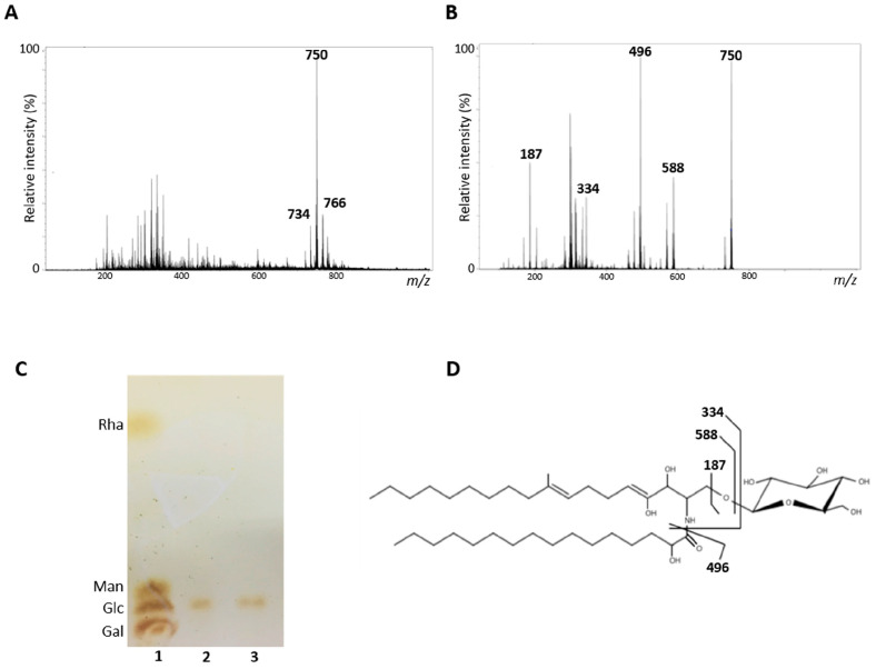
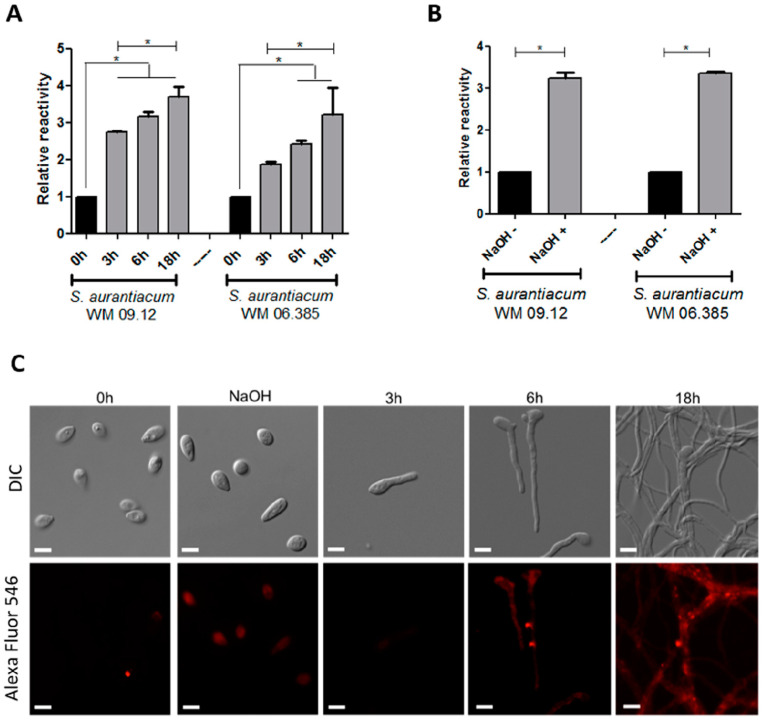
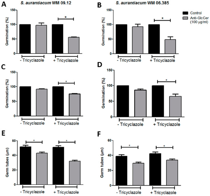
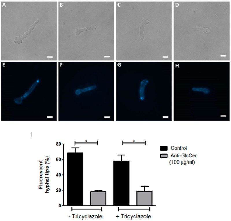
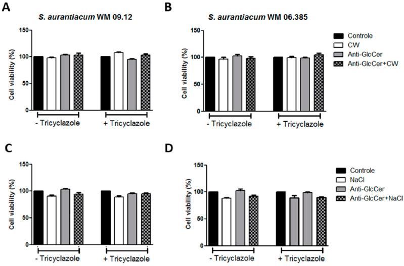
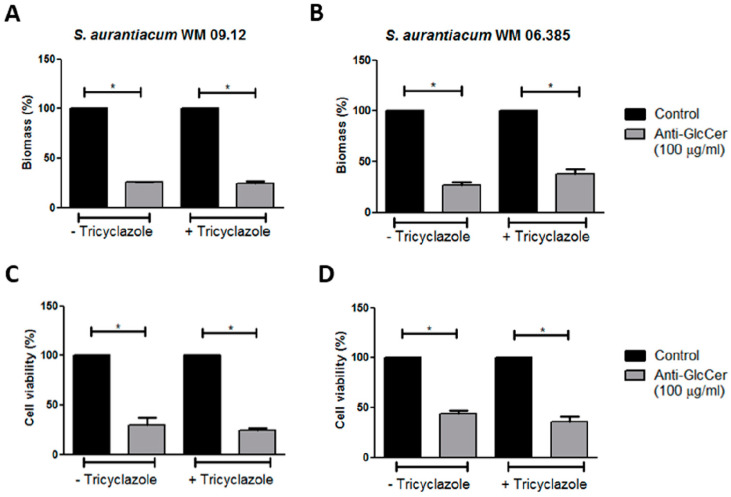
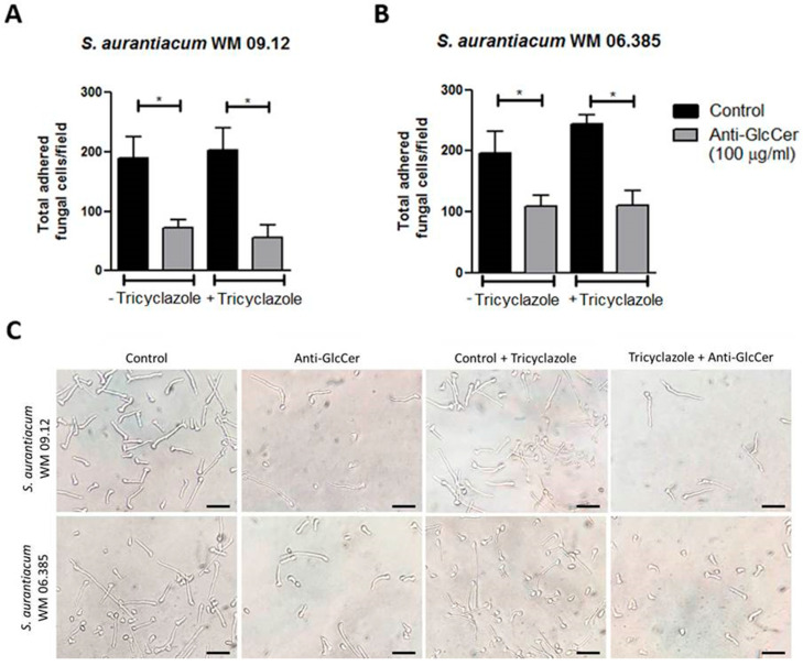
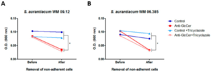
Infections caused by Scedosporium species present a wide range of clinical manifestations, from superficial to disseminated, especially in immunocompromised patients. Glucosylceramides (GlcCer) are glycosphingolipids found on the fungal cell surface and play an important role in growth and pathogenicity processes in different fungi. The present study aimed to evaluate the structure of GlcCer and its role during growth in two S. aurantiacum isolates. Purified GlcCer from both isolates were obtained and its chemical structure identified by mass spectrometry. Using ELISA and immunofluorescence techniques it was observed that germination and NaOH-treatment of conidia favor GlcCer exposure. Monoclonal anti-GlcCer antibody reduced germination when cultivated with the inhibitor of melanin synthesis tricyclazole and also reduced germ tube length of conidia, both cultivated or not with tricyclazole. It was also demonstrated that anti-GlcCer altered lipid rafts organization, as shown by using the fluorescent stain filipin, but did not affect the susceptibility of the cell surface to damaging agents. Anti-GlcCer reduced total biomass and viability in biofilms formed on polystyrene plates. In the presence of anti-GlcCer, germinated S. aurantiacum conidia and biofilms could not adhere to polystyrene with the same efficacy as control cells. These results highlight the relevance of GlcCer in growth processes of S. aurantiacum.

Keywords: Scedosporium; biofilm; fungal growth; glucosylceramide.


Conflict of interest statement

The authors declare no conflict of interest.

Figures

Figure 1
Figure 1
Structural analysis of glucosylceramides (GlcCer) obtained from S. aurantiacum isolates. (A) Electrospray ionization mass spectrometry ESI-MS (ESI-MS1) of purified GlcCer. (B) ESI-MS2 of ion species m/z 750 observed in ESI-MS1. (C) High performance thin layer chromatography of monosaccharides from S. aurantiacum GlcCer. 1. Rhamnose (Rha), mannose (Man), glucose (Glc) and galactose (Gal) standards; 2. Hydrolyzed GlcCer of S. aurantiacum WM 09.12; 3. Hydrolyzed GlcCer of S. aurantiacum WM 06.385. (D) Proposed structure for the major GlcCer species found in S. aurantiacum WM 09.12 and WM 06.385.
Figure 2
Figure 2
GlcCer exposure on S. aurantiacum WM 09.12 and WM 06.385 cell surface detected by monoclonal anti-GlcCer antibody. (A) ELISA of anti-GlcCer (100 µg/mL) bound to S. aurantiacum cells during different germination time points and (B) to conidia after treatment with NaOH 1M. (C) Immunofluorescence analysis of anti-GlcCer (100 µg/mL) reactivity to S. aurantiacum WM 06.385. Scale bar: 5 µm. * p < 0.05.
Figure 3
Figure 3
Anti-GlcCer effect in germination of S. aurantiacum WM 09.12 (A,C,E) and WM 06.385 (B,D,F). Germination rates of S. aurantiacum conidia in the presence of anti-GlcCer (100 µg/mL) for 3 h (A,B) and 6 h (C,D). Germ tube lengths were measured after 6 h of germination with anti-GlcCer (E,F). Conidia were cultivated in the absence (−Tricyclazole) or presence (+Tricyclazole) of tricyclazole (16 µg/mL). * p < 0.05.
Figure 4
Figure 4
Organization of lipid rafts in S. aurantiacum WM 06.385 after 3 h of germination. Filipin staining was performed in untreated (E,F) and tricyclazole-treated (G,H) conidia, in the absence (E,G) or presence of anti-GlcCer (100 µg/mL) (F,H). In (A–D) differential interferential contrast microscopy is presented. Scale bar: 5 µm. The graph in (I) represents the quantification of cells that displayed fluorescent hyphal tips. * p < 0.05.
Figure 5
Figure 5
S. aurantiacum WM 09.12 (A,C) and WM 06.385 (B,D) susceptibility to NaCl and Calcofluor White in the presence of anti-GlcCer (100 µg/mL). S. aurantiacum cells were pre-incubated with anti-GlcCer for 3 h and then cell viability was determined by XTT-reduction assay after 24 h incubation with Calcofluor White (10 µg/mL) (A,B) or NaCl (3%) (C,D). Conidia were cultivated either with (+) or without (−) tricyclazole (16 µg/mL). CW, Calcofluor White.
Figure 6
Figure 6
Influence of anti-GlcCer in S. aurantiacum WM 09.12 (A,C) and WM 06.385 (B,D) biofilm formation. S. aurantiacum biofilms formed in the presence of anti-GlcCer (100 µg/mL) for 24 h had total biomass analyzed by crystal violet staining (A,B) and cell viability determined by XTT-reduction assay (C,D). Conidia were cultivated in the absence (−) or presence (+) of tricyclazole (16 µg/mL). * p < 0.01.
Figure 7
Figure 7
Adhesion of germinated conidia from S. aurantiacum to polystyrene after 6 h incubation with anti-GlcCer (100 µg/mL). Total adhered cells of S. aurantiacum WM 09.12 (A) and WM 06.385 (B) were counted in an inverted microscope. (C) Representative images of adhered fungal cells to polystyrene. Conidia were cultivated in the absence (−) or presence (+) of tricyclazole (16 µg/mL). * p < 0.01. Scale bar: 10 μm.
Figure 8
Figure 8
Anti-GlcCer effect in S. aurantiacum WM 09.12 (A) and WM 06.385 (B) biofilm adhesion to polystyrene. Biofilm growth was measured by determining the optical density at 660 nm before and after removal of non-adherent cells. Conidia cultivated in the absence or presence of tricyclazole (16 µg/mL) were used and grown for 24 h in the presence of anti-GlcCer (100 µg/mL). * p < 0.05.

References

    1. Cortez K.J., Roilides E., Quiroz-Telles F., Meletiadis J., Antachopoulos C., Knudsen T., Buchanan W., Milanovich J., Sutton D.A., Fothergill A., et al. Infections caused by Scedosporium spp. Clin. Microbiol. Rev. 2008;21:157–197. doi: 10.1128/CMR.00039-07. - DOI - PMC - PubMed
    1. Kaltseis J., Rainer J., De Hoog G.S. Ecology of Pseudallescheria and Scedosporium species in human-dominated and natural environments and their distribution in clinical samples. Med. Mycol. 2009;47:398–405. doi: 10.1080/13693780802585317. - DOI - PubMed
    1. Luplertlop N. Pseudallescheria/Scedosporium complex species: From saprobic to pathogenic fungus. J. Mycol. Med. 2018;28:249–256. doi: 10.1016/j.mycmed.2018.02.015. - DOI - PubMed
    1. Blyth C.C., Middleton P.G., Harun A., Sorrell T.C., Meyer W., Chen S.C. Clinical associations and prevalence of Scedosporium spp. in Australian cystic fibrosis patients: Identification of novel risk factors? Med. Mycol. 2010;48(Suppl. 1):S37–S44. doi: 10.3109/13693786.2010.500627. - DOI - PubMed
    1. Lackner M., de Hoog G.S., Verweij P.E., Najafzadeh M.J., Curfs-Breuker I., Klaassen C.H., Meis J.F. Species-specific antifungal susceptibility patterns of Scedosporium and Pseudallescheria species. Antimicrob. Agents Chemother. 2012;56:2635–2642. doi: 10.1128/AAC.05910-11. - DOI - PMC - PubMed

LinkOut - more resources