Review
. 2020 Dec 8;9(12):1244.
doi: 10.3390/antiox9121244.

Metabolic Dysregulation and Neurovascular Dysfunction in Diabetic Retinopathy

Affiliations
Review

Metabolic Dysregulation and Neurovascular Dysfunction in Diabetic Retinopathy

Thangal Yumnamcha et al. Antioxidants (Basel). .

Abstract

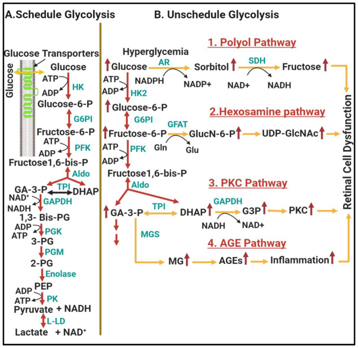
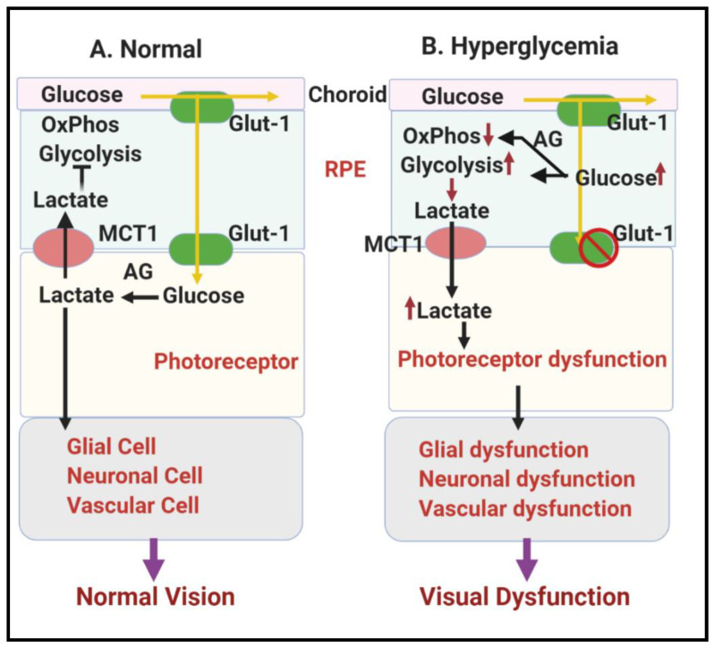
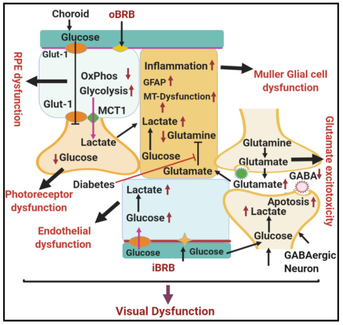
Diabetic retinopathy is a major cause of ocular complications in patients with type 1 and type 2 diabetes in developed countries. Due to the continued increase in the number of people with obesity and diabetes in the United States of America and globally, the incidence of diabetic retinopathy is expected to increase significantly in the coming years. Diabetic retinopathy is widely accepted as a combination of neurodegenerative and microvascular changes; however, which change occurs first is not yet understood. Although the pathogenesis of diabetic retinopathy is very complex, regulated by numerous signaling pathways and cellular processes, maintaining glucose homeostasis is still an essential component for normal physiological functioning of retinal cells. The maintenance of glucose homeostasis is finely regulated by coordinated interplay between glycolysis, Krebs cycle, and oxidative phosphorylation. Glycolysis is the most conserved metabolic pathway in biology and is tightly regulated to maintain a steady-state concentration of glycolytic intermediates; this regulation is called scheduled or regulated glycolysis. However, an abnormal increase in glycolytic flux generates large amounts of intermediate metabolites that can be shunted into different damaging pathways including the polyol pathway, hexosamine pathway, diacylglycerol-dependent activation of the protein kinase C pathway, and Amadori/advanced glycation end products (AGEs) pathway. In addition, disrupting the balance between glycolysis and oxidative phosphorylation leads to other biochemical and molecular changes observed in diabetic retinopathy including endoplasmic reticulum-mitochondria miscommunication and mitophagy dysregulation. This review will focus on how dysregulation of glycolysis contributes to diabetic retinopathy.

Keywords: diabetic retinopathy; endothelial cell; glycolytic overload; glycolytic pathway; hyperglycemia; metabolic deregulation; neurovascular dysfunction.


Conflict of interest statement

The authors declare no conflict of interest

Figures

Figure 1
Figure 1
Schematic diagram depicting the interaction among different cell types in the retina.
Figure 2
Figure 2
The contribution of unscheduled glycolysis to diabetic retinopathy. (A) represents key regulatory steps of scheduled glycolysis, while (B) represents hyperglycemia-linked glycolytic overload and metabolic dysfunction in unscheduled glycolysis resulting from activation of the polyol, hexosamine, PKC, and AGEs pathways, which further cause oxidative stress and inflammation leading to retinal cell dysfunction. Yellow arrows depict metabolic dysfunction in unscheduled glycolysis. Abbreviations: Glucose-6-P, glucose-6-phosphate; Fructose-6-P, fructose-6-phosphate; Fructose 1,6-bis-P, fructose 1,6-bis-Phosphate; GA-3-P, glyceraldehyde-3-phosphate; DHAP, dihydroxyacetone phosphate; 1,3 bis PG, 1,3 diphosphoglycerate; 3-PG, 3-phosphoglycerate; 2-PG; 2-phosphoglycerate; PEP, phosphoenolpyruvate; HK, hexokinase; G6PI, glucose 6-phosphate isomerase; PFK, phosphofructokinase; TPI, triose phosphate isomerase; Aldo, aldolase; GAPDH, glyceraldehyde-3-phosphate dehydrogenase; PGK, phosphoglycerate kinase; PGM, phosphoglycerate mutase; PK, pyruvate kinase; L-LD, L-lactic dehydrogenase; HK2, hexokinase2; OS, osmotic stress; AR, aldolase reductase; SDH, sorbitol dehydrogenase; GlucN-6-P, glucosamine-6-P; UDP-GlcNAc, uridine diphosphate-N-acetylhexosamine; GFAT, glutamine fructose-6-P amidotransferase; Gln, glutamine; Glu, glutamate; G3P, glycerol-3-P; PKC, protein kinase C; MG, methylglycoxal; MGS, methylglycoxal synthase AGE, advance glycation end-products. The figure is adopted with modification from [43].
Figure 3
Figure 3
Schematic representation showing glucose metabolism in RPE and photoreceptors in the RPE-retinal ecosystem. (A) Normal glucose metabolism in RPE and photoreceptors in the RPE-retinal ecosystem under normal physiology leading to normal vision. (B) Hyperglycemia linked metabolic dysfunctions in RPE and photoreceptors in RPE-retinal ecosystem causing visual dysfunction in DR. Abbreviations: RPE, retinal pigment epithelium; Glut-1, Glucose transporter 1; MCT1, monocarboxylate transporter 1; AG, aerobic glycolysis.
Figure 4
Figure 4
Schematic diagram showing normal glucose metabolism in different cells of the neurovascular unit in the retina. Different cell types in the neurovascular unit of the in retina form a metabolic ecosystem to perform normal vision. Abbreviations: RPE, retinal pigmented epithelium; Glut-1, glucose transporter 1; AG, aerobic glycolysis; OxPhos, oxidative phosphorylation; iBRB, inner blood–retinal barrier; oBRB, outer blood–retinal barrier; MCT1, monocarboxylate transporter.
Figure 5
Figure 5
Schematic diagram showing the effect of hyperglycemia on metabolic dysfunction in the diabetic retina. Hyperglycemia-induced dysfunction of the different cell types in the neurovascular unit of the retina disrupts the entire metabolic ecosystem, leading to visual dysfunction in DR. Abbreviations: RPE, retinal pigmented epithelium; Glut-1, glucose transporter 1; AG, aerobic glycolysis; OxPhos, oxidative phosphorylation; iBRB, inner blood–retinal barrier; oBRB, outer blood–retinal barrier; MCT1, monocarboxylate transporter; GFAP, glial fibrillary acidic protein; GABA, gamma-aminobutyric acid.

References

    1. Harris Nwanyanwu K., Talwar N., Gardner T.W., Wrobel J.S., Herman W.H., Stein J.D. Predicting development of proliferative diabetic retinopathy. Diabetes Care. 2013;36:1562–1568. doi: 10.2337/dc12-0790. - DOI - PMC - PubMed
    1. Klein R., Knudtson M.D., Lee K.E., Gangnon R., Klein B.E. The Wisconsin Epidemiologic Study of Diabetic Retinopathy XXIII: The twenty-five-year incidence of macular edema in persons with type 1 diabetes. Ophthalmology. 2009;116:497–503. doi: 10.1016/j.ophtha.2008.10.016. - DOI - PMC - PubMed
    1. Resnikoff S., Pascolini D., Etya’ale D., Kocur I., Pararajasegaram R., Pokharel G.P., Mariotti S.P. Global data on visual impairment in the year 2002. Bull. World Health Organ. 2004;82:844–851. - PMC - PubMed
    1. Sivaprasad S., Gupta B., Gulliford M.C., Dodhia H., Mohamed M., Nagi D., Evans J.R. Ethnic variations in the prevalence of diabetic retinopathy in people with diabetes attending screening in the United Kingdom (DRIVE UK) PLoS ONE. 2012;7:e32182. doi: 10.1371/journal.pone.0032182. - DOI - PMC - PubMed
    1. Simó-Servat O., Hernández C., Simó R. Genetics in diabetic retinopathy: Current concepts and new insights. Curr. Genom. 2013;14:289–299. doi: 10.2174/13892029113149990008. - DOI - PMC - PubMed

LinkOut - more resources