. 2020 Dec 8;21(24):9352.
doi: 10.3390/ijms21249352.

The Inhibitory Role of Rab11b in Osteoclastogenesis through Triggering Lysosome-Induced Degradation of c-Fms and RANK Surface Receptors

Affiliations

The Inhibitory Role of Rab11b in Osteoclastogenesis through Triggering Lysosome-Induced Degradation of c-Fms and RANK Surface Receptors

Manh Tien Tran et al. Int J Mol Sci. .

Abstract

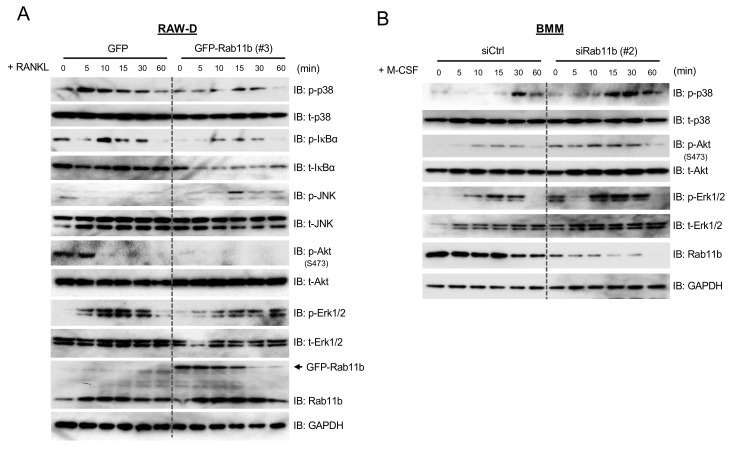
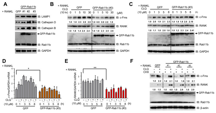
Rab11b, abundantly enriched in endocytic recycling compartments, is required for the establishment of the machinery of vesicle trafficking. Yet, no report has so far characterized the biological function of Rab11b in osteoclastogenesis. Using in vitro model of osteoclasts differentiated from murine macrophages like RAW-D cells or bone marrow-derived macrophages, we elucidated that Rab11b served as an inhibitory regulator of osteoclast differentiation sequentially via (i) abolishing surface abundance of RANK and c-Fms receptors; and (ii) attenuating nuclear factor of activated T-cells c1 (NFATc-1) upstream signaling cascades, following RANKL stimulation. Rab11b was localized in early and late endosomes, Golgi complex, and endoplasmic reticulum; moreover, its overexpression enlarged early and late endosomes. Upon inhibition of lysosomal function by a specific blocker, chloroquine (CLQ), we comprehensively clarified a novel function of lysosomes on mediating proteolytic degradation of c-Fms and RANK surface receptors, drastically ameliorated by Rab11b overexpression in RAW-D cell-derived osteoclasts. These findings highlight the key role of Rab11b as an inhibitor of osteoclastogenesis by directing the transport of c-Fms and RANK surface receptors to lysosomes for degradation via the axis of early endosomes-late endosomes-lysosomes, thereby contributing towards the systemic equilibrium of the bone resorption phase.

Keywords: NFATc-1; RANK; Rab11b; c-Fms; osteoclasts; vesicular transport.


Conflict of interest statement

The authors declare no conflict of interest.

Figures

Figure 1
Figure 1
Rab11b upregulation at the late stage of osteoclastogenesis and the effect of RANKL (300 ng/mL) on osteoclast and monocyte/macrophage colony stimulating factor (M-CSF) (30 ng/mL) for a time course. (A,B) Total RNA was extracted, and cDNA was prepared from RAW-D cells or bone marrow-derived macrophages (BMMs) following a time course of RANKL (300 ng/mL) stimulation. Rab11b mRNA expression levels were analyzed by quantitative Polymerase Chain Reaction (qPCR). Mean ± SD of three independent repeats; n.s, nonsignificant (Student’s t-test), n = 3. (C,D) RAW-D cells or BMMs were treated with RANKL (300 ng/mL) over the indicated time course. Total expression levels of c-Fos, NFATc-1, CTSK, and Rab11b were assessed by immunoblotting and, GAPDH was used as a loading control. (E,F) TRAP-staining was carried out to assess osteoclast formation derived from RAW-D cells (E) and BMMs (F), following the indicated time course of RANKL stimulation. Bars 200 µm.
Figure 2
Figure 2
The effects of Rab11b knockdown and overexpression on osteoclastogenesis. (A) RAW-D cells were transfected with 10 pmol non-targeting siRNA (siCtrl) or two different types of Rab11b-specific siRNA (siRab11b #1 and #2) for 24 h. Then, the cells were cultured for an additional 3 days with media containing RANKL (300 ng/mL). TRAP-staining was performed to evaluate osteoclast formation. Bars 200 µm. (B) The number of TRAP-positive osteoclasts simultaneously having more than 3 nuclei and less than 10 nuclei per viewing field was counted. ** p < 0.01; n.s, nonsignificant (Student’s t-test); compared to the control. (C) The number of TRAP-positive osteoclasts having 10 or more nuclei per viewing field was counted. * p < 0.05; ** p < 0.01 (Student’s t-test); compared to the control. Mean ± SD of three independent repeats. (D) The images of the bone resorption area induced by RAW-D cell-derived osteoclasts transfected with siCtrl or siRab11b before seeded and cultured on the Osteo Assay StripWell upon RANKL (500 ng/mL) stimulation for 7 days. (E) The bone resorption area was measured and analyzed using Image J software. Mean ± SD of triplicate samples. ** p < 0.01 (Student’s t-test), n = 3. (F) After siRNA transfection for 24 h, RAW-D cells and BMMs were differentiated into osteoclasts by being cultured with RANKL (300 ng/mL) and a combination of RANKL (300 ng/mL) + M-CSF (30 ng/mL), respectively, for an additional 72 h. The total expression levels of c-Fms, RANK, NFATc-1, CTSK, and Rab11b were evaluated by immunoblotting, and GAPDH was used as a loading control. (F) The shown data were representative from two independent repeats. (G) RAW-D cells were transfected with retrovirus vectors either encoding Green Fluorescent Protein (GFP) as control or three different clones of GFP-Rab11b (abbreviated by #1, #2, and #3). The cultured cells were incubated with RANKL (300 ng/mL) for 3 days, and subsequently subjected to TRAP-staining analysis to assess the osteoclast formation. Bars 200 µm. (H) The number of TRAP-positive osteoclasts having simultaneously more than 3 and less than 10 nuclei was counted. Mean ± SD of three independent repeats. * p < 0.05, ** p < 0.01 (Student’s t-test); compared to the control. Bars 200 µm. (I) The number of TRAP-positive osteoclasts having 10 or more nuclei per viewing field was counted. Mean ± SD of three independent repeats. ** p < 0.01. (J) The bone-resorbing activities of the RAW-D-derived osteoclasts expressing GFP or GFP-Rab11b (clone #3). The cells were seeded and cultured on the Osteo Assay Stripwell Plates upon RANKL (500 ng/mL) stimulation for 10 days. (K) The images of the bone-resorption area were measured and analyzed using Image J software. Mean ± SD of the triplicate repeats. ** p < 0.01 (Student’s t-test) for the indicated comparisons. (L) The total expression levels of c-Fms, RANK, NFATc-1, CTSK, and Rab11b were evaluated by immunoblotting, and GAPDH was used as a loading control. The shown data were the representative from three independent repeats.
Figure 3
Figure 3
The effects of exogenous expression of Rab11b on NFATc-1 stabilization in response to (Ca2+i) elevation. (A) RAW-D cells expressing GFP or GFP-Rab11b (clone #3) was incubated with RANKL (300 ng/mL) over a time course, and subsequently harvested. The cell lysates were subjected to SDS-PAGE followed by immunoblotting for the detection of CTSK, NFATc-1, Rab11b, and GAPDH was used as a loading control. The representative data were obtained from two independent repeats. (B) Cytosolic and nuclear fractions were prepared from the RAW-D cell-derived osteoclasts expressing GFP or one of three different clones of GFP-Rab11b (#1, #2, and #3) upon RANKL (300 ng/mL) stimulation for 3 days. Cytosolic and nuclear fractions were subjected to SDS-PAGE and immunoblot analysis for detection of NFATc-1, Rab11b, and Histone H3 and GAPDH was used as the cytosolic and nuclear markers, respectively. The shown data were representative of two independent repeats. (C) (Ca2+i) oscillation in RAW-D cells expressing GFP or GFP-Rab11b (clone #3) in response to RANKL stimulation. After RANKL (300 ng/mL) addition for 2 days, the cells were washed by the serum (−/−) media, and subsequently loaded with 1 μM Cal-590 for 1 h. The cells were then washed and analyzed by Olympus Uplsapo 10X. Bars 100 µm. (D) The (Ca2+i) fluorescent intensities were measured and analyzed by Image J software. Mean ± SD of two independent repeats. (E) Quantitative Real Time-PCR (RT-PCR) analysis of NFATc-1 mRNA expression levels obtained from RAW-D cell-derived osteoclasts expressing GFP or one of two different clones of GFP-Rab11b (#2 and #3) after 3 days of RANKL (300 ng/mL) treatment. Mean ± SD of triplicate repeats. ** p < 0.01 (Student’s t-test), compared to the control (GFP). (F) Osteoclasts differentiated from RAW-D cell expressing GFP or one of two different clones of GFP-Rab11b (#2 and #3) upon RANKL (300 ng/mL) for 3 days. The cells were incubated with MG132 (10 μM for 4 h) before being lysed and subjected to anti-IgG or anti-NFATc-1 antibody IP followed by anti-Ub or anti-Ub48 with immunoblot analysis. The densitometer reading of polyubiquitinated NFATc-1 levels was measured underneath from 5th to 6th (for #2) or from 7th to 8th (for #3) lanes. The 5th or 7th lane was arbitrarily set as 1.0. The given results were obtained from two independent repeats.
Figure 4
Figure 4
The effects of Rab11b on NFATc-1 upstream signaling cascades in macrophages stimulated by RANKL and M-CSF, respectively. (A) RAW-D cells expressing GFP or GFP-Rab11b (clone #3) were incubated with serum (−/−) culture media in the absence of RANKL. After RANKL (300 ng/mL) supplementation, the cells were incubated for the indicated times, and subsequently collected. The cell lysates were subjected to SDS-PAGE followed by immunoblotting for the detection of total expression levels of p-p38, t-p38, p-IκBα, t-IκBα, p-JNK, t-JNK, p-Akt (S473), t-Akt, p-Erk 1/2, t-Erk 1/2, Rab11b, and GAPDH was used as a loading control. The shown data were obtained from two independent repeats. (B) BMMs transfected with non-targeting siRNA (siCtrl) or Rab11b siRNA (siRab11b, type 2) were pre-incubated with serum (−/−) culture media in the absence of M-CSF. Then, the cells were incubated with M-CSF (50 ng/mL) for the indicated periods, and subsequently harvested. The cell lysates were subjected to SDS-PAGE followed by immunoblotting for the detection of p-p38, t-p38, p-Akt (S473), t-Akt, p-Erk 1/2, t-Erk 1/2, Rab11b, and GAPDH was used as a loading control.
Figure 5
Figure 5
Subcellular localization of Rab11b in RAW-D cell-derived osteoclasts, and the effects of Rab11b overexpression on size-based modification of organelles. (A) The osteoclasts derived from RAW-D cells stably expressing GFP-Rab11b (clone #1) (green) were seeded on cover glasses with fixation and permeabilization of 0.2% Triton X-100 in PBS, and subsequently reacted with one of the antibodies against Rab5, Rab7, MG130, LAMP1, or KDEL (red, as indicated) that are specific markers for early endosomes, late endosomes, Golgi complex, lysosomes, or endoplasmic reticulum, respectively. Osteoclast DNA was stained with 4′,6-diamidino-2-phenylindole (DAPI) (blue). Specific regions of interest within a field are shown by yellow boxes in a magnified form on the left-hand top of each image it was taken from. Specific regions of interest within a field, designated by yellow boxes, are magnified on the right side of each image it was taken from. Mean ± SD of three independent repeats. Scale bar: 10 μm. (B,C) GFP-Rab11b co-localization with Rab5, Rab7, MG130, LAMP1 and KDEL was determined and evaluated by Pearson coefficient (B) or by color threshold analysis (C) using Fiji/ImageJ on at least 4 cells. (DF) RAW-D cells stably expressing GFP or GFP-Rab11b were fixed and permeabilized with 0.2% Triton X-100, and stained with the specific antibodies to detect (D) Rab5 (red), (E) Rab7 (red), and (F) MG130 (red). The images were captured by confocal laser microscopy. The particle size was measured by (μm2) using ImageJ (shown on the right side). Mean ± SD of at least four independent repeats. * p < 0.05, n.s., nonsignificant (Student’s t-test).
Figure 6
Figure 6
The effects of CLQ-mediated lysosomal function on endogenous levels of c-Fms and RANK in RAW-D cell-derived osteoclasts. (A) RAW-D cells expressing GFP or one of three different types of GFP-Rab11b (#1, #2, and #3) were treated with RANKL (300 ng/mL) for 3 days. The immunoblotting analysis of endogenous levels of LAMP1, a specific lysosomal receptor, and two lysosomal enzymes, Cathepsins B and D was done. GAPDH was used as a loading control. The shown data was representative of two independent repeats. (B) RAW-D cells expressing GFP or GFP-Rab11b (type #3) was pre-treated with RANKL (300 ng/mL) for 3 days before incubation with CLQ (0, 1, 5, 10, and 30 μM) for 10 h. The WB analysis of endogenous levels of c-Fms, RANK, and Rab11b was done. GAPDH was used as a loading control. The shown data was representative of two independent repeats. (C) RAW-D cells expressing GFP or GFP-Rab11b (type #3) were pre-treated with RANKL (300 ng/mL) for 3 days before incubation with CLQ (10 μM) over a time course (0, 1, 3, 5, and 8 h). The WB analysis of endogenous levels of c-Fms, RANK, and Rab11b was done. GAPDH was used as a loading control. (D,E) Total RNA was extracted, and cDNA was prepared from osteoclasts differentiated from RAW-D cells expressing GFP or GFP-Rab11b (type #3), following RANKL (300 ng/mL) stimulation for 3 days. Expression levels of c-Fms (D) and RANK (E) mRNAs were analyzed by qRT-PCR. Mean ± SD of two independent repeats. * p < 0.05, ** p < 0.01 (Student’s t-test). (F) RAW-D cells expressing GFP or one of three different types of GFP-Rab11b (#1, #2, and #3) were pre-treated with RANKL (300 ng/mL) for 3 days. The cells were subsequently treated with CHX (20 μg/mL) and simultaneously with or without CLQ (10 μM) for 5 h. The WB analysis of endogenous levels of c-Fms, RANK, and Rab11b was done. β-actin was used as a loading control. The shown data was the representative of two independent repeats.
Figure 7
Figure 7
The effects of Rab11b overexpression on lysosome-mediated proteolysis of c-Fms and RANK receptors in osteoclasts. (A) RAW-D cells expressing GFP or three different types of GFP-Rab11b (#1, #2, and #3) were stimulated with RANKL (300 ng/mL) for 3 days. The cells were fractionated into two separate fractions including biotinylated fraction and total fraction as the protocol indicated in the “Material and Methods” section. The surface and total levels of c-Fms and RANK were evaluated by biotinylation pulldown and immunoblotting, respectively. (B) The densitometry quantification of bands indicating surface expression of c-Fms and RANK receptors was shown in the presence of GFP-Rab11b as a percentage of control (GFP). Mean ± SD of three independent repeats; * p < 0.05, ** p < 0.01 (Student’s t-test). (C) RAW-D cells were transfected with scrambled siRNA (siCtrl) or one of two different Rab11b siRNAs (Rab11b siRNA #1, Rab11b siRNA #2), followed by RANKL (300 ng/mL) stimulation for 3 days. The cells were fractionated into two separate fractions including biotinylated fraction and total fraction as the above protocol. The surface and total levels of c-Fms and RANK were evaluated by biotinylation pulldown and immunoblotting, respectively. (D) The densitometry quantification of bands indicating surface expression of c-Fms and RANK receptors was shown in the presence of Rab11b siRNAs as a percentage of control (siCtrl). Mean ± SD of three independent repeats; * p < 0.05, ** p < 0.01 (Student’s t-test). (E) RAW-D cells expressing GFP or GFP-Rab11b (#1) were stimulated with RANKL (300 ng/mL) for 3 days, followed by treatment with a combination of MG132 (20 μM) and CHX (20 μg/mL) for 0, 1, 3, and 5 h. Total levels of c-Fms and RANK were evaluated by immunoblotting. (F,G) The densitometry quantification of bands indicating total expression of c-Fms (F) and RANK (G) was shown as a percentage of the 1st lane for the GFP group referred to as the control (Ctrl), and the 5th lane for the GFP-Rab11b group referred to as the control (Ctrl). Mean ± SD of three independent repeats. ** p < 0.01, *** p < 0.001 (Student’s t-test). (H) Cell viability was assessed by the cellular ATP content measurement using the Cell Titer Glo Assay system. After stimulated with RANKL (300 ng/mL) for 3 days, RAW-D cells were co-treated with CHX (20 μg/mL) and CLQ (10 μM) over a time course. The values were the average of triplicate determinations with SD indicated by error bars. n.s., nonsignificant (Student’s t-test). The experiments were repeated thrice.
Figure 8
Figure 8
The model epitomizing the findings of our current works. c-Fms and RANK receptors were early internalized into early endosomes, and transported to late endosomes by the transport vesicles, which was subsequently fused with lysosomes for proteolysis of c-Fms and RANK surface receptors. Both homologs, Rab11a and b, upregulated at a late stage of osteoclast differentiation dictated the transport of c-Fms and RANK surface receptors to the lysosome via the axis of early and late endosomes-lysosomes. Rab11-mediated lysosomal proteolysis of c-Fms and RANK receptors sequentially weakened the osteoclastogenic signaling cascades, inhibited the nuclear translocation of the transcription factor, NFATc-1, reduced the expression level of CTSK, and eventually abolished osteoclastogenesis (bone resorption).

Similar articles

Cited by

References

    1. Boyle W.J., Simonet W.S., Lacey D.L. Osteoclast differentiation and activation. Nature. 2003;423:337–342. doi: 10.1038/nature01658. - DOI - PubMed
    1. Väänänen H.K., Zhao H., Mulari M., Halleen J.M. The cell biology of osteoclast function. (Pt 3)J. Cell Sci. 2000;113:377–381. - PubMed
    1. Kim J.H., Kim N. Signaling Pathways in Osteoclast Differentiation. Chonnam Med. J. 2016;52:12–17. doi: 10.4068/cmj.2016.52.1.12. - DOI - PMC - PubMed
    1. Ballanti P., Minisola S., Pacitti M.T., Scarnecchia L., Rosso R., Mazzuoli G.F., Bonucci E. Tartrate-resistant acid phosphate activity as osteoclastic marker: Sensitivity of cytochemical assessment and serum assay in comparison with standardized osteoclast histomorphometry. Osteoporos. Int. 1997;7:39–43. doi: 10.1007/bf01623458. - DOI - PubMed
    1. Everts V., Korper W., Hoeben K.A., Jansen I.D., Bromme D., Cleutjens K.B., Heeneman S., Peters C., Reinheckel T., Saftig P., et al. Osteoclastic Bone Degradation and the Role of Different Cysteine Proteinases and Matrix Metalloproteinases: Differences Between Calvaria and Long Bone. J. Bone Miner. Res. 2006;21:1399–1408. doi: 10.1359/jbmr.060614. - DOI - PubMed

Substances

LinkOut - more resources