. 2020 Dec 10;20(1):484.
doi: 10.1186/s12886-020-01734-0.

Increased Association of Deamidated αA-N101D with Lens membrane of transgenic αAN101D vs. wild type αA mice: potential effects on intracellular ionic imbalance and membrane disorganization

Affiliations

Increased Association of Deamidated αA-N101D with Lens membrane of transgenic αAN101D vs. wild type αA mice: potential effects on intracellular ionic imbalance and membrane disorganization

Om Srivastava et al. BMC Ophthalmol. .

Abstract

We have generated two mouse models, in one by inserting the human lens αAN101D transgene in CRYαAN101D mice, and in the other by inserting human wild-type αA-transgene in CRYαAWT mice. The CRYαAN101D mice developed cortical cataract at about 7-months of age relative to CRYαAWT mice. The objective of the study was to determine the following relative changes in the lenses of CRYαAN101D- vs. CRYαAWT mice: age-related changes with specific emphasis on protein insolubilization, relative membrane-association of αAN101D vs. WTαA proteins, and changes in intracellular ionic imbalance and membrane organization.

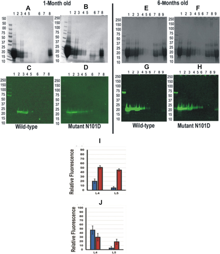
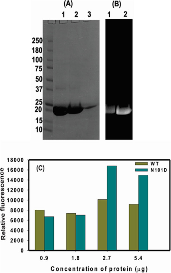
Methods: Lenses of varying ages from CRYαAWT and CRYαAN101D mice were compared for an age-related protein insolubilization. The relative lens membrane-association of the αAN101D- and WTαA proteins in the two types of mice was determined by immunohistochemical-, immunogold-labeling-, and western blot analyses. The relative levels of membrane-binding of recombinant αAN101D- and WTαA proteins was determined by an in vitro assay, and the levels of intracellular Ca2+ uptake and Na, K-ATPase mRNA were determined in the cultured epithelial cells from lenses of the two types of mice.

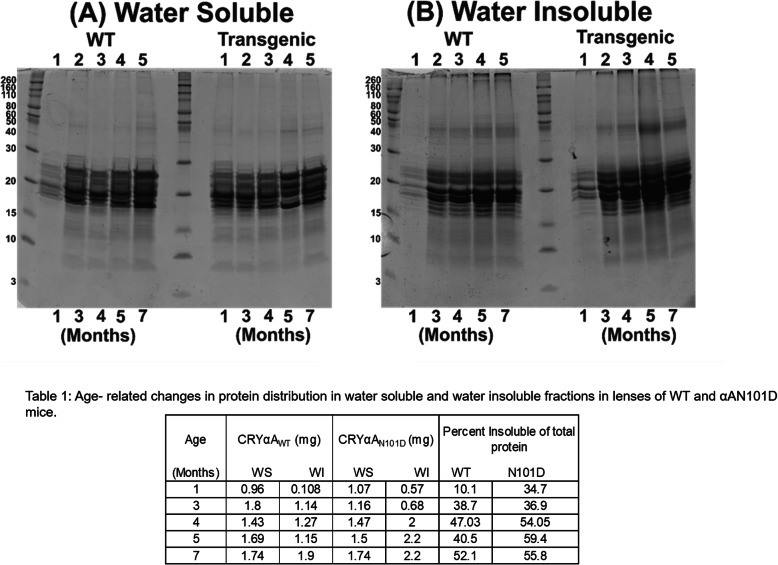
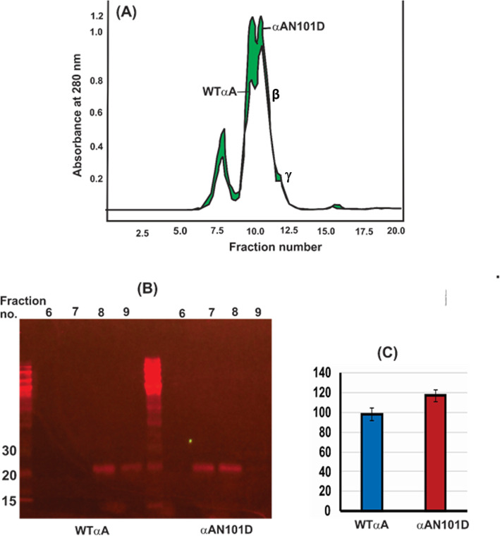
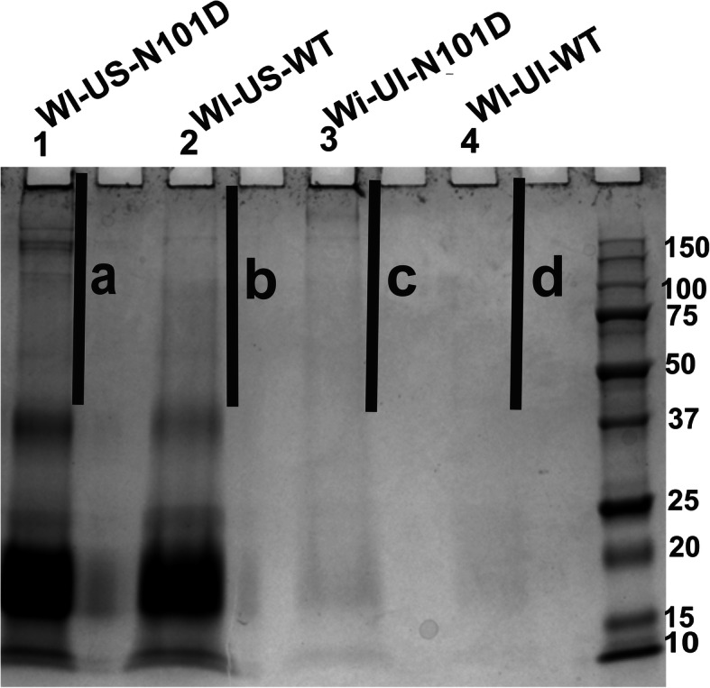
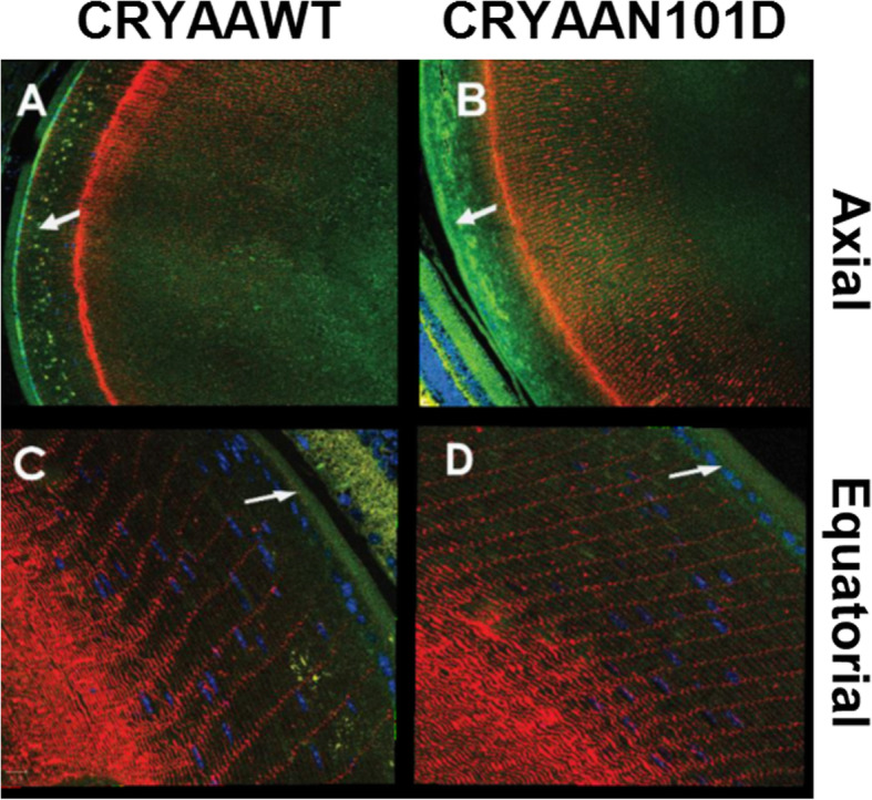
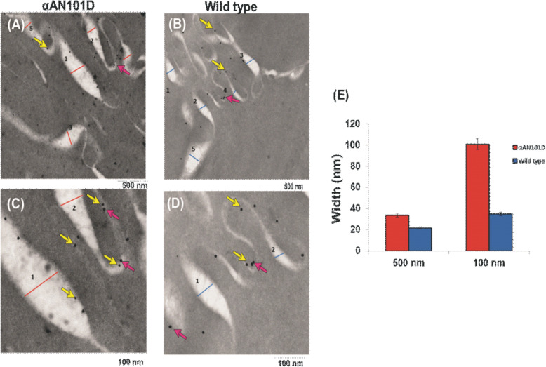
Results: Compared to the lenses of CRYαAWT, the lenses of CRYαAN101D mice exhibited: (A) An increase in age-related protein insolubilization beginning at about 4-months of age. (B) A greater lens membrane-association of αAN101D- relative to WTαA protein during immunogold-labeling- and western blot analyses, including relatively a greater membrane swelling in the CRYαAN101D lenses. (C) During in vitro assay, the greater levels of binding αAN101D- relative to WTαA protein to membranes was observed. (D) The 75% lower level of Na, K-ATPase mRNA but 1.5X greater Ca2+ uptake were observed in cultured lens epithelial cells of CRYαAN101D- than those of CRYαAWT mice.

Conclusions: The results show that an increased lens membrane association of αAN101D--relative WTαA protein in CRYαAN101D mice than CRYαAWT mice occurs, which causes intracellular ionic imbalance, and in turn, membrane swelling that potentially leads to cortical opacity.

Keywords: Cataract; Crystallins; Deamidation; Lens; Post-translational modifications; Transgenic mice.


Conflict of interest statement

No competing interest.

Figures

Fig. 1
Fig. 1
SDS-PAGE analysis of WS- and WI-proteins from lenses of CRYαAWT- and CRYαAN101D mice at different ages. a. Coomassie blue stained gel of WS protein from both WT and Transgenic mice at different ages as indicated at the bottom of the gel (months). And the numbers 1–5 at the top of the gel indicate lanes. b Coomassie blue stained gel of WI protein from both WT and Transgenic mice at different ages as indicated at the bottom of the gel (months).). Note that a greater insolubilization and aggregated proteins (Mr > 30 kDa) were seen in WI-proteins of lenses of 4-month and older CRYαAN101Dmice compared to age-matched lenses from CRYαAWT mice. The Table 1. shows quantification of protein levels in the WS- and WI-protein fractions of lenses of different ages from CRYαAN101D and CRYαAWT mice. Gel images are not cropped
Fig. 2
Fig. 2
Size-exclusion HPLC (using a G-4000PWXL column) and SDS-PAGE analysis of WS-HMW proteins eluted in the void volume. a HPLC-protein elution profiles at 280 nm of WS-proteins from lenses of 5-month-old CRYαAN101D - and CRYαAWT mice. The green region shows the difference in the A280 profiles of WS-proteins from the two type of lenses. b Western blot analysis of the void volume peaks (constituted by the fraction no. 6 to 9 in [A]) following HPLC separation of WS-proteins from lenses of CRYαAN101D - and CRYαAWT mice. Note that in the CRYαAWT lenses, the αA-immunoreactive bands were in fractions no. 8 and 9 whereas it were in fraction no. 7 and 8 in CRYαAN101D lenses, suggesting a higher Mr of HMW proteins in the latter. c Quantification of the Western Blot using Image J. Gel images are not cropped
Fig. 3
Fig. 3
SDS-PAGE analysis of WI-US- and WI-UI-protein fractions of 5-month-old lenses from CRYαAN101D and CRYαAWT mice. To normalize the protein analyses, the protein fractions were isolated under identical conditions and with identical buffer volumes. Equal volumes of protein fractions from lenses of two types of mice were used during the analysis. The four fractions numbers as 1 to 4 (containing total proteins in WI-US- and WI-UI-fractions) and four fractions numbered as a to d (containing aggregated proteins with Mr. > 30 kDa) were analyzed by mass-spectrometric methods, and the results are shown in Supplementary Tables A, B, C and D. Gel images are not cropped
Fig. 4
Fig. 4
a Confocal-immunohistochemical analysis of 5-month old lenses from CRYαAN101D and CRYαAWT mice by using anti-His monoclonal (green, for αA detection)- and polyclonal anti-aquaporin 0 (red, for membrane detection)-antibodies. a and b: The axial sections at 10X magnification showed an irregular deposition of His-tagged αA (Green) in the lens outer cortex of CRYαAN101D mice (in B, shown by an arrow) relative to CRYαAWT mice (in A, shown by an arrow). The equatorial sections (at 40X magnification) show a greater deposit of green fluorescence in the outer cortex of CRYαAN101D lens relative to CRYαAWT (shown by arrows in c and d)
Fig. 5
Fig. 5
Relative Membrane-Association of WTαA- and αAN101D in Lenses of CRYαAN101D and CRYαAWT Mice. a, b, e and f: the relative levels of association of WTαA and αAN101D with the purified membrane preparations at different membrane purification steps analyzed using SDS-PAGE analysis at two different age groups (1 and 6 months) (c, d, g and h): Similarly the samples were analyzed by Western blot using anti-His antibody at two different age groups (1and 6 months). Additionally, in both left and right panels, the lanes 1, 2 and 3 show the WS-protein fractions recovered after 1st, 2nd and 3rd consecutive washes in buffer B to solubilize WS-proteins, respectively. Lanes 4 and 5 represent the urea soluble-protein fractions recovered during two consecutive washes of WI-protein pellet with buffer B containing 8 M urea, similarly, lanes 7 and 8 from 6-month old lenses (E and F) represent purified membrane preparation. Lane 9 of 6-month old lenses represents crude WS-homogenate. i & j: Quantification of immunoreactive bands of αA- recovered in urea-soluble fractions (Lane 4 [L4] and lane 5 [L5] represent two consecutive urea wash of WI proteins during membrane isolation from lenses of CRYαAWT and CRYαAN101Dmice as shown in Western blot analysis in Fig. 5. i Relative levels of immunoreactive WTαA lenses (blue) and αA-N101D αA- (red) during membrane purification from 1-month old lenses. j Relative levels of immunoreactive WTαA lenses (blue) and αAN101D αA- (red) during membrane purification from 6-months old lenses. Note that relatively higher levels of αAN101D than WTαA was associated with purified membranes in lanes 4 (in 1-month old) and lane 5 (in both 1- and 6- month-old) of the two types of lenses. Gel images are not cropped
Fig. 6
Fig. 6
Relative in vitro binding of recombinant αAN101D and WTαA proteins to lens membrane. a The recombinant WT αA- and αA-N101D proteins were labeled with Alexa 350, purified by a size-exclusion HPLC column and analyzed by SDS-PAGE. Lane 1: Coomassie blue-stained WTαA, lane 2: αAN101D mutant protein, and lane 3: purified lens membrane from non-transgenic C57 mice. b Images of labeled αAN101D and WTαA proteins. Lane 1: Alexa 350-labeled WT αA, and lane 2: αAN101D protein. c Binding of a WT αA, and αAN101D with purified lens membrane (2.5 mg protein; isolated from 1- to 3-month old non-transgenic C57 mice). During the binding assay, the protein mixtures were incubated with increasing but identical concentrations of either Alexa-labelled WTαA- or αA-N101D at 37οC for 6 h, centrifuged at 14,000Xg and the supernatant and pellet (membrane fraction) recovered. After washing the membrane fraction with water and centrifugation as above, the relative fluorescence of membranes incubated with WT αA- and αA-N101D mutant proteins was determined. The values reported are the average of triplicate assays
Fig. 7
Fig. 7
Immunogold-labeling to determine relative localization of αA-WT and αAN101D in lens membranes of CRYαAN101D and CRYαAWT mice. a and c show membranes of lenses from CRYαAN101D (at 500 nm and 100 nm magnification respectively) and (b) and (d) from CRYαAWT (at 500 nm and 100 nm magnification respectively). The bigger particles (25 nm, red arrows) represented the aquaporin 0 whereas the smaller gold particles (10 nm, yellow arrows) represented the His-tagged αAN101D and WTαA-. As shown in the representative images in (a) to (d), both 10 nm and the 25 nm gold particles were bound to membranes.e: Quantification of width of membranes from lenses of CRYαAN101D and CRYαAWT mice. Note that the lens membranes of CRYαAN101D mice were about 2X wider that those from CRYαAN101D mice, suggesting membrane swelling of the former lenses
Fig. 8
Fig. 8
Determination of levels of intracellular Ca2+ and Na, K-ATPase mRNA in cultured epithelial cells from lenses of CRYαAN101D and CRYαAWT mice. a Left and right panels show intracellular Ca2+ staining following uptake from calcium orange in cells from CRYαAWT- and CRYαAN101D mice, respectively. b Quantification by Image J of the number of cells that showed positive intracellular Ca2+-staining following uptake from calcium orange in CRYαAWT - and CRYαAN101D cells. c Relative levels of Na, K-ATPase mRNA in epithelial cells from CRYαAWT- and CRYαAN101D mice as determined by the QRT-PCR method

Similar articles

Cited by

References

    1. Mathias RT, White TW, Gong X. Lens gap junctions in growth, differentiation, and homeostasis. Physiol Rev. 2010;90:179–206. doi: 10.1152/physrev.00034.2009. - DOI - PMC - PubMed
    1. Wride MA. Lens fibre cell differentiation, and organelle loss: many paths lead to clarity. Philos Trans R Soc Lond Ser B Biol Sci. 2011;366:1219–1233. doi: 10.1098/rstb.2010.0324. - DOI - PMC - PubMed
    1. Sharma KK, Santhoshkumar P. Lens aging: effects of crystallins. Biochim Biophys Acta. 1790;2009:1095–1108. - PMC - PubMed
    1. Stewart DN, Lango J, Nambiar KP, Falso MJS, FitzGerald PG, Rocke DM, Hammock BD, Buchholz BA. Carbon turnover in the water-soluble protein of the adult human lens. Mol Vis. 2013;19:463–475. - PMC - PubMed
    1. Beebe DC, Holekamp NM, Siegfried C, Y-Bo S. Vitreoretinal influences on lens function and cataract. Philos Trans R Soc Lond Ser B Biol Sci. 2011;366:1293–1300. doi: 10.1098/rstb.2010.0228. - DOI - PMC - PubMed