. 2020 Dec 10;10(1):21729.
doi: 10.1038/s41598-020-77747-y.

A fluorescence immunoassay for a rapid detection of Listeria monocytogenes on working surfaces

Affiliations

A fluorescence immunoassay for a rapid detection of Listeria monocytogenes on working surfaces

Alessandro Capo et al. Sci Rep. .

Abstract

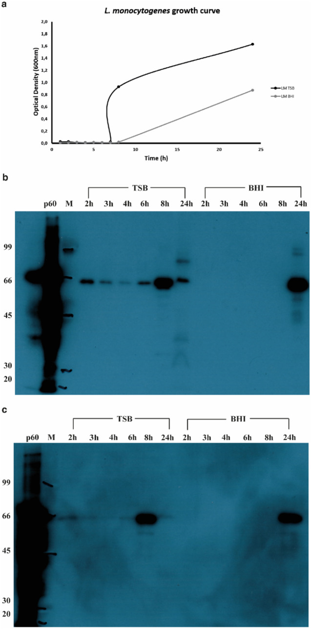
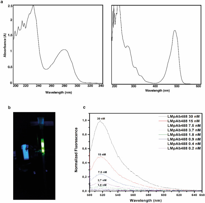
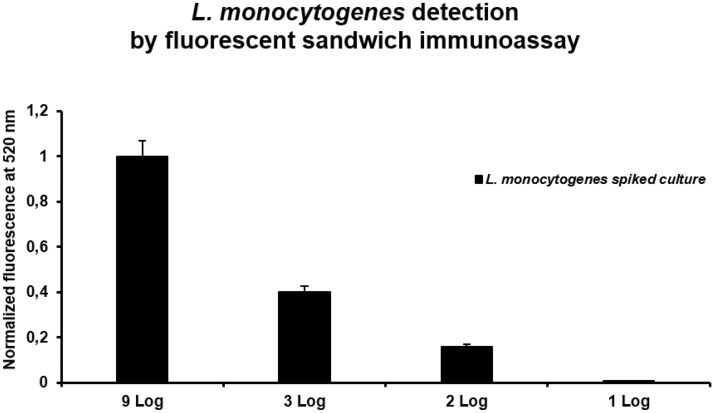
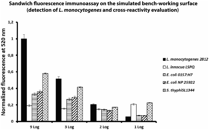
Listeria monocytogenes is a foodborne pathogen responsible for human listeriosis. The increasing incidence of listeriosis induced governments and food manufacturing enterprises to act to diminish the problem. Several methods for the detection of Listeria monocytogenes in food industries were developed. However, they are time-consuming and require the use of specialized equipment. To reduce the detection time of Listeria monocytogenes in food, in this work we developed a fluorescence sandwich immunoassay based on the use of an innovative chitosan-cellulose nanocrystal (CNC) membrane that improves the antigen capture during bacterial growth. The combined use of CNC film for the capture of p60 protein-specific antigen together with the use of fluorescence detection reduced the time of analysis from 24 to 12 h with a limit of detection (LOD) of the assay of 102 CFU/mL (2 Log). In addition, the use of monoclonal anti-PepD covalently immobilized to a CNC membrane assured a high specificity of the assay. Interestingly, the obtained results show no cross-reactivity with the five most diffused pathogen bacteria strains tested.


Conflict of interest statement

The authors declare no competing interests.

Figures

Figure 1
Figure 1
Sampling procedure performed on simulated bench working surface. Sampling procedure performed on simulated bench working surface (alumina foil). The alumina foil was been contaminated with LM cultures, following the procedure described in the “Materials and methods” section.
Figure 2
Figure 2
Bacterial growth curve and Western-blot analysis of the p60 production. (a) The kinetic bacterial growth curves—black colour—the results obtained with the TSB medium; grey colour—the results obtained with the BHI medium and (b) Western-blot with the LM pAb and (c) Western-blot with the LM mAb. p60 is the recombinant protein; M: molecular size marker; 2 h, 3 h, 4 h, 6 h, 8 h and 24 h was the sample collected during the growth grouped by culture medium used (TSB and BHI).
Figure 3
Figure 3
Labelling of the LM pAb with Alexa Fluor 488 dye and its fluorescence emission analysis. (a) Absorbance of LM pAb -on the left- and LMpAb488 -on the right-. After the labelling procedure, appeared an absorbance peak at 495 nm that confirmed the presence of dye bound to the antibody. (b) Exclusion chromatography phase: two peaks are present, -bottom- labelled antibody fraction and -top- the unreacted probe. (c) Normalized fluorescence emission spectra of LMpAb488 excited at 495 nm. The emission peak centered at 520 nm.
Figure 4
Figure 4
Multiwell microplate preparation: CNC membrane deposition and immobilization of capture antibody. (a) F16MOD microplate details: left side—clean wells and right side—CNC membrane coated wells. (b) Absorbance emission at 450 nm for the different LM mAb immobilization protocols performed. The best immobilization was been obtained at 10 mg/mL of monoclonal IgG, following protocol number two. (c) Normalized background fluorescence emission of each performed protocols.
Figure 5
Figure 5
Sandwich fluorescence immunoassay: spike test. Sandwich fluorescence immunoassay of LM spiked samples (8 h of growth).
Figure 6
Figure 6
Sandwich fluorescence immunoassay on the simulated bench-working surface (detection of LM and cross-reactivity evaluation). Sandwich fluorescence immunoassay: four dilutions were been tested (9 Log, 3 Log, 2 Log and 1 Log) of five different bacteria strains. The developed assay shows a LOD value of 2 Log.

References

    1. Buchanan R, et al. Risk assessment of Listeria monocytogenes in ready-to-eat foods. Geneva: Food and Agriculture Organization of the United Nations; 2004.
    1. Schuchat A, Swaminathan B, Broome CV. Epidemiology of human listeriosis. Clin. Microbiol. Rev. 1991;4:169–183. doi: 10.1128/CMR.4.2.169. - DOI - PMC - PubMed
    1. Vázquez-Boland JA, Domínguez-Bernal G, González-Zorna B, Kreft J, Goebel W. Pathogenicity islands and virulence evolution in Listeria. Microbes Infect. 2001;3:571–584. doi: 10.1016/S1286-4579(01)01413-7. - DOI - PubMed
    1. Public Health Agency of Canada. Food Directorate Bureau of Microbial Hazards, Health Products and Food Branch (2011).
    1. Møretrø T, Langsrud S. Listeria monocytogenes: biofilm formation and persistence in food-processing environments. Biofilms. 2004;1:107–121. doi: 10.1017/S1479050504001322. - DOI

Publication types

MeSH terms