. 2020 Dec 10;10(1):21693.
doi: 10.1038/s41598-020-78383-2.

DNA damage in lens epithelial cells exposed to occupationally-relevant X-ray doses and role in cataract formation

Affiliations

DNA damage in lens epithelial cells exposed to occupationally-relevant X-ray doses and role in cataract formation

Ion Udroiu et al. Sci Rep. .

Abstract

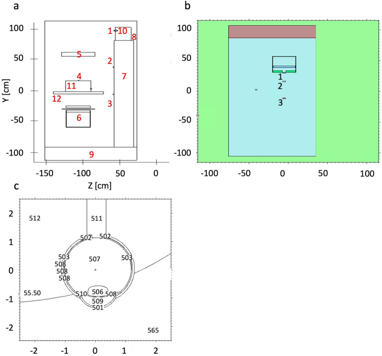
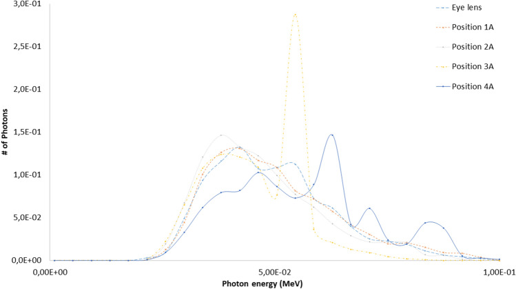
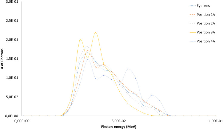
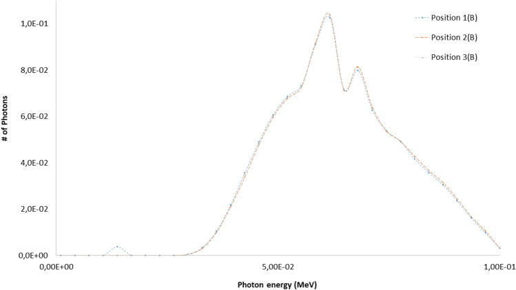
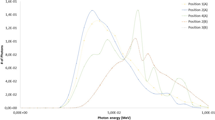
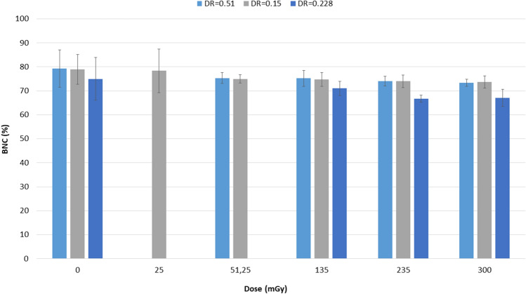
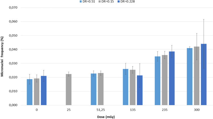
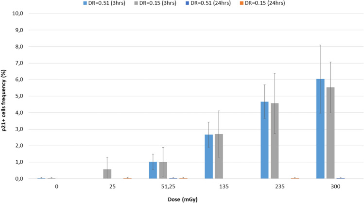
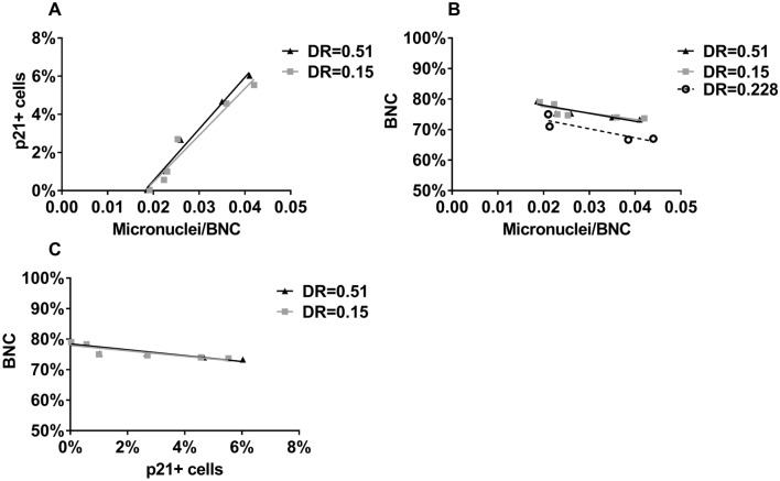
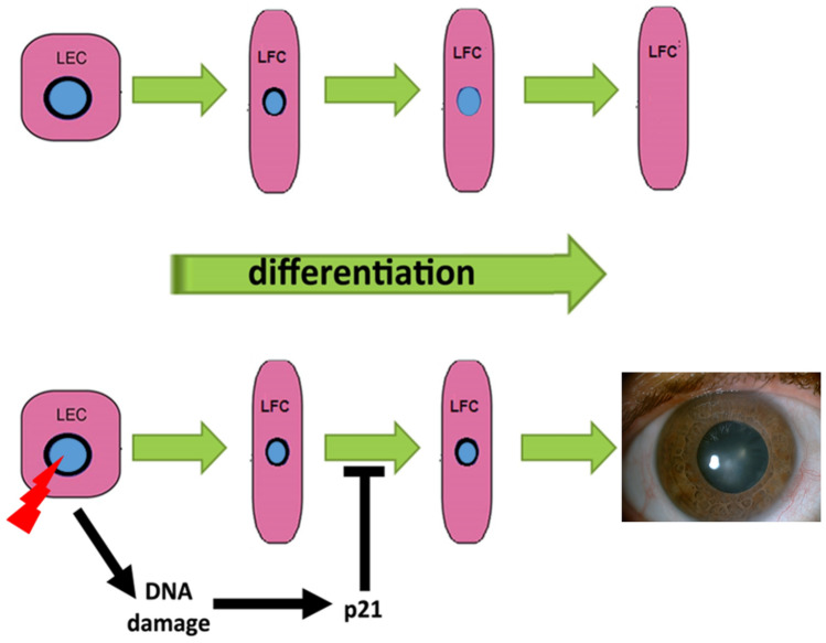
The current framework of radiological protection of occupational exposed medical workers reduced the eye-lens equivalent dose limit from 150 to 20 mSv per year requiring an accurate dosimetric evaluation and an increase understanding of radiation induced effects on Lens cells considering the typical scenario of occupational exposed medical operators. Indeed, it is widely accepted that genomic damage of Lens epithelial cells (LEC) is a key mechanism of cataractogenesis. However, the relationship between apoptosis and cataractogenesis is still controversial. In this study biological and physical data are combined to improve the understanding of radiation induced effects on LEC. To characterize the occupational exposure of medical workers during angiographic procedures an INNOVA 4100 (General Electric Healthcare) equipment was used (scenario A). Additional experiments were conducted using a research tube (scenario B). For both scenarios, the frequencies of binucleated cells, micronuclei, p21-positive cells were assessed with different doses and dose rates. A Monte-Carlo study was conducted using a model for the photon generation with the X-ray tubes and with the Petri dishes considering the two different scenarios (A and B) to reproduce the experimental conditions and validate the irradiation setups to the cells. The simulation results have been tallied using the Monte Carlo code MCNP6. The spectral characteristics of the different X-ray beams have been estimated. All irradiated samples showed frequencies of micronuclei and p21-positive cells higher than the unirradiated controls. Differences in frequencies increased with the delivered dose measured with Gafchromic films XR-RV3. The spectrum incident on eye lens and Petri, as estimated with MCNP6, was in good agreement in the scenario A (confirming the experimental setup), while the mean energy spectrum was higher in the scenario B. Nevertheless, the response of LEC seemed mainly related to the measured absorbed dose. No effects on viability were detected. Our results support the hypothesis that apoptosis is not responsible for cataract induced by low doses of X-ray (i.e. 25 mGy) while the induction of transient p21 may interfere with the disassembly of the nuclear envelop in differentiating LEC, leading to cataract formation. Further studies are needed to better clarify the relationship we suggested between DNA damage, transient p21 induction and the inability of LEC enucleation.


Conflict of interest statement

The authors declare no competing interests.

Figures

Figure 1
Figure 1
(a) experimental setup, (b) example of dose map measured using the Gafchromic films XR-RV3 and (c) used calibration curve.
Figure 2
Figure 2
(a) Scenario A. MCNP geometry. Left, X-ray exposure setup in Z-Y view; 1, detector simulating the Petri dish at the eye lens level; 2, detector simulating the Petri dish at the chest level; 3, detector for the spectra estimation at the gonads level; 4, detector simulating the Petri dish in the attenuated primary beam; 5, X-ray detector; 6, X-ray system; 7, water phantom simulating the operator body; 8, water phantom simulating the operator head; 9, concrete floor; 10, operator eye (see right part of this figure for details); 11, PMMA phantom; 12, patient couch. (b) Scenario B. MCNP geometry visualization. Background color representative of the zero-importance region outside the “simulation box”. The visualized MCNP cells related to the concrete, the X-ray tube model, the Monte Carlo detector and the 2 Petri dishes are shown. Different colors for distinguishing the different MCNP cells. Tally regions 2(B) and 3(B) related to the Petri dish positions. Tally region 1(B) referred to a numerical detector. (c) Operator eye in X–Y view as plotted by the MCNP intrinsic viewer. 501, Cornea; 502, Sclera; 503, Retina; 506, Eye lens; 507, Vitreous body; 508, Choroid; 509, Anterior chamber 510, Iris; 511, Optic nerve. Eye model volume, 7.89 cm3; Axial length, 2.58 cm; Lens Volume, 0.163 cm3; Equatorial length, 0.410 cm; Lens depth, 0.390 cm.
Figure 3
Figure 3
Typical Weight Window generation chain for MCNP, calculated with the ADVANTG discrete ordinates code. Left, forward flux values map interpreted by ADVANTG from the MCNP input file used as basis or the WW creation. Right, adjoint flux reflecting the importance function of the problem. ADVANTG simulations on a rectangular mesh of 5.0E+ 05 elements, Legendre polynomial order for scattering anisotropy modeling equal to 3, 8azimuthal and polar angles per octant, DPLUS MG photon library. See Fig. 2a–c for geometry details.
Figure 4
Figure 4
Typical WW distributions. Left, YZ view. Right-up, XY view, chest detector quote (2A). Right-low, XY view, eye lens quote (left eye, 1A). The slight asymmetry is due to the source centered in the vertical axis of the reference system and eye lens shifted by 3.5 cm. See Fig. 2a–c for geometry details.
Figure 5
Figure 5
Importance function evaluated by the ADVANTG tool. Left, adjoint flux. Centre, Weight Window typical behavior in YZ view. Right, WW at the detector levels in XY view. ADVANTG simulations on a rectangular mesh of 5.0E+05 elements, Legendre polynomial order equal to 1 (linear anisotropy), 2azimuthal and polar angles per octant, DPLUS multigroup photon library. See Fig. 2b for geometry details.
Figure 6
Figure 6
Scenario A. Photon spectra at the detectors (see Fig. 2 for details of position and characteristics) estimated with the MCNP code with the 100 kV endpoint X-ray source (see Fig. 2 for details). Photon energy scale in MeV.
Figure 7
Figure 7
Scenario A. Photon spectra at the detectors (see Fig. 2 for details of position and characteristics) estimated with the MCNP code with the 80 kV endpoint X-ray source. Photon energy scale in MeV.
Figure 8
Figure 8
Scenario B. Photon spectra evaluation at the three MCNP detectors (see Fig. 2 and Table 5 for details). Photon energy scale in MeV.
Figure 9
Figure 9
Scenario A and B spectral comparison at the various Petri dishes as estimated with MCNP (see Fig. 2, and Tables 1, 2, 3, 4, 5 for details). Photon energy scale in MeV.
Figure 10
Figure 10
Frequencies of binucleated cells (BNC). In all irradiated samples the frequencies of BNC were lower than the unirradiated controls although not statistically significant. Dose rate (DR) is expressed in Gy/min.
Figure 11
Figure 11
Frequencies of micronuclei (MN) in BNC. Dose rate (DR) is expressed in Gy/min.
Figure 12
Figure 12
Frequencies of p21-positive cells. After 3 h, all irradiated samples showed frequencies of p21-positive cells higher than the unirradiated control. After 24 h, the level of p21-positive cells was the same as in unirradiated samples. Dose rate (DR) is expressed in Gy/min.
Figure 13
Figure 13
Frequencies of (a) p21-positive cells and BNC (b) versus frequencies of MN. Frequencies of (c) BNC versus those of p21-positive cells. Dose rate (DR) is expressed in Gy/min.
Figure 14
Figure 14
Proposed model of DNA damage-induced cataract. In the upper panel, the normal process of differentiation from LEC to lens fiber cells (LFC) comprises loss of the nuclear envelope (in black) and consequent degradation of the nucleus (in light blue color). In the lower panel, X-ray-induced DNA damage causes an increase of p21, which impedes the disassembly of the nuclear envelope, thus impairing differentiation and leading to cataract formation.

References

    1. Council Directive 2013/59/Euratom of 5 December 2013 laying down basic safety standards for protection against the dangers arising from exposure to ionizing radiation https://eur-lex.europa.eu/eli/dir/2013/59/2014-01-17 (2013).
    1. Stewart FA, et al. ICRP publication 118: ICRP statement on tissue reactions and early and late effects of radiation in normal tissues and organs–threshold doses for tissue reactions in a radiation protection context. Ann ICRP. 2012;41:1–322. doi: 10.1016/j.icrp.2012.02.001. - DOI - PubMed
    1. Haqqani OP, Agarwal PK, Halin NM, Iafrati MD. Defining the radiation "scatter cloud" in the interventional suite. J. Vasc. Surg. 2013;58:1339–1345. doi: 10.1016/j.jvs.2013.01.025. - DOI - PubMed
    1. Nikjoo H, Lindborg L. RBE of low energy electrons and photons. Phys. Med. Biol. 2010;55:65–109. doi: 10.1088/0031-9155/55/10/R01. - DOI - PubMed
    1. White SA, Reniers B, de Jong EE, Rusch T, Verhaegen F. A comparison of the relative biological effectiveness of low energy electronic brachytherapy sources in breast tissue: A Monte Carlo study. Phys. Med. Biol. 2016;61:383–399. doi: 10.1088/0031-9155/61/1/383. - DOI - PubMed

Publication types