. 2020 Dec 10;10(1):21637.
doi: 10.1038/s41598-020-78481-1.

A novel nerve transection and repair method in mice: histomorphometric analysis of nerves, blood vessels, and muscles with functional recovery

Affiliations

A novel nerve transection and repair method in mice: histomorphometric analysis of nerves, blood vessels, and muscles with functional recovery

Jung Il Lee et al. Sci Rep. .

Abstract

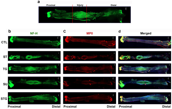
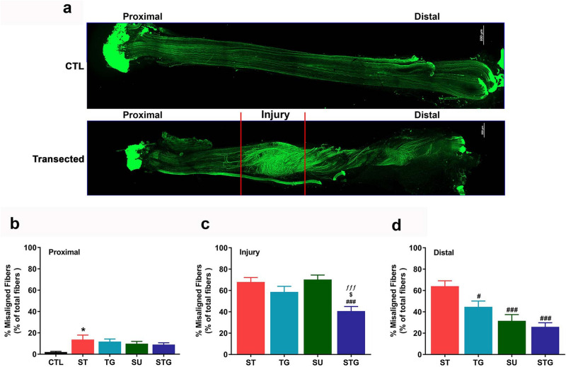
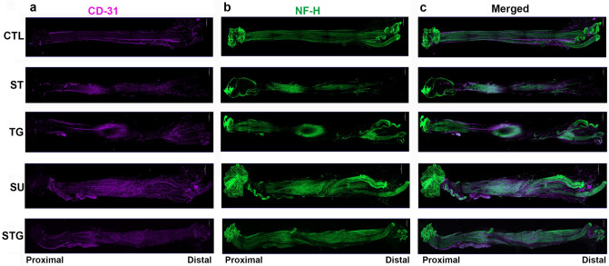
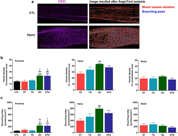
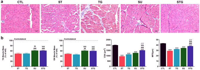
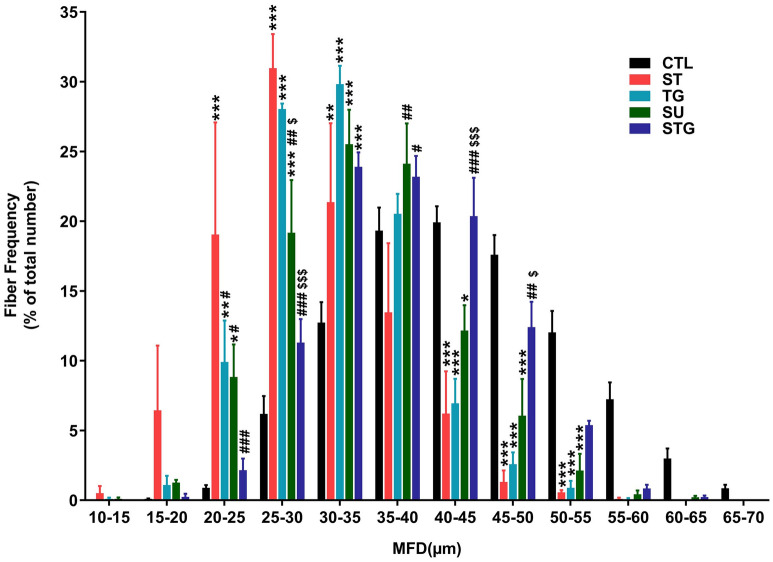
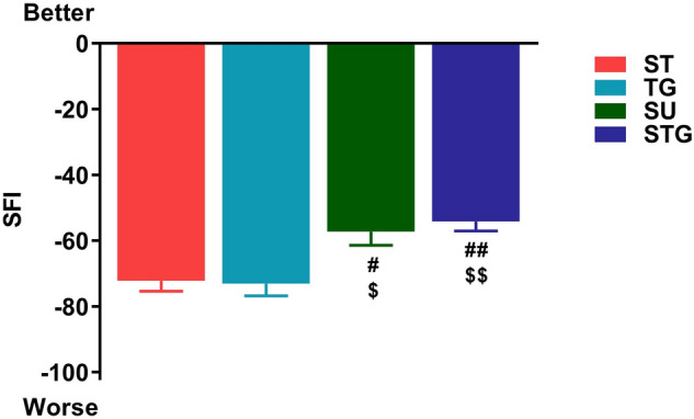
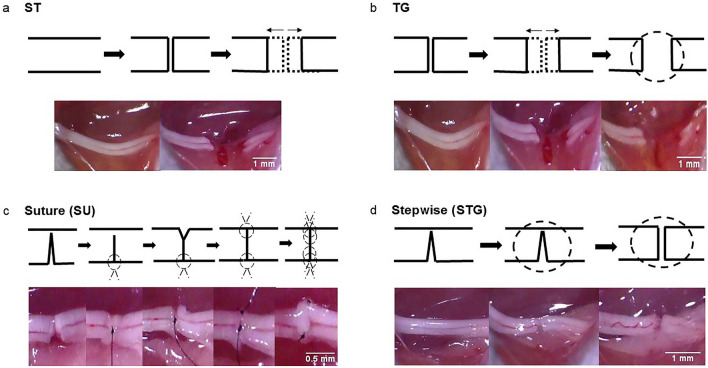
Peripheral nerve transection is associated with permanent functional deficit even after advanced microsurgical repair. While it is difficult to investigate the reasons of poor functional outcomes of microsurgical repairs in humans, we developed a novel pre-clinical nerve transection method that allows reliable evaluation of nerve regeneration, neural angiogenesis, muscle atrophy, and functional recovery. Adult male C57BL/6 mice were randomly assigned to four different types of sciatic nerve transection: Simple Transection (ST), Simple Transection & Glue (TG), Stepwise Transection and Sutures (SU), and Stepwise Transection and Glue (STG). Mice were followed for 28 days for sciatic function index (SFI), and sciatic nerves and hind limb muscles were harvested for histomorphological and cellular analyses. Immunohistochemistry revealed more directional nerve fiber growth in SU and STG groups compared with ST and TG groups. Compared to ST and TG groups, optimal neural vessel density and branching index in SU and STG groups were associated with significantly decreased muscle atrophy, increased myofiber diameter, and improved SFI. In conclusion, our novel STG method represents an easily reproducible and reliable model with close resemblance to the pathophysiological characteristics of SU model, and this can be easily reproduced by any lab.


Conflict of interest statement

The authors declare no competing interests.

Figures

Figure 1
Figure 1
Evaluation of whole mount immunostaining of the transected nerve at post-injury day 28 for nerve fiber distribution. (a) For the purpose of evaluation and quantification, imaged whole nerve was divided into 3 zones: proximal, injury and distal. Representative compact images of immunofluorescence staining of the whole nerve for NF-H (b) in green, MP0 (c) in red, and the merged view (d). Scale bar, 500 µm (at upper right corner); magnification, × 5. For other details, see Fig. 8.
Figure 2
Figure 2
(a) Representative images from an uninjured and a transected nerve to show the orientation of neurofilaments at different zones. Scale bar, 500 µm (at upper right corner). Bar graph showing the quantification of misaligned fibers at proximal (b), injury (c) and distal (d) zones of transected nerves. Misaligned fibers are shown as the percentage of total number of fibers in each zone. n = 3/group, *P < 0.05, ***P < 0.001 vs. uninjured (CTL), #P < 0.05, ###P < 0.001 vs. ST, $P < 0.05 vs. TG, and ƒƒƒP < 0.001 vs. SU. For other details, see Fig. 8.
Figure 3
Figure 3
Evaluation of whole mount immunostaining of the transected nerve at post-injury day 28 for angiogenesis. Representative compact images of immunofluorescence staining of the whole nerve for CD31 (a) in purple, NF-H (b) in green, and the merged view (c). Scale bar, 500 µm (at upper right corner); magnification, × 5. For other details, see Fig. 8.
Figure 4
Figure 4
(a) Representative images from an uninjured and a transected nerve to show the blood vessels (left) and AngioTool reconstruction result (right) images. AngioTool images clearly depict the blood vessel architecture as red lines and their branching points as blue dots. Bar graph showing the quantification of blood vessels at proximal, injury, and distal zones of transected nerves. (b) Blood vessel density is shown as the percentage of number of blood vessel in total area. (c) Branching index of blood vessels is shown as the number of blood vessel junctions/mm2. n = 3/group, **P < 0.01, ***P < 0.001 vs. uninjured (CTL), #P < 0.05, ##P < 0.01 vs. ST, and $$P < 0.05 vs. TG. For other details, see Fig. 8.
Figure 5
Figure 5
Effect of different transection models on TA muscle histology, muscle mass, and quantitative measurements of the muscle fibers. (a) Representative images of TA muscle cross sections stained with H&E. Scale bar, 50 µm (at lower right corner); magnification 20 ×. (b) TA muscle mass as % of left side, n = 10–12/group; CSA of TA muscle as µm2, n = 3/group; and MFD of TA muscle as µm, n = 3/group. ***P < 0.001 vs. CTL, ###P < 0.001 vs. ST, $$P < 0.01, $$$P < 0.001 vs. TG, and ƒƒƒP < 0.001 vs. SU. For other details, see Fig. 8.
Figure 6
Figure 6
Effect of different transection models on muscle fiber distribution. Quantitative muscle fiber size distribution from the MFD value (µm) in each group as a percentage of total fiber number. n = 3/group; *P < 0.05, **P < 0.01, ***P < 0.001 vs. uninjured (CTL), #P < 0.05, ##P < 0.01, ###P < 0.001 vs. ST, and $$$P < 0.001 vs. TG. For other details, see Fig. 8.
Figure 7
Figure 7
Effect of different transection models on the functional recovery (SFI) at post-injury day 28. n = 10–12/group. #P < 0.05, ##P < 0.01 vs. ST, and $P < 0.05, $$P < 0.01 vs. TG. For other details, see Fig. 8.
Figure 8
Figure 8
Experimental groups with schematic surgical steps (top panel) and representative gross nerve images (lower panel) of surgical procedures in different nerve transection methods. Experimental groups are (a) Simple Transection (ST), (b) Transection & Glue (TG), (c) Stepwise Transection and Sutures (SU), and (d) Stepwise Transection and Glue (STG). The complete transection of the nerve was confirmed under direct microscopic vision.

References

    1. Robinson LR. Traumatic injury to peripheral nerves. Muscle Nerve. 2000;23(6):863–873. doi: 10.1002/(SICI)1097-4598(200006)23:6<863::AID-MUS4>3.0.CO;2-0. - DOI - PubMed
    1. Taylor CA, Braza D, Rice JB, Dillingham T. The incidence of peripheral nerve injury in extremity trauma. Am. J. Phys. Med. Rehabil. 2008;87(5):381–385. doi: 10.1097/PHM.0b013e31815e6370. - DOI - PubMed
    1. Evans GR. Peripheral nerve injury: a review and approach to tissue engineered constructs. Anat. Rec. 2001;263(4):396–404. doi: 10.1002/ar.1120. - DOI - PubMed
    1. Robinson LR. Traumatic injury to peripheral nerves. Suppl. Clin. Neurophysiol. 2004;57:173–186. doi: 10.1016/S1567-424X(09)70355-1. - DOI - PubMed
    1. Campbell WW. Evaluation and management of peripheral nerve injury. Clin. Neurophysiol. 2008;119(9):1951–1965. doi: 10.1016/j.clinph.2008.03.018. - DOI - PubMed

Publication types

LinkOut - more resources