. 2020 Dec 10;10(1):21763.
doi: 10.1038/s41598-020-78694-4.

Mechanical and structural characterisation of the dural venous sinuses

Affiliations

Mechanical and structural characterisation of the dural venous sinuses

Darragh R Walsh et al. Sci Rep. .

Abstract

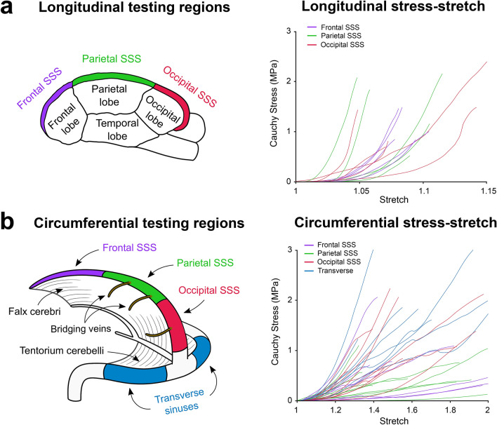
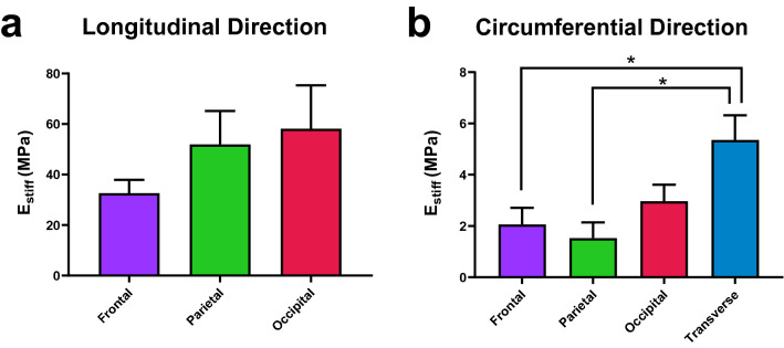
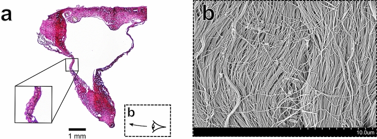
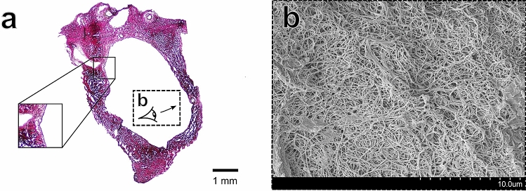
The dural venous sinuses play an integral role in draining venous blood from the cranial cavity. As a result of the sinuses anatomical location, they are of significant importance when evaluating the mechanopathology of traumatic brain injury (TBI). Despite the importance of the dural venous sinuses in normal neurophysiology, no mechanical analyses have been conducted on the tissues. In this study, we conduct mechanical and structural analysis on porcine dural venous sinus tissue to help elucidate the tissues' function in healthy and diseased conditions. With longitudinal elastic moduli values ranging from 33 to 58 MPa, we demonstrate that the sinuses exhibit higher mechanical stiffness than that of native dural tissue, which may be of interest to the field of TBI modelling. Furthermore, by employing histological staining and a colour deconvolution protocol, we show that the sinuses have a collagen-dominant extracellular matrix, with collagen area fractions ranging from 84 to 94%, which likely explains the tissue's large mechanical stiffness. In summary, we provide the first investigation of the dural venous sinus mechanical behaviour with accompanying structural analysis, which may aid in understanding TBI mechanopathology.


Conflict of interest statement

The authors declare no competing interests.

Figures

Figure 1
Figure 1
Illustration of the regions tested with the accompanying plots of the Cauchy stress-stretch curves of the dural venous sinus samples tested in (a) the vessel’s longitudinal test direction and (b) the vessel’s circumferential test direction. Note the different scales on the x-axes of the stress-stretch graphs.
Figure 2
Figure 2
(a) Mean Estiff results for the samples tested in the longitudinal test direction utilising uniaxial tensile testing. (b) Mean Estiff results for the samples tested in the circumferential test direction utilising ring testing. *Statistical significance (p<0.05), error bars represent standard error about the mean. Note that the y-axis scale differs between (a) and (b).
Figure 3
Figure 3
Structural images of the frontal region of the SSS (a) Verhoeff–Van Gieson stained sample of the frontal region. The solid-line inset image shows what appears to be a meningeal artery (b) SEM imaging of the bone surface layer (as illustrated in dashed-line inset of (a)) showing disorganised focal aggregations of collagen fibres.
Figure 4
Figure 4
Structural images of the parietal region of the SSS (a) Verhoeff–Van Gieson stained sample. The solid-line inset image shows the thin, collagenic wall of the sinus with a thin endothelial layer on the lumen wall (b) The arachnoid surface of the sinus (as illustrated in dashed-line inset of (a)) demonstrating collagenic alignment along the longitudinal direction of the sinus.
Figure 5
Figure 5
Structural images of the occipital region of the SSS (a) Verhoeff–Van Gieson stained sample. The solid-line inset image again demonstrates the thin endothelial later of the dural sinus tissue (b) The surface of the occipital region lumen wall (as illustrated in dashed-line inset of (a)) depicting seemingly random collagenic alignment, with the endothelial layer removed from the maceration protocol.
Figure 6
Figure 6
Structural images of the transverse sinus (a) Verhoeff–Van Gieson stained sample. (b) The cross-section of the dural sinus wall with the lumen (*) and arachnoid surface (#) highlighted. Distinct layers appear to constitute the wall of the dural sinus tissue.

References

    1. Balik V, et al. Variability in wall thickness and related structures of major dural sinuses in posterior cranial fossa: a microscopic anatomical study and clinical implications. Operative neurosurgery (Hagerstown, Md.) 2019;17:88–96. doi: 10.1093/ons/opz086. - DOI - PubMed
    1. Sindou M, Auque J. The intracranial venous system as a neurosurgeon’s perspective. Adv. Tech. Stand. Neurosurg. 2000;26:131–216. doi: 10.1007/978-3-7091-6323-8_5. - DOI - PubMed
    1. Mortazavi, M. M. et al. Chapter 27—surgical nuances in management of intracranial venous sinus injuries. In Anatomy 231–236 (Elsevier, Philadelphia, 2020).
    1. Patchana T, Zampella B, Berry JA, Lawandy S, Sweiss RB. Superior sagittal sinus: a review of the history, surgical considerations, and pathology. Cureus. 2019;11:e4597–e4597. - PMC - PubMed
    1. Behera SK, Senapati SB, Mishra SS, Das S. Management of superior sagittal sinus injury encountered in traumatic head injury patients: analysis of 15 cases. Asian J. Neurosurg. 2015;10:17–20. doi: 10.4103/1793-5482.151503. - DOI - PMC - PubMed

Publication types

MeSH terms