

Ultraviolet A irradiation induces ultraweak photon emission with characteristic spectral patterns from biomolecules present in human skin
- PMID: 33303911
- PMCID: PMC7728812
- DOI: 10.1038/s41598-020-78884-0
Ultraviolet A irradiation induces ultraweak photon emission with characteristic spectral patterns from biomolecules present in human skin
Abstract
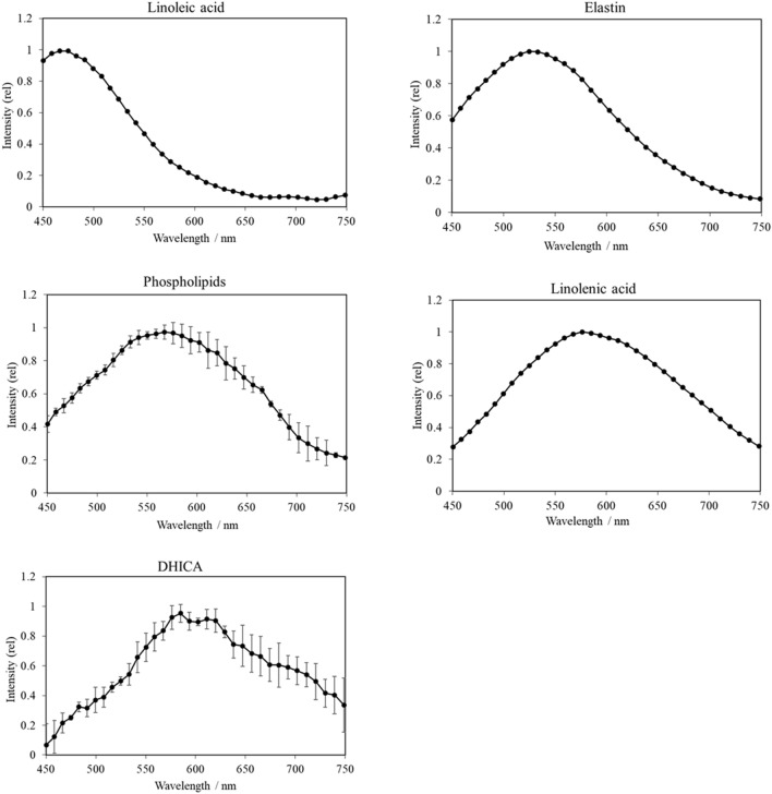
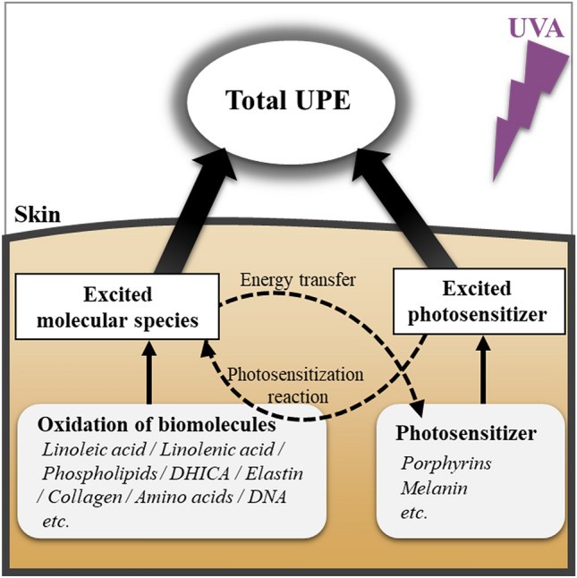
Oxidative stress is associated with photoaging of the skin as well as with skin cancer, and is therefore, critical to monitor. Ultraweak photon emission (UPE) is extremely weak light generated during the oxidative process in the living body and has been used as a non-invasive and label-free marker for the evaluation of oxidative stress. However, the mechanism of UPE generation is not clear. Therefore, we aimed to elucidate the molecular mechanism underlying UPE generation by analyzing the spectra of UPE generated from biomolecules in the skin during ultraviolet A (UVA) exposure. The spectra of UVA-induced UPE generated from linoleic acid, linolenic acid, elastin, phospholipids, and 5,6-dihydroxyindole-2-carboxylic acid were measured, and the spectrum of human skin tissue was also obtained. The spectral patterns varied for the different biomolecules and the peaks were distinct from those of the skin tissue. These results suggested that the UPE generated from skin tissue is a collection of light emitted by biomolecules. Moreover, we proposed that UPE is generated through a photosensitization reaction and energy transfer. The identified characteristic spectral patterns of UPE can be useful to elucidate UVA-induced oxidative stress in the skin, with implications for prevention and treatment of photoaging and skin diseases.
Conflict of interest statement
The authors declare no competing interests.
Figures


Similar articles
-
Imaging of ultraweak photon emission for evaluating the oxidative stress of human skin.J Photochem Photobiol B. 2019 Sep;198:111562. doi: 10.1016/j.jphotobiol.2019.111562. Epub 2019 Jul 17. J Photochem Photobiol B. 2019. PMID: 31349151
-
Non-invasive monitoring of oxidative skin stress by ultraweak photon emission measurement. II: biological validation on ultraviolet A-stressed skin.Skin Res Technol. 2008 Feb;14(1):112-20. doi: 10.1111/j.1600-0846.2007.00207.x. Skin Res Technol. 2008. PMID: 18211609
-
Skin lightness affects ultraviolet A-induced oxidative stress: Evaluation using ultraweak photon emission measurement.Exp Dermatol. 2023 Feb;32(2):146-153. doi: 10.1111/exd.14690. Epub 2022 Oct 31. Exp Dermatol. 2023. PMID: 36256509
-
The application of ultra-weak photon emission in dermatology.J Photochem Photobiol B. 2014 Oct 5;139:63-70. doi: 10.1016/j.jphotobiol.2013.10.003. Epub 2013 Oct 26. J Photochem Photobiol B. 2014. PMID: 24275519 Review.
-
Ultraviolet A radiation-induced biological effects in human skin: relevance for photoaging and photodermatosis.J Dermatol Sci. 2000 Mar;23 Suppl 1:S22-6. doi: 10.1016/s0923-1811(99)00077-8. J Dermatol Sci. 2000. PMID: 10764987 Review.
Cited by
-
Imaging of Lipid Peroxidation-Associated Chemiluminescence in Plants: Spectral Features, Regulation and Origin of the Signal in Leaves and Roots.Antioxidants (Basel). 2022 Jul 6;11(7):1333. doi: 10.3390/antiox11071333. Antioxidants (Basel). 2022. PMID: 35883824 Free PMC article.
-
Open quantum systems theory of ultraweak ultraviolet photon emissions: Revisiting Gurwitsch's onion experiment as a prototype for quantum biology.Comput Struct Biotechnol J. 2024 Nov 29;26:78-91. doi: 10.1016/j.csbj.2024.11.030. eCollection 2024 Dec. Comput Struct Biotechnol J. 2024. PMID: 39717158 Free PMC article. Review.
-
Extracellular vesicles derived from mesenchymal stem cells: the wine in Hebe's hands to treat skin aging.Precis Clin Med. 2024 Feb 24;7(1):pbae004. doi: 10.1093/pcmedi/pbae004. eCollection 2024 Mar. Precis Clin Med. 2024. PMID: 38516531 Free PMC article. Review.
-
RNAs m6A modification facilitates UVB-induced photoaging.Heliyon. 2024 Oct 18;10(21):e39532. doi: 10.1016/j.heliyon.2024.e39532. eCollection 2024 Nov 15. Heliyon. 2024. PMID: 39512467 Free PMC article.
-
Blue light-induced lipid oxidation and the antioxidant property of hypotaurine: evaluation via measuring ultraweak photon emission.Photochem Photobiol Sci. 2023 Feb;22(2):345-356. doi: 10.1007/s43630-022-00319-8. Epub 2022 Oct 22. Photochem Photobiol Sci. 2023. PMID: 36271182
References
-
- Bartosz G. Oxidative stress in plants. Acta Physiol. Plant. 1997;19:47–64. doi: 10.1007/s11738-997-0022-9. - DOI
MeSH terms
Substances
LinkOut - more resources
Full Text Sources
Medical
