. 2021 May;28(5):1669-1687.
doi: 10.1038/s41418-020-00693-9. Epub 2020 Dec 10.

E6-mediated activation of JNK drives EGFR signalling to promote proliferation and viral oncoprotein expression in cervical cancer

Affiliations

E6-mediated activation of JNK drives EGFR signalling to promote proliferation and viral oncoprotein expression in cervical cancer

Ethan L Morgan et al. Cell Death Differ. 2021 May.

Abstract

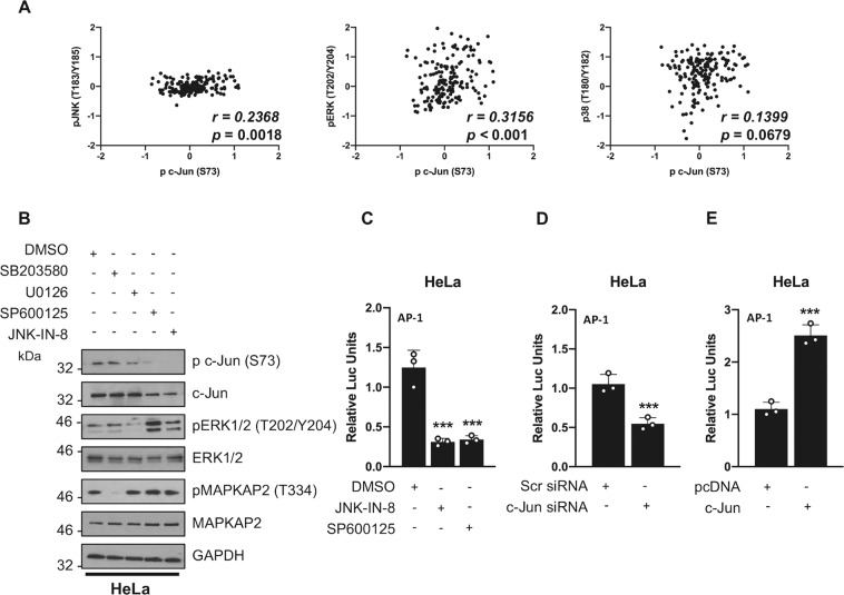
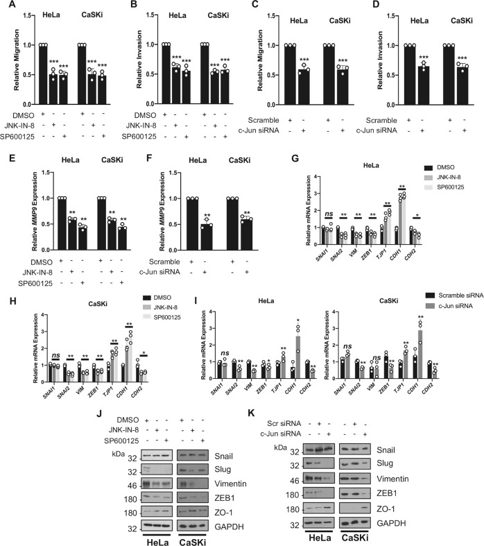
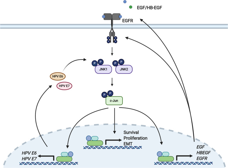
Human papillomaviruses (HPV) are a major cause of malignancy worldwide, contributing to ~5% of all human cancers including almost all cases of cervical cancer and a growing number of ano-genital and oral cancers. HPV-induced malignancy is primarily driven by the viral oncogenes, E6 and E7, which manipulate host cellular pathways to increase cell proliferation and enhance cell survival, ultimately predisposing infected cells to malignant transformation. Consequently, a more detailed understanding of viral-host interactions in HPV-associated disease offers the potential to identify novel therapeutic targets. Here, we identify that the c-Jun N-terminal kinase (JNK) signalling pathway is activated in cervical disease and in cervical cancer. The HPV E6 oncogene induces JNK1/2 phosphorylation in a manner that requires the E6 PDZ binding motif. We show that blockade of JNK1/2 signalling using small molecule inhibitors, or knockdown of the canonical JNK substrate c-Jun, reduces cell proliferation and induces apoptosis in cervical cancer cells. We further demonstrate that this phenotype is at least partially driven by JNK-dependent activation of EGFR signalling via increased expression of EGFR and the EGFR ligands EGF and HB-EGF. JNK/c-Jun signalling promoted the invasive potential of cervical cancer cells and was required for the expression of the epithelial to mesenchymal transition (EMT)-associated transcription factor Slug and the mesenchymal marker Vimentin. Furthermore, JNK/c-Jun signalling is required for the constitutive expression of HPV E6 and E7, which are essential for cervical cancer cell growth and survival. Together, these data demonstrate a positive feedback loop between the EGFR signalling pathway and HPV E6/E7 expression, identifying a regulatory mechanism in which HPV drives EGFR signalling to promote proliferation, survival and EMT. Thus, our study has identified a novel therapeutic target that may be beneficial for the treatment of cervical cancer.


Conflict of interest statement

The authors declare that they have no conflict of interest.

Figures

Fig. 1
Fig. 1. c-Jun N-terminal kinases are significantly phosphorylated in HPV+ cervical cancer.
A Representative western blot of normal human keratinocytes (NHK) and NHKs containing the HPV18 genome (HPV18) analysed for phosphorylated JNK1/2 and total JNK expression. GAPDH served as a loading control. Data shown are representative of at least three independent experiments. B Representative western blot from cytology samples of CIN lesions of increasing grade analysed for phosphorylated JNK1/2 and total JNK expression. GAPDH served as a loading control. C Scatter plot of densitometry analysis of a panel of cytology samples. Twenty samples from each clinical grade (Neg, CIN 1–3) were analysed by western blot and densitometry analysis was performed for phosphorylated JNK1/2 and total JNK expression. D Representative western blot analysis of a panel of six cervical cancer cell lines—two HPV− (C33A and DoTc2 4510), two HPV16+ (SiHa and CaSKi) and HPV18+ (SW756 and HeLa)—and NHK cells for phosphorylated JNK1/2 and total JNK. GAPDH served as a loading control. Densitometry analysis of phosphorylated JNK1/2, normalised to total JNK and GAPDH expression, from three independent experiments is shown below. E Representative results of immunohistochemical staining for phosphorylated JNK1/2 expression in cervical cancer tissues and adjacent non-tumour tissues. Images were acquired using identical exposure times. Scale bar, 50 μm. Statistical analysis for phosphorylated JNK1/2 expression in cervical cancer tissues (n = 39) compared with non-tumour tissues (n = 9) is shown on the right. Each section was stained and analysed in duplicate. Phosphorylated JNK1/2 was quantified in an automated fashion using the IHC Profiler Plug-in for ImageJ. Error bars represent the mean +/− standard deviation. *P < 0.05, **P < 0.01, ***P < 0.001 (Student’s t-test).
Fig. 2
Fig. 2. JNK activity is required for c-Jun/AP-1 activity in cervical cancer.
A Scatter plot of expression levels of c-Jun phosphorylation and JNK phosphorylation, ERK phosphorylation and p38 phosphorylation as analysed using the MD Anderson public RPPA data set. The correlation coefficient (r) was calculated using Spearman’s rank analysis. B Representative western blot of HeLa cells treated with the p38 inhibitor SB203580 (10 µM), the MEK1/2 inhibitor U0126 (10 µM), or the JNK inhibitors SP600125 (10 µM) and JNK-IN-8 (3 µM). Lysates were analysed for phosphorylated and total c-Jun, phosphorylated and total ERK and phosphorylated and total MAPKAP2. GAPDH served as a loading control. Data shown are representative of at least three independent experiments. C Representative luciferase reporter assay from HeLa cells transfected with an AP-1 luciferase reporter, in the presence or absence of JNK-IN-8 (3 µM) or SP600125 (10 µM). Promoter activity was measured using a dual-luciferase system. Data are presented as relative to the DMSO control. D Luciferase reporter assay from HeLa cells co-transfected with an AP-1 luciferase reporter and pool of siRNA against the JNK substrate c-Jun. Promoter activity was measured using a dual-luciferase system. Data are presented as relative to the scrambled siRNA transfection control. E Representative luciferase reporter assay from HeLa cells co-transfected with an AP-1 luciferase reporter and c-Jun. Promoter activity was measured using a dual-luciferase system. Data are presented as relative to the pcDNA transfection control. Error bars represent the mean +/− standard deviation of a minimum of three biological repeats. *P < 0.05, **P < 0.01, ***P < 0.001 (Student’s t-test).
Fig. 3
Fig. 3. HPV E6 induces JNK phosphorylation via its PDZ-binding domain.
A Representative western blot of C33A and NHK cells transiently transfected with GFP or GFP tagged HPV18 E6, E7 and E6/E7 and analysed for phosphorylated JNK1/2 and total JNK expression. Expression of HPV oncoproteins was confirmed by fluorescence microscopy for GFP expression (not shown) and HPV18 E6 and HPV18 E7 specific antibodies. GAPDH served as a loading control. Data shown are representative of at least three independent experiments. B Representative western blot of HeLa and CaSKi cells transfected with a pool of specific siRNAs against HPV18 E6/E7 or HPV16 E6/E7 respecitvely and analysed for phosphorylated JNK1/2 and total JNK expression. GAPDH served as a loading control. Data shown are representative of at least three independent experiments. C Representative western blot of C33A cells co-transfected with GFP or GFP tagged E6, in the presence or absence of JNK-IN-8 (3 µM). Lysates were analysed for phosphorylated c-Jun and total c-Jun expression. GAPDH served as a loading control. Data shown are representative of at least three independent experiments. D Luciferase reporter assay from C33A cells co-transfected with GFP or GFP tagged E6, and an AP-1 luciferase reporter, in the presence or absence of JNK-IN-8 (3 µM) or SP600125 (10 µM). Promoter activity was measured using a dual-luciferase system. Data are presented as relative to the GFP transfection control. E Representative western blot of C33A cells transiently transfected with GFP tagged E6 and E6 mutants and analysed for phosphorylated JNK and total JNK expression. Expression of HPV E6 and mutants was confirmed using a GFP antibody and GAPDH served as a loading control. Data shown are representative of at least three independent experiments. F Representative western blot of C33A cells co-transfected with GFP tagged E6 from different HPV types and analysed for phosphorylated JNK and total JNK expression. Expression of HPV E6 proteins was confirmed using a GFP antibody and GAPDH served as a loading control. Data shown are representative of at least three independent experiments. Error bars represent the mean +/− standard deviation of a minimum of three biological repeats. *P < 0.05, **P < 0.01, ***P < 0.001 (Student’s t-test).
Fig. 4
Fig. 4. Inhibition of JNK/c-Jun signalling impaires the proliferation of HPV+ cervical cancer cells.
A Representative western blots of HeLa and CaSKi cells treated with increasing doses of JNK-IN-8. Lysates were analysed for the phosphorylation and expression of the JNK substrate c-Jun. GAPDH was used as a loading control. Data shown are representative of at least three independent experiments. B Growth curve analysis of HeLa and CaSKi cells treated with JNK-IN-8 (3 µM) or SP600125 (10 µM). C Colony formation assay (anchorage-dependent growth) of HeLa and CaSKi cells treated with JNK-IN-8 (3 µM) or SP600125 (10 µM). D Soft agar assay (anchorage-independent growth) of HeLa and CaSKi cells treated with JNK-IN-8 (3 µM) or SP600125 (10 µM). E Representative western blots of HeLa and CaSKi transfected with a pool of siRNA against c-Jun. Lysates were analysed for the expression of c-Jun. GAPDH was used as a loading control. Data shown are representative of at least three independent experiments. F Growth curve analysis of HeLa and CaSKi cells transfected with a pool of siRNA against c-Jun. G Colony formation assay (anchorage-dependent growth) of HeLa and CaSKi cells transfected with a pool of siRNA against the JNK substrate c-Jun. H Soft agar assay (anchorage-independent growth) of HeLa and CaSKi cells transfected with a pool of siRNA against c-Jun. Error bars represent the mean +/− standard deviation of a minimum of three biological repeats. *P < 0.05, **P < 0.01, ***P < 0.001 (Student’s t-test).
Fig. 5
Fig. 5. JNK/c-Jun activity is required for cell migration/invasion and regulates epithelial to mesenchymal transition (EMT).
A Transwell® migration assay of HeLa and CaSKi cells treated with JNK-IN-8 (3 µM) or SP600125 (10 µM). The average number of invaded cells per field was calculated from five representative fields per experiment. B Transwell® invasion assay of HeLa and CaSKi cells treated with JNK-IN-8 (3 µM) or SP600125 (10 µM). The average number of invaded cells per field was calculated from five representative fields per experiment. C Transwell® migration assay of HeLa and CaSKi cells after transfection of a pool of siRNA against c-Jun. The average number of invaded cells per field was calculated from five representative fields per experiment. D Transwell® invasion assay of HeLa and CaSKi cells after transfection of a pool of siRNA against c-Jun. The average number of invaded cells per field was calculated from five representative fields per experiment. E qPCR analysis of MMP9 mRNA expression HeLa and CaSKi cells treated with JNK-IN-8 (3 µM) or SP600125 (10 µM). U6 was used as a loading control. F qPCR analysis of MMP9 mRNA expression HeLa and CaSKi cells cells after transfection of a pool of siRNA against c-Jun. U6 was used as a loading control. G qPCR analysis of genes involved in EMT in HeLa cells treated with JNK-IN-8 (3 µM) or SP600125 (10 µM). U6 was used as a loading control. H qPCR analysis of genes involved in EMT in CaSKi cells treated with JNK-IN-8 (3 µM) or SP600125 (10 µM). U6 was used as a loading control. I qPCR analysis of genes involved in EMT in HeLa and CaSKi cells after transfection of a pool of siRNA against c-Jun. U6 was used as a loading control. J Representative western blots of HeLa and CaSKi treated JNK-IN-8 (3 µM) or SP600125 (10 µM). Lysates were probed for the expression of Slug, Snail, Vimentin, ZO-1 and ZEB1. GAPDH was used as a loading control. Data shown are representative of at least three independent experiments. K Representative western blots of HeLa and CaSKi after transfection of a pool of siRNA against c-Jun. Lysates were probed for the expression of Slug, Snail, Vimentin, ZO-1 and ZEB1. GAPDH was used as a loading control. Data shown are representative of at least three independent experiments. Error bars represent the mean +/− standard deviation of a minimum of three biological repeats. *P < 0.05, **P < 0.01, ***P < 0.001 (Student’s t-test).
Fig. 6
Fig. 6. Regulation of EGFR signalling contributes to JNK mediated proliferation.
A qPCR analysis of EGFR and EGFR ligand expression in HeLa and CaSKi cells treated with JNK-IN-8 (3 µM) or SP600125 (10 µM) for 48 h. U6 was used as a loading control. B) Representative western blots of HeLa and CaSKi cells treated with JNK-IN-8 (3 µM) or SP600125 (10 µM) for the expression of EGFR for 48 h. Data shown are representative of at least three independent experiments. C qPCR analysis of EGFR and EGFR ligand expression in HeLa and CaSKi cells after transfection of a pool of siRNA against c-Jun for 72 h. U6 was used as a loading control. D Representative western blot of HeLa and CaSKi cells after transfection of a pool of siRNA against c-Jun for the expression of EGFR for 72 h. Data shown are representative of at least three independent experiments. E Scatter plot of expression levels of JUN and EGFR, EGF and HBEGF as analysed using the MD Anderson public RPPA data set. The correlation coefficient (r) was calculated using Spearman’s rank analysis. F Schematic for conditioned media rescue assay. G Colony formation assay (anchorage-dependent growth) of HeLa and CaSKi treated with SP600125 (10 µM) or DMSO control for 48 h. SP600125 was subsequently washed out and control or SP600125-treated cells were grown in conditioned media from cells treated with DMSO or SP600125. In addition, cells were also grown in SP600125 conditioned media containing EGF, HB-EGF, or DMSO conditioned media with or without neutralising EGFR, EGF or HB-EGF antibodies. Colonies were stained and counted after 14 days. Error bars represent the mean +/− standard deviation of a minimum of three biological repeats. *P < 0.05, **P < 0.01, ***P < 0.001 (Student’s t-test).
Fig. 7
Fig. 7. JNK mediated c-Jun activation plays a significant role in HPV oncogene expression.
A Luciferase reporter assay from HeLa cells transfected with a HPV18 URR luciferase reporter with or without treatment with JNK-IN-8 (3 µM). Promoter activity was measured using a dual-luciferase system. Data are presented as relative to the DMSO control. B qPCR analysis of HPV 16/18 E6 and E7 mRNA expression in HeLa and CaSKi cells treated with JNK-IN-8 (3 µM). U6 was used as a loading control. C qPCR analysis of HPV 16/18 E6 and E7 mRNA expression in HeLa and CaSKi cells after transfection of a pool of siRNA against c-Jun. U6 was used as a loading control. D Representative western blots of HeLa and CaSKi treated with increasing doses of JNK-IN-8 (3 µM) or SP600125 (10 µM). Lysates were analysed for the expression of HPV 16/18 E6 and E7. GAPDH was used as a loading control. Data shown are representative of at least three independent experiments. E Representative western blots of HeLa and CaSKi after transfection of a pool of siRNA against c-Jun. Lysates were analysed for the expression of HPV 16/18 E6 and E7. GAPDH was used as a loading control. Data shown are representative of at least three independent experiments. F ChIP-qPCR analysis of c-Jun binding to the HPV18 URR in HeLa cells with or without JNK-IN-8 (3 µM). Chromatin was prepared from HeLa cells and c-Jun was immunoprecipitated using an anti-c-Jun antibody, followed by RT-qPCR using primers specific to AP-1 binding sites in the HPV18 URR enhancer or promoter region. c-Jun binding is presented as percentage of input chromatin. G Luciferase reporter assay from HeLa cells transfected with a HPV18 URR luciferase reporter and treated with EGF (50 ng/mL, 8 h) with or without treatment with JNK-IN-8 (3 µM). Promoter activity was measured using a dual-luciferase system. Data are presented as relative to the GFP transfected control. H qPCR analysis of HPV 16/18 E6 and E7 mRNA expression in HeLa and CaSKi cells treated with EGF (50 ng/mL, 8 h) with or without JNK-IN-8 (3 µM). U6 was used as a loading control. I Representative western blots of HeLa and CaSKi treated with EGF (50 ng/mL, 8 h) with or without JNK-IN-8 (3 µM). Lysates were analysed for the expression of HPV 16/18 E6 and E7. GAPDH was used as a loading control. Data shown are representative of at least three independent experiments. J ChIP-qPCR analysis of c-Jun binding to the HPV18 URR in HeLa cells treated with EGF (50 ng/mL, 8 h), with or without JNK-IN-8 (3 µM). Chromatin was prepared from HeLa cells and c-Jun was immunoprecipitated using an anti-c-Jun antibody, followed by RT-qPCR using primers specific to AP-1 binding sites in the HPV18 URR enhancer or promoter region. c-Jun binding is presented as percentage of input chromatin. K qPCR analysis of HPV 16/18 E6 and E7 mRNA expression in HeLa and CaSKi cells treated with EGF (50 ng/mL, 8 h) after transfection of a pool of siRNA against c-Jun. U6 was used as a loading control. L Representative western blots of HeLa and CaSKi treated with EGF (50 ng/mL, 8 h) after transfection of a pool of siRNA against c-Jun. Lysates were analysed for the expression of HPV E6 and E7. GAPDH was used as a loading control. Data shown are representative of at least three independent experiments. Error bars represent the mean +/− standard deviation of a minimum of three biological repeats. *P < 0.05, **P < 0.01, ***P < 0.001 (Student’s t-test).
Fig. 8
Fig. 8. Schematic demonstrating E6-mediated JNK activation and downstream signalling.
HPV E6 induces JNK1/2 phosphorylation and subsequent c-Jun/AP-1 activation. This drives the activation of genes involved in cell proliferation, such as EGFR and EGF ligands, which subsequently forms a positive feedback loop, driving JNK activation and proliferation. JNK signalling also drives the expression of genes involved in cell migration, invasion and EMT. Furthermore, the JNK/c-Jun/AP-1 signalling axis is required for basal and EGF-induced viral oncogene expression. Figure was created using BioRender.com.

References

    1. Hausen zur H. Papillomaviruses and cancer: from basic studies to clinical application. Nat Rev Cancer. 2002;2:342–50. - PubMed
    1. Crosbie EJ, Einstein MH, Franceschi S, Kitchener HC. Human papillomavirus and cervical cancer. Lancet. 2013;382:889–99. - PubMed
    1. Zhang B, Srirangam A, Potter DA, Roman A. HPV16 E5 protein disrupts the c-Cbl-EGFR interaction and EGFR ubiquitination in human foreskin keratinocytes. Oncogene. 2005;24:2585–8. - PMC - PubMed
    1. Purpura V, Belleudi F, Caputo S, Torrisi MR. HPV16 E5 and KGFR/FGFR2b interplay in differentiating epithelial cells. Oncotarget. 2013;4:192–205. - PMC - PubMed
    1. Wasson CW, Morgan EL, Müller M, Ross RL, Hartley M, Roberts S, et al. Human papillomavirus type 18 E5 oncogene supports cell cycle progression and impairs epithelial differentiation by modulating growth factor receptor signalling during the virus life cycle. Oncotarget. 2017;8:103581–600. - PMC - PubMed

Publication types

Substances