. 2021 May;28(5):1688-1704.
doi: 10.1038/s41418-020-00694-8. Epub 2020 Dec 10.

Kdm6a suppresses the alternative activation of macrophages and impairs energy expenditure in obesity

Affiliations

Kdm6a suppresses the alternative activation of macrophages and impairs energy expenditure in obesity

Jun Chen et al. Cell Death Differ. 2021 May.

Abstract

Histone lysine demethylase 6a (Kdm6a) mediates the removal of repressive trimethylation from histone H3 lysine 27 (H3K27me3) to activate target gene expression. Obesity is associated with metabolic inflammation, and adipose tissue macrophages (ATMs) are key players orchestrating metabolic inflammation. However, it is still unclear whether the Kdm6a pathway in ATMs regulates energy homeostasis. Here, we identified Kdm6a as a critical epigenetic switch that modulates macrophage polarisation and further disrupts energy balance. Myeloid-specific Kdm6a knockout in Kdm6aF/Y;Lyz2-Cre mice significantly reversed the high-fat diet (HFD)-induced M1-M2 imbalance in white adipose tissue (WAT) and blocked HFD-induced obesity. The brown adipose tissue (BAT) activity, WAT browning and energy expenditure were significantly increased in Kdm6aF/Y;Lyz2-Cre mice. Furthermore, Kdm6a regulated the Ire1α expression in a demethylase activity-dependent manner and augmented the M2 polarisation of macrophages. Macrophage with higher Kdm6a significantly promotes adipogenesis in white adipocyte and inhibits thermogenesis in beige adipocytes. These results suggest that the Kdm6a in macrophages drives obesity and metabolic syndrome by impairing BAT activity and WAT differentiation.


Conflict of interest statement

The authors declare that they have no conflict of interest.

Figures
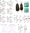
Fig. 1
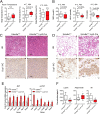
Fig. 1. The myeloid-specific knockout of Kdm6a blocks high fat diet-induced obesity.
A The body weight after the Kdm6aF/Y mice and Kdm6aF/Y;Lyz2-Cre mice were fed a high fat diet (HFD) or normal diet (ND) for 16 weeks (n = 10 in each group). ***p < 0.001; **p < 0.005; *p < 0.05. N.S., not significant. B Representative images of HFD-fed wild-type and myeloid-specific knockout mice. The epididymal and subcutaneous adipose tissues are also presented. C The OGTT assay was used to detect blood glucose homeostasis in HFD- and ND-fed mice. D The ITT assay was used to detect blood glucose homeostasis in HFD- and ND-fed mice. E Serum leptin and adiponectin concentrations of mice fed with HFD or ND. F Representative images of H&E-stained WAT in the HFD-fed mice, bar = 100 μm. G The body temperature was recorded.
Fig. 2
Fig. 2. The myeloid Kdm6a ablation increases energy expenditure of DIO mice but not lean mice.
A The mice were fed with HFD or ND for 16 weeks. The oxygen consumption (VO2) monitored over a 24-h period is shown as averaged values (n = 8 in each group). B Carbon dioxide production (VCO2) monitored over a 24-h period, shown as averaged values. C The RER was analysed for 24 h. D The locomotor activity were measured.
Fig. 3
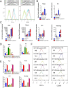
Fig. 3. Kdm6a deficiency reverses the ATM M1–M2 imbalance.
A Representative flow cytometry histograms and the quantitative data from the analysis of CD11b+ F4/80+ cells in SVFs of the epWAT and scWAT. B The percentage of CD11c+ cells gated from the CD11b+ F4/80+ population in epWAT was decreased in the Kdm6aF/Y;Lyz2-Cre mice. The percentage of CD206+ cells gated from CD11b+ F4/80+ population in epWAT was increased in the Kdm6aF/Y;Lyz2-Cre mice. C Realtime PCR assays to detect the abundance of the indicated mRNAs in the CD11b+ cells and adipocytes isolated from the epWAT. D The serum concentration of Il1β in the DIO mice. E The H&E-stained sections of HFD-fed mice indicating lipid droplets in the BAT. Arrows indicate the focal enhancement of Ucp1 expression in the BAT, bar = 100 μm. F Realtime PCR assays to detect the abundance of the indicated mRNAs in the BAT.
Fig. 4
Fig. 4. Myeloid Kdm6a ablation improves adaptive thermogenesis.
A Body temperature and body weight of mice at room temperature and cold exposure at 4 °C for 48 h. B The concentration of nonesterified fatty acid (NEFA), low-density lipoprotein-cholesterol (LDL-C) and total cholesterol (TC) in the mouse serum after 48 h cold exposure. C H&E staining and IHC images of BAT in the Kdm6aF/Y and Kdm6aF/Y;Lyz2-Cre mice, bar = 100 μm. D H&E staining and IHC images of scWAT in the mice, bar = 100 μm. E Realtime PCR assays to detect the abundance of the indicated mRNAs in the BAT and scWAT. F The serum concentrations of leptin, adiponectin and Il6 were measured.
Fig. 5
Fig. 5. Kdm6a enhances the polarisation of macrophages via demethylase activity.
A Representative flow cytometry histograms from the analysis of CD11c+ LPS-stimulated BMDMs and CD206+ Il4-stimulated BMDMs. B Realtime PCR assays to detect the abundance of the indicated mRNAs in LPS-stimulated BMDMs. C Realtime PCR assays to detect the abundance of the indicated mRNAs in the Il4-stimulated BMDMs. D Realtime PCR assays to detect the abundance of the indicated mRNAs in the LPS-stimulated upon ectopic expression of the full-length Kdm6a(Kdm6a) and Kdm6a truncation lacking the JmjC domain (del JmjC) in wild-type (WT) or Kdm6a-deficient (KO) BMDMs. E Realtime PCR assays to detect the abundance of the indicated mRNAs in the Il4-stimulated upon ectopic expression of the full-length Kdm6a (Kdm6a) and Kdm6a truncation lacking the JmjC domain (del JmjC) in wild-type (WT) or Kdm6a-deficient (KO) BMDMs. F Representative flow cytometry histograms from the analysis of CD11c+ LPS-stimulated BMDMs and CD206+ Il4-stimulated BMDMs. The median numbers of histogram were indicated.
Fig. 6
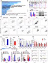
Fig. 6. Kdm6a regulates M1–M2 polarisation through Ire1α expression.
A The enrichment of KEGG pathway between OE-Kdm6a and OE-Vector BMDMs in the ChIP-Seq assay. B Genome browser view of normalised ChIP-Seq signals of H3K27me3 at the Ire1α locus in vector, OE-Kdm6a and OE-del JmjC BMDMs. C Realtime PCR assays and immunoblotting assays indicated Ire1α expression in Kdm6a-deficient BMDMs compared to the counterparts. D The expressions of Kdm6a and Ire1α mRNA are positively correlated in multiple human organs from the GTEx database. E Realtime PCR assays to detect the abundance of the indicated mRNAs in the Il4-stimulated BMDMs after overexpression of Kdm6a or knocking down with specific siRNA against Kdm6a. F ChIP-qPCR analysis of H3K27me3 modifications at the Ire1α loci upon ectopic expression of Kdm6a in BMDMs. G ChIP-qPCR analysis of H3K27me3 modifications at the Ire1α loci upon si-Kdm6a or si-NC treatment in BMDMs. H Realtime PCR assays to detect the abundance of indicated mRNAs in BMDMs. The cells were transfected with control group (vector + si-NC), Ire1α (OE-Ire1α + si-NC), siRNAs against Kdm6a (vector + si-Kdm6a) or Ire1α overexpression plus si-Kdm6a (OE-Ire1α + si-Kdm6a). ***p < 0.001; **p < 0.005; *p < 0.05. N.S., not significant. I Representative flow cytometry histograms to analyse the median number of LPS-stimulated CD11c+ BMDMs and Il4-stimulated CD206+ BMDMs.
Fig. 7
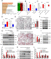
Fig. 7. Kdm6a deficiency regulates differentiation of adipocyte through Il10.
A The enrichment of KEGG pathway indicates the Il10 signalling gained highest score in Kdm6a-deficient BMDMs compared to the wild-type cells. B Realtime PCR assays to detect the abundance of Il10 mRNA upon ectopic expression of Kdm6a or Kdm6a-deficient BMDMs. C Concentrations of Il10 in the serum of wild-type and Kdm6a-deficient mice fed with ND or after exposed to 4 °C for 48 h. D Concentrations of Il10 in culture medium of Kdm6a-deficient BMDMs or upon ectopic expression of Kdm6a. E The representative oil red staining images of 3T3-L1 differentiation, bar = 200 μm. F Immunoblotting assays to ACC, C/ebpβ, Pparγ and Fabp4 in wild-type or Kdm6a-deficient BMDMs medium-treated 3T3-L1 cells in the presence or absence of Il10 neutralising antibody. G Immunoblotting assays to phosphorylated insulin receptor and phosphorylated Akt levels in differentiated 3T3-L1 cells, which were incubated with indicated BMDMs medium. H The representative oil red images and quantitative data of differentiation in C3H10-T1/2 cells after received the indicated conditional medium from BMDMs, bar = 200 μm. I Immunoblotting assays to examine the Ucp1 and Prdm16 proteins in the C3H10-T1/2 cells. During the differentiation, cells were incubated with indicated BMDMs medium. J Relative mRNA abundance of differentiation markers was detected with Realtime PCR assay. During the differentiation, C3H10-T1/2 cells were incubated with indicated BMDMs medium. K Immunoblotting assays to indicate the thermogenesis markers in the differentiated C3H10-T1/2 cells, after the already differentiated cells treated with indicated BMDMs medium. L Relative mRNA abundance of the thermogenesis markers in the differentiated C3H10-T1/2 cells was detected with Realtime PCR, after the already differentiated cells were treated with conditional medium from wild-type or Kdm6a-deficient BMDMs.
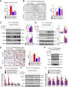
Fig. 8
Fig. 8. Kdm6a-deficient BMDM regulates differentiation of adipocyte via Ire1α.
A Concentrations of Il10 in culture medium from indicated BMDMs. B The representative images of oil red staining of 3T3-L1 adipocytes and the quantitative data. During the differentiation, cells were treated with conditional medium from indicated BMDMs, bar = 200 μm. C Immunoblotting assays to detect ACC, C/ebpβ, Pparγ and Fabp4 proteins in 3T3-L1 cells after received the treatment of indicated BMDMs conditional medium. D Immunoblotting assays to examine the p-Insulin Receptor and p-Akt levels in 3T3-L1 cells in response to insulin incubation, after cells received the indicated BMDMs conditional medium. E The representative images of oil red staining of C3H10-T1/2 adipocytes and the quantitative data. During the differentiation, cells were treated with conditional medium from indicated BMDMs, bar = 200 μm. F Immunoblotting assays to examine the Ucp1 and Prdm16 proteins in the C3H10-T1/2 cells. During the differentiation, cells were incubated with conditional medium from indicated BMDMs. G Relative mRNA abundance of shown mRNAs in the C3H10-T1/2 cells. During the differentiation, cells were incubated with conditional medium from indicated BMDMs. H Immunoblotting assays to indicate the thermogenesis markers in the already differentiated C3H10-T1/2 cells, after the differentiated cells were treated with indicated BMDMs medium. I Relative mRNA abundance of thermogenesis markers in the differentiated C3H10-T1/2 cells was detected with Realtime PCR, after the differentiated cells were treated with indicated BMDMs medium.

Similar articles

Cited by

References

    1. Liu X, Wang C, Liu W, Li J, Li C, Kou X, et al. Distinct features of H3K4me3 and H3K27me3 chromatin domains in pre-implantation embryos. Nature. 2016;537:558–62. doi: 10.1038/nature19362. - DOI - PubMed
    1. Wang L, Zhao Z, Ozark PA, Fantini D, Marshall SA, Rendleman EJ, et al. Resetting the epigenetic balance of Polycomb and COMPASS function at enhancers for cancer therapy. Nat Med. 2018;24:758–69. doi: 10.1038/s41591-018-0034-6. - DOI - PMC - PubMed
    1. Kruidenier L, Chung CW, Cheng Z, Liddle J, Che K, Joberty G, et al. A selective jumonji H3K27 demethylase inhibitor modulates the proinflammatory macrophage response. Nature. 2012;488:404–8. doi: 10.1038/nature11262. - DOI - PMC - PubMed
    1. Wang X, Cao W, Zhang J, Yan M, Xu Q, Wu X, et al. A covalently bound inhibitor triggers EZH2 degradation through CHIP-mediated ubiquitination. EMBO J. 2017;36:1243–60. doi: 10.15252/embj.201694058. - DOI - PMC - PubMed
    1. Sen GL, Webster DE, Barragan DI, Chang HY, Khavari PA. Control of differentiation in a self-renewing mammalian tissue by the histone demethylase JMJD3. Genes Dev. 2008;22:1865–70. doi: 10.1101/gad.1673508. - DOI - PMC - PubMed

Publication types