. 2020 Nov-Dec;53(6):413-418.
doi: 10.1590/0100-3984.2019.0131.

Anatomical variations and congenital anomalies of the ribs revisited by multidetector computed tomography

Affiliations

Anatomical variations and congenital anomalies of the ribs revisited by multidetector computed tomography

Lucas de Pádua Gomes de Farias et al. Radiol Bras. 2020 Nov-Dec.

Abstract

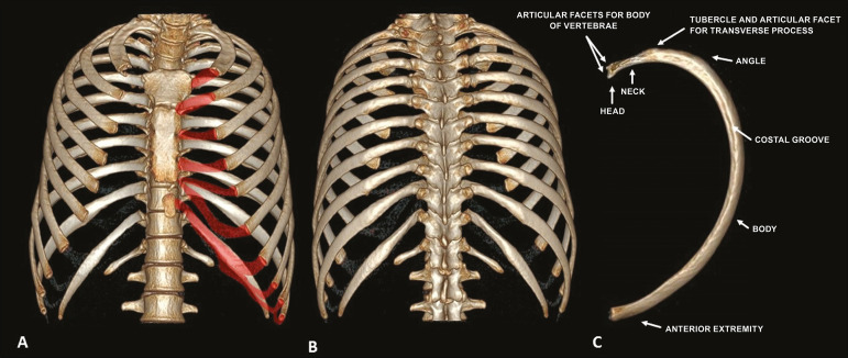
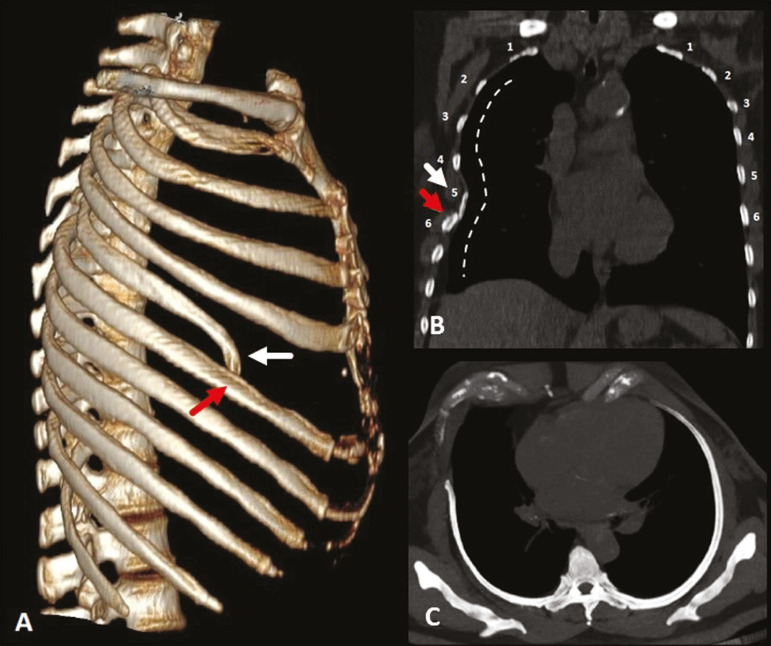
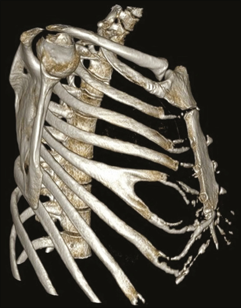
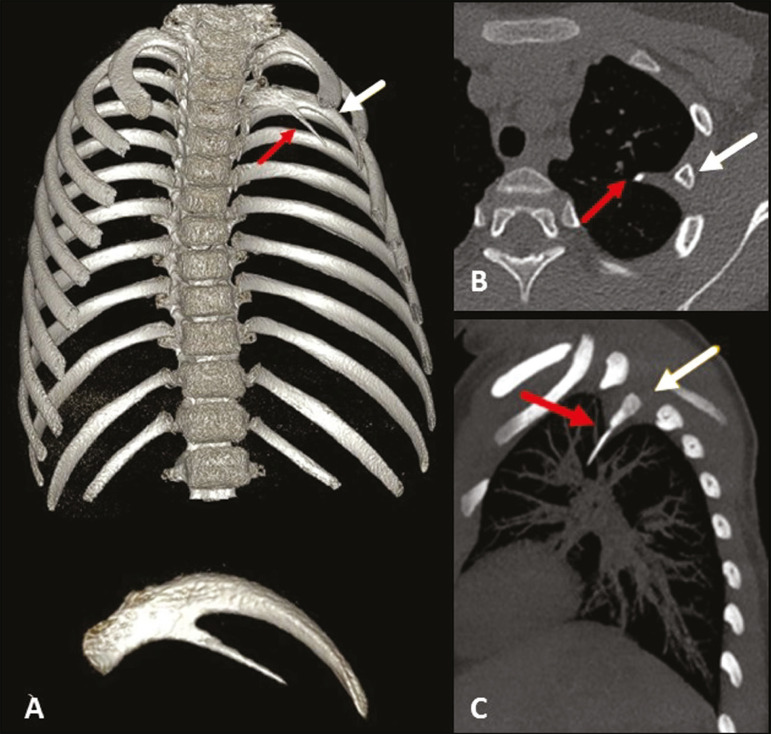
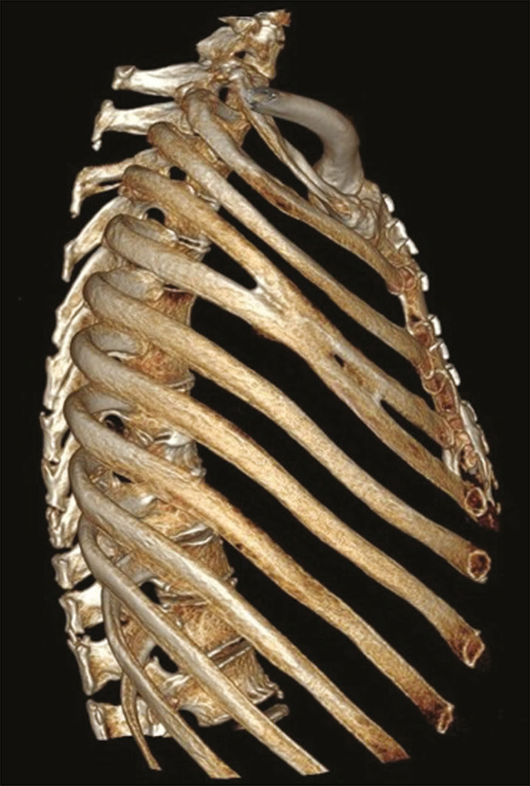
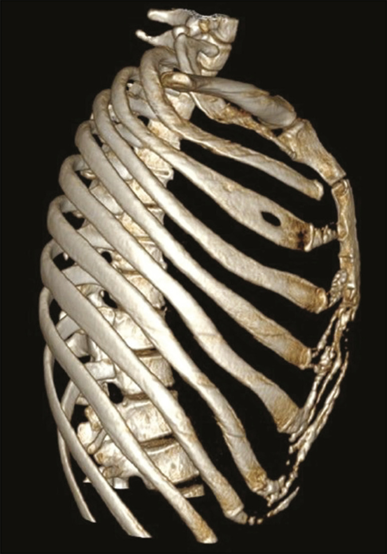
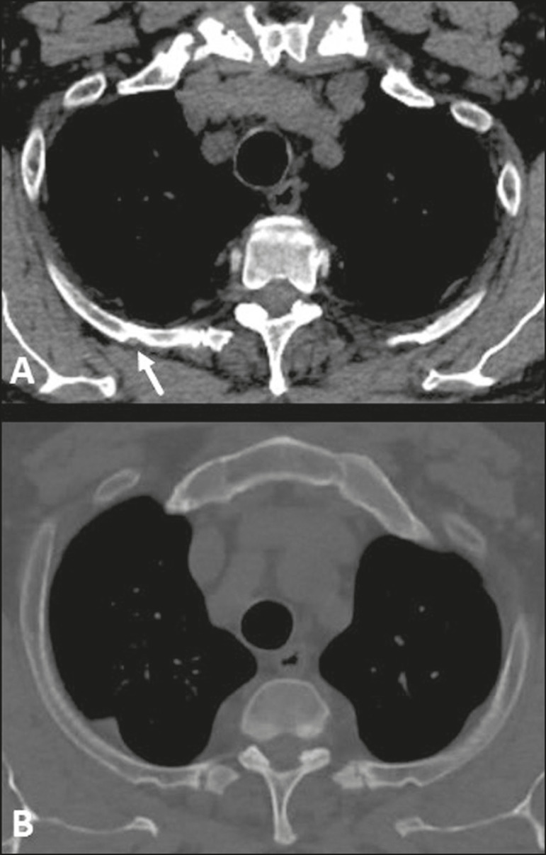
As they are asymptomatic or have a nonspecific, anatomical variations of the ribs are usually detected as incidental findings on imaging studies. They may be isolated changes or can be related to anomalies or clinical syndromes. Such variations are easily overlooked on conventional radiography and computed tomography if they are not actively investigated, mainly because most indications for a chest X-ray studies aim to evaluate the lung parenchyma and mediastinal structures. The objective of this pictorial essay was to use multislice computed tomography images to illustrate the imaging aspects of the main anatomical variations and congenital anomalies of the ribs.

As variantes anatômicas dos arcos costais são, geralmente, achados incidentais nos exames de imagem, em razão do seu comportamento assintomático ou quadro inespecífico, podendo constituir alterações isoladas ou relacionadas a anomalias e síndromes clínicas. Essas alterações são facilmente negligenciadas na radiografia convencional e na tomografia computadorizada se não forem investigadas ativamente, principalmente quando a maioria das indicações do estudo radiológico do tórax tem como objetivo a avaliação do parênquima pulmonar e das estruturas mediastinais. O objetivo deste artigo é demonstrar, por meio da tomografia computadorizada multidetectores, os aspectos de imagem das principais variantes anatômicas e anomalias congênitas dos arcos costais.

Keywords: Anatomical variation; Multislice computed tomography; Ribs.


Figures

Figure 1
Figure 1
Anatomy of the ribs. MDCT (A,B,C - three-dimensional reconstructions) showing the usual anatomy of the ribs in a young adult characterized by 12 ribs articulated posteriorly to the respective vertebral bodies (T1 to T12). The first to seventh ribs (the true ribs) articulate directly to the sternum through the costal cartilages, whereas the eighth to the tenth ribs (false ribs) articulate indirectly to the sternum through the superior costal cartilage of the seventh ipsilateral rib and the eleventh and twelfth ribs (floating ribs) are points of origin and insertion of the myotendinous junction, without connecting to the sternum. Note the anatomical components of the rib, through its lower view (in C), and the projection of the hyaline costal cartilages (red transparency).
Figure 2
Figure 2
Pseudarthrosis and physiological costochondral calcifications. MDCT (three-dimensional reconstruction) showing pseudarthrosis in the first bilateral rib (arrows) in a 45-year-old female patient. Note the pattern of nodular and peripheral calcification of bilateral sternocostal cartilage.
Figure 3
Figure 3
Physiological costochondral calcifications. MDCT (A - three-dimensional reconstruction; B - coronal view) showing fusion of the first rib to the sternal manubrium (arrows) in an 83-year-old male patient, together with calcification of the other sternocostal and bilateral chondrocostal cartilages. Note the tramtrack pattern of calcification.
Figure 4
Figure 4
Cervical rib. MDCT (A - three-dimensional reconstruction; B - axial maximum intensity projection) showing a supernumerary rib on the right (arrows), articulating posteriorly with the C7 vertebral body, with no articular structure in its aspect anterior to the sternum or any bony structure (floating ). Note its close relationship with the first thoracic rib and the ipsilateral clavicle, which can be related, in some cases, to vascular compression syndrome.
Figure 5
Figure 5
Short rib. MDCT (A - threedimensional reconstruction; B - coronal view; C - axial oblique maximum intensity projection on the longitudinal axis of the rib) showing hypoplasia of the right fifth rib that presents a lower inclination at its anterior end (white arrows), juxtaposed with the middle aspect of the ipsilateral sixth rib (red arrows). Note the asymmetry when compared with the contralateral rib in the coronal and axial images, together with the locoregional deformity of the rib cage (dotted line).
Figure 6
Figure 6
Bifid rib. MDCT (three-dimensional reconstruction) showing the bifid aspect of the right fifth rib in its anterior portion, including the sternocostal hyaline cartilage, although without causing significant deformities in the rib cage. Bilateral sternocostal and chondrocostal calcifications.
Figure 7
Figure 7
Intrathoracic rib (type Ib). MDCT (A - three-dimensional reconstruction; B - axial view; C - sagittal maximum intensity projection) showing a supernumerary rib (red arrows) originating in the posterior portion of a short left third rib (white arrows), with a lateral, inferior oblique path to the pulmonary parenchyma. Note that the rib has no visible medullary cavity. No changes were identified in the posterior vertebral bodies or ribs.
Figure 8
Figure 8
Rib fusion. MDCT (three-dimensional reconstruction) showing fusion of the middle body portions of the fourth and fifth ribs on the right, although without causing significant deformities in the rib cage.
Figure 9
Figure 9
Rib foramen. MDCT (three-dimensional reconstruction) showing a well-circumscribed foramen, with corticated contours, in the anterior region of the right third rib.
Figure 10
Figure 10
Rib notching. MDCT (A,B - axial oblique view) showing bone deformation in the posterior aspects of the ribs on both sides, most evident in the upper ribs on the right, which follow the arterial vascular path (arrow).

References

    1. Guttentag AR, Salwen JK. Keep your eyes on the ribs: the spectrum of normal variants and diseases that involve the ribs. Radiographics. 1999;19:1125–1142. - PubMed
    1. Glass RBJ, Norton KI, Mitre SA, et al. Pediatric ribs: a spectrum of abnormalities. Radiographics. 2002;22:87–104. - PubMed
    1. Talbot BS, Gange CP Jr, Chaturvedi A, et al. Traumatic rib injury: patterns, imaging pitfalls, complications, and treatment. Radiographics. 2017;37:628–651. - PubMed
    1. Levine BD, Motamedi K, Chow K, et al. CT of rib lesions. AJR Am J Roentgenol. 2009;193:5–13. - PubMed
    1. Ontell FK, Moore EH, Shepard JA, et al. The costal cartilages in health and disease. Radiographics. 1997;17:571–577. - PubMed

LinkOut - more resources