Review
. 2020 Dec 4:15:2277-2289.
doi: 10.2147/CIA.S280931. eCollection 2020.

Case Report and Literature Review on Low-Osmolar, Non-Ionic Iodine-Based Contrast-Induced Encephalopathy

Affiliations
Review

Case Report and Literature Review on Low-Osmolar, Non-Ionic Iodine-Based Contrast-Induced Encephalopathy

Meng-Ru Liu et al. Clin Interv Aging. .

Abstract

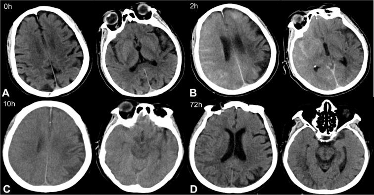
Contrast-induced encephalopathy (CIE) is a rare complication following percutaneous carotid and coronary interventions, and important diagnostic radiological signs include brain edema and cortical enhancement. In this report, we detail a case of probable CIE in an 84-year-old woman following a normal diagnostic coronary angiography (CAG) that involved 20 mL of the low-osmolar, non-ionic monomeric, iodine-based contrast agent iopromide (Ultravist 370). The patient was unconscious and presented with hemiparesis, hemianopia, recurrent seizures, and cardiac and respiratory arrest within minutes to hours following the procedure. Non-contrast computed tomography (CT) of the head showed increased subarachnoid density, cortical enhancement, and brain edema in the right hemisphere. Three days of rehydration, reduction in cranial pressure, and treatment with an anticonvulsant and dexamethasone resulted in a gradual recovery with no neurological deficits. This case highlights that severe neurotoxic symptoms may occur in response to low doses of low-osmolar, non-ionic, monomeric contrast agents. This finding is of importance to interventional cardiologists for diagnostic considerations and development of treatment plans.

Keywords: contrast-induced encephalopathy; coronary angiography; percutaneous carotid and coronary interventions.


Conflict of interest statement

The authors report no conflicts of interest in this work.

Figures

Figure 1
Figure 1
Contrast-induced encephalopathy in brain computed tomography (CT) scans. (A) Emergency brain CT showed no acute pathological findings. (B) Brain CT 2 hours after surgery indicated multiple high-density regions in the subarachnoid space. (C) Brain CT 10 hours after surgery showed significantly swollen brain tissue, particularly in the right hemisphere, the left frontal lobe, and the left occipital lobe. The high-density shading in the subarachnoid cavity was significantly denser than in previous scans. (D) Brain CT 72 hours after surgery showed the sulci of the right hemisphere became shallow, and low-density shadows could be seen in the frontal parietal lobe. But cerebral edema was significantly improved.
Figure 2
Figure 2
Computed tomography angiography (CTA) showed no evidence of macrovascular embolism in (A) posteroanterior (PA), (B) posterior (P) and (C) left anterior oblique (LAO) view.
Figure 3
Figure 3
Brain magnetic resonance imaging (MRI) 2 hours after surgery showed hyperintense areas in the right cortex in (A) T2-weighted image and (B) fluid-attenuated inversion recovery (FLAIR) images, and no clear signs of subarachnoid hemorrhage were observed on (C) diffusion-weighted imaging (DWI).

References

    1. Fang HY, Kuo YL, Wu CJ. Transient contrast encephalopathy after carotid artery stenting mimicking diffuse subarachnoid hemorrhage: a case report. Catheter Cardiovasc Interv. 2009;73(1):123–126. doi: 10.1002/ccd.21779 - DOI - PubMed
    1. Kocabay G, Karabay C, Kalayci A, et al. Contrast-induced neurotoxicity after coronary angiography. Herz. 2014;39(4):522–527. doi: 10.1007/s00059-013-3871-6 - DOI - PubMed
    1. Dattani A, Au L, Tay KH, Davey P. Contrast-induced encephalopathy following coronary angiography with no radiological features: a case report and literature review. Cardiology. 2018;139(3):197–201. doi: 10.1159/000486636 - DOI - PubMed
    1. Leong S, Fanning NF. Persistent neurological deficit from iodinated contrast encephalopathy following intracranial aneurysm coiling. Interv Neuroradiol. 2012;18(1):33–41. doi: 10.1177/159101991201800105 - DOI - PMC - PubMed
    1. Sawaya RA, Hammoud R, Arnaout S, Alam S. Contrast-induced encephalopathy following coronary angioplasty with iohexol. South Med J. 2007;100(10):1054–1055. doi: 10.1097/SMJ.0b013e3181540086 - DOI - PubMed

MeSH terms