Review
. 2020 Nov 16:14:602796.
doi: 10.3389/fnins.2020.602796. eCollection 2020.

Exposure to Artificial Light at Night and the Consequences for Flora, Fauna, and Ecosystems

Affiliations
Review

Exposure to Artificial Light at Night and the Consequences for Flora, Fauna, and Ecosystems

Jack Falcón et al. Front Neurosci. .

Abstract

The present review draws together wide-ranging studies performed over the last decades that catalogue the effects of artificial-light-at-night (ALAN) upon living species and their environment. We provide an overview of the tremendous variety of light-detection strategies which have evolved in living organisms - unicellular, plants and animals, covering chloroplasts (plants), and the plethora of ocular and extra-ocular organs (animals). We describe the visual pigments which permit photo-detection, paying attention to their spectral characteristics, which extend from the ultraviolet into infrared. We discuss how organisms use light information in a way crucial for their development, growth and survival: phototropism, phototaxis, photoperiodism, and synchronization of circadian clocks. These aspects are treated in depth, as their perturbation underlies much of the disruptive effects of ALAN. The review goes into detail on circadian networks in living organisms, since these fundamental features are of critical importance in regulating the interface between environment and body. Especially, hormonal synthesis and secretion are often under circadian and circannual control, hence perturbation of the clock will lead to hormonal imbalance. The review addresses how the ubiquitous introduction of light-emitting diode technology may exacerbate, or in some cases reduce, the generalized ever-increasing light pollution. Numerous examples are given of how widespread exposure to ALAN is perturbing many aspects of plant and animal behaviour and survival: foraging, orientation, migration, seasonal reproduction, colonization and more. We examine the potential problems at the level of individual species and populations and extend the debate to the consequences for ecosystems. We stress, through a few examples, the synergistic harmful effects resulting from the impacts of ALAN combined with other anthropogenic pressures, which often impact the neuroendocrine loops in vertebrates. The article concludes by debating how these anthropogenic changes could be mitigated by more reasonable use of available technology - for example by restricting illumination to more essential areas and hours, directing lighting to avoid wasteful radiation and selecting spectral emissions, to reduce impact on circadian clocks. We end by discussing how society should take into account the potentially major consequences that ALAN has on the natural world and the repercussions for ongoing human health and welfare.

Keywords: anthropogenic impact; artificial-light-at-night; biological clocks; ecosystems; light-emitting-diodes; photoreception.


Figures

FIGURE 1
FIGURE 1
(A) Illuminance measured in the horizontal plane from a typical street light (Phillips Cosmopolis, metal halide lamp). The illuminance level decays rapidly with distance to the lamp. (B) Comparison of measured illuminance from natural sources of light to artificial light sources – axis is on a logarithmic scale, and bars present approximate ranges based on field measurements. From Bennie et al. (2016). No special permission required.
FIGURE 2
FIGURE 2
The chloroplast of plants and photosynthetic algae absorbs basic elements and uses sunlight to produce sugar and other organic molecules to fulfil their needs (Kirchhoff, 2019) @JackFalcón.
FIGURE 3
FIGURE 3
(A) Rhabdomeric microvilli-based (invertebrates) and cilia-based (vertebrates) photoreceptors display conserved cell polarity and topology. They evolved most probably from a common ancestor in early Bilateria. The photosensory pole is made of stacks of plasma membrane separated from the baso-lateral membrane by a zonula adherens. N, nucleus. (B) The main optical designs of eyes: (a) The pinhole eye; light (yellow arrow) falls directly upon the photoreceptors (brown layer). (b) The concave-mirror eye; light crosses the retina, and is then focused back onto the retina upon reflection from a hemispheric reflective mirror (tapetum, grey zone). (c) The camera type eye; light is focused by the lens to form an image on the retina. (d and e) The compound eyes; light reaches the photoreceptors exclusively from the small corneal lens (d type) located directly above, or focused through a large number of corneal facets and cones to be directed towards single rhabdoms (e type). Redrawn from Warrant (2019).
FIGURE 4
FIGURE 4
Extraretinal photoreception in vertebrates. (A) Dorsal view of the head of the Polar Cod Boreogadus saida; the pineal organ (PO) is located in the sagittal axis just behind the eyes in an area with unpigmented meninges (@JackFalcón). (B) Dorsal view of the brains of the Red Mullet Mullus surmulletus showing the location of the pineal organ (thick arrow), located in between the two cerebral hemispheres (Ch); OT, optic tectum; Cer, cerebellum; from Baudelot (1883) (no permission required). (C) Schematic sagittal sections through the epithalamus area of, from top to bottom, lampreys, chondrichtyens and teleost fish; from Studnicka (1905). Note that the skull above the pineal organ is thinner, as also seen in panel (D) (no permission required). The histological sagittal section is from the Sea Bream Sparus aurata; the pineal is located in a kind of large pit below the skull (note that the tegument above also appears thinner) (gift from Professor J.A. Muñoz Cueto, Cadiz, Spain). (E,F) Head dorsal views showing the spot position of the frontal organ in the American Bullfrog Rana catesbeiana (E) and the parietal eye of the Zebra-tailed Lizard Callisaurus draconoides (F) (arrows) (@JackFalcón). (G,H) Schematic sagittal sections through the epithalamus areas of frogs (G) and lizards (H); the pineal organs are located below the skull, while the frontal/parietal eyes are located in the skin connected to the brain by a stalk (Studnicka, 1905) (no permission required). (I) Dorsal fossil skull of the ancestral amphibian Thoosuchus jakovlevi showing the location of the frontal organ hole just equidistant from the eyes (with permission from https://commons.wikimedia.org/wiki/File:Thoosuchus_jakovlevi.JPG). (J) The pineal eye of the tuatara Sphenodon punctatus resembles a simplified retina with an eye cup and a lens-like structure; sagittal section from Dendy (1911) (no permission required). (K) In the avian brain the pineal organ form a gland in between the cerebral hemispheres and the cerebellum (gift from Professor J.P. Collin). (L) In humans the gland is located deep in the brain (@JackFalcón).
FIGURE 5
FIGURE 5
Extra-ocular light perception in various insect species (A-E) and eyes of a spider (F). Arrows point to ocellar structures as found in Netelia sp. (A), Heptagenia sp. (B), grasshopper Locusta migratoria (C), Eristalinus sepulchralis (D), Vespa cabro (E), and Philodromus dispar (F). Photo credits: P. Falatico (A,B,D,E; @ http://aramel.free.fr/), J Falcón (C), D. Vaudoré (F; @https://www.galerie-insecte.org/galerie/ref-183890.htm). No special permissions required.
FIGURE 6
FIGURE 6
(A) The spectral sensitivity of plants. See text and (Huche-Thelier et al., 2016) for details. (B) Different states of the flavoquinone cofactor of Cry and corresponding photosensitivity (see text for details). (C) Phylogenetic tree of the photolyase/cryptochrome family. Modified from Du et al. (2014), with permission.
FIGURE 7
FIGURE 7
The family of opsins in the tree of evolution. C-opsin family includes the vertebrates visual and brain opsins (Rh1, Rh2, SWS1, SWS2, M/LWS, pinopsins, parapinopsins, vertebrate ancient and parietal opsins), the chordates’ brain opsins (teleost multiple tissue opsins (TMTs), encephalopsins and uncharacterized amphioxus and urchin opsins), the arthropod opsins (honeybee ptersopsin, and uncharacterized insect and Daphnia pulex opsins), and the annelids group (uncharacterized Platynereis brain and urchin opsins). Cnidops family includes ctenophore and cnidiarian opsins. R-type opsins include the arthropod visual pigments (M, LWS, and SWS), the annelid, Platyhelminthes and mollusc visual pigments, the melanopsins (vertebrates’ melanopsin 1 and 2, and amphioxus sequences) and uncharacterized tunicate, amphioxus and mollusc opsins. Group 4 Opsins include neuropsins (four separate clades), amphioxus, sea urchin and scallop opsins, RGR (uncharacterized mollusc opsins) and peropsins (amphioxus and hemichordate opsins). See text and (Porter et al., 2012) for more details. Modified from Porter et al. (2012). No special permission required.
FIGURE 8
FIGURE 8
Spectral sensitivity curves of selected vertebrate and invertebrate representatives, illustrating the wide variety of light detection systems encountered. Vertebrates: human Homo sapiens, mouse Mus musculus, chicken Gallus domesticus, Salamander Salamandra, goldfish Carassius auratus. Invertebrates: elephant hawk moth Deilephila elpenor, dragonfly Hemicordulia tau, butterfly Papilio xuthus, annelid worm Torrea candida, nocturnal spider Cupiennius salei. Adapted and modified from Imamoto and Shichida (2014), Warrant (2019).
FIGURE 9
FIGURE 9
(A) Penetration of light into the water column and (B) illustration of the depth at which different colours of light penetrate ocean waters. (B is modified from the NOAA Office of Ocean Exploration and Research, with permission).
FIGURE 10
FIGURE 10
Simplified schematic representation of the circadian clock in (A) mammals, (B) insects, (C) Cyanobacteria, (D) fungi, and (E) plants. For details see Saini R. et al. (2019). Abbreviations: CCA1, circadian clock associated 1; CCG, clock controlled genes; Clk, clock; CRY, cryptochrome; CYC, cycle; ELF, early flowering; FRH, FRQ-interacting RNA, helicase; FRQ, frequency; GI, gigantea; LHY, late elongated hypocotyl; LUX, lux arrhythmo; PER, period; Rev-Erbβ (orphan nuclear receptor family 1); PRR, pseudo-response regulator; RORα, retinoic acid receptor (RAR)-related orphan receptors; TIM, timeless; TOC1, timing of cab expression 1; VVD, vivid; WC, white collar; WCC, white collar complex. Modified from Saini R. et al. (2019) No special permission required.
FIGURE 11
FIGURE 11
Schematic representation of the photoneuroendocrine organization in the non-mammalian brain. The drawing pictures a frontal section of the brain diencephalic area. Light information is captured by the lateral eyes and the pineal organ. Photosensitive units, expressing different types of opsins, have also been identified along the 3rd ventricle (3rd V; yellow and green circles). Major circadian clock machineries formula image 
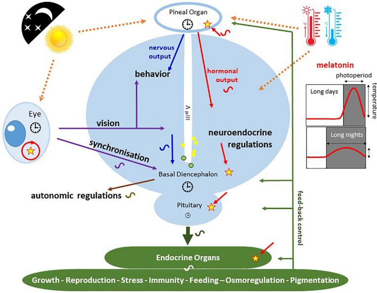
are present in the pineal and retinal photoreceptors as well as in the basal diencephalon (preoptic area [POA] and suprachiasmatic nuclei [SCN]) of lizards and birds. The pineal gland of fish and lizards also integrates temperature information from the external environment. The concomitant action of light, temperature and other internal factors, shapes the rhythmic nervous (blue) and hormonal (red; melatonin) outputs (see text for details), providing a temporal message transmitted to the neuroendocrine axis and downstream targets (peripheral endocrine organs). Melatonin acts through specific receptors (stars) distributed in different tissues and organs. While the main retinal output subserves visual function, a few other fibres also terminate in different parts of the basal diencephalon, where some converge with fibres originating from the pineal gland. Some of the targeted areas also express melatonin receptors. This double or triple input contributes to synchronizing the neuronal activity of the basal diencephalon. In sauropsids the POA and SCN neurons also relay retinal information to the pineal gland. The entire neuroendocrine axis is targeted by ALAN together with multiple other disruptors including temperature rises and pollutants [e.g., endocrine disruptors] acting directly or indirectly at different levels of the loop.
FIGURE 12
FIGURE 12
(A) Observed abundances in fish populations from the harbour of Sydney (Australia) under a 12L/12D cycle plotted in 15 min bins. Under the natural LD cycle the number of fish is higher during night (black line) than during day (blue line); they were sedentary at night with low predation activity (P↓), while displaying a predatory behaviour during day (P↑). ALAN (40-50 lx, warm LED light), transformed the nocturnal pattern into a diurnal one. Modified and adapted from Bolton et al. (2017). (B) Orientation response of 4 species of sea turtles hatchlings to coloured light sources. Olive Ridley Sea Turtle Lepidochelys olivacea, Green Sea Turtle Chelonia mydas, Hawksbill Sea Turtle Eretmochelys imbricata, were attracted when illuminated with UV-A to yellow wavelengths. The Loggerhead Sea Turtle Caretta differed in that UV-A to green lights were attractive, but yellow wavelengths were repulsive, an effect reversed by red illumination. For details see (Witherington and Martin, 2003) from which the figure was modified and adapted.
FIGURE 13
FIGURE 13
(A) The impact of alternative LED lighting strategies on the total numbers of individual grassland spiders (Araneae) (a) and beetles (Coleoptera) (b) caught in each year, respectively. LED lighting was equivalent to that experienced at ground level under LED street lighting for HIW (high-intensity white 29.6 ± 1.2 lx), under dimmed street lighting for DW (dimmed white, 14.6 ± 0.3 lx), or under timed dimmed street lighting for DWT (14.4 ± 0.8 lx, switched off between 00:00 and 04:00 GMT). AMB was amber lighting (18.2 ± 1.3 lx, λmax = 588 nm). Controls (CON) experienced total darkness. Bar heights and error bars denote means 95% confidence intervals. Stars denote differences with the controls that were significant with 95% (*), 99% (**), and 99.9% or greater (***) confidence. From Davies et al. (2017). No special permission required. (B) Effects of artificial lighting on parameters of overall quantified nocturnal plant-flower visitor networks of seven dark sites (above) and seven experimentally illuminated sites (below). The rectangles represent insect species (top) and plant species (bottom), and the connecting lines represent interactions among species. Species codes for the plants and a list of insect species are given in Knop et al. (2017). The study was run in 14 sites of the Swiss Alps; illumination was using neutral white LED street lamps (4,000K) that provided 52.0 ± 4.2 lx on the ground. Adapted from Knop et al. (2017). More details in the original publication. With permission.
FIGURE 14
FIGURE 14
The mosaic plot illustrates the proportion of moth flight responses under four different conditions: absence or presence of bats (Nyctalus sp.) under total darkness or white LED illumination, in the area of Bristol (United Kingdom). Moths respond to the presence of bats under unlit conditions at night by escape movements. This escape behaviour is markedly affected in the presence of white LED. Column width is proportional to sample size. From Wakefield et al. (2015). No special permission required.
FIGURE 15
FIGURE 15
Migration is a crucial event in the Atlantic salmon, Salmo salar. In the Loire/Allier basin a ∼800 km downstream migration brings young smolts from their hatching area to the sea, where they feed and mature. In the journey they have to face light pollution (ALAN) when crossing cities or areas of active human activities (nuclear plants, industrial areas, harbours) as well as a series of other threats of anthropogenic origin, including physical barriers, overfishing, water temperature rise, physical (noise) and chemical (e.g., endocrine disruptors) pollution. They must run another 800 km back when returning to the spawning grounds. Altogether this addition of threats impacts on metabolic reactions and physiological regulation, including their rhythmic components, which have put the species in danger of extinction.

References

    1. Acharya J., Rechner O., Neugart S., Schreiner M., Poehling H. M. (2016). Effects of light-emitting diode treatments on Brevicoryne brassicae performance mediated by secondary metabolites in Brussels sprouts. J. Plant Dis. Protec. 123 321–330. 10.1007/s41348-016-0029-9 - DOI
    1. Alsanius B. W., Karlsson M., Rosberg A. K., Dorais M., Naznin M. T., Khalil S., et al. (2019). Light and microbial lifestyle: the impact of light quality on plant-microbe interactions in horticultural production systems-a review. Horticulturae 5:41 10.3390/horticulturae5020041 - DOI
    1. Altringham J., Kerth G. (2015). “Bats and Roads,” in Bats in the Anthropocene: Conservation of Bats in a Changing World, eds Voigt C., Kingston T. (Cham: Springer; ), 35–62.
    1. Arboleda E., Zurl M., Waldherr M., Tessmar-Raible K. (2019). Differential impacts of the head on Platynereis dumerilii peripheral circadian rhythms. Front. Physiol. 10:900. 10.3389/fphys.2019.00900 - DOI - PMC - PubMed
    1. Arikawa K., Iwanaga T., Wakakuwa M., Kinoshita M. (2017). Unique temporal expression of triplicated long-wavelength opsins in developing butterfly eyes. Front. Neural Circ. 11:96. 10.3389/fncir.2017.00096 - DOI - PMC - PubMed

LinkOut - more resources