Review
. 2020 Jun;16(2):200047.
doi: 10.1183/20734735.0047-2020.

Primary ciliary dyskinesia: a major player in a bigger game

Affiliations
Review

Primary ciliary dyskinesia: a major player in a bigger game

Reena Bhatt et al. Breathe (Sheff). 2020 Jun.

Abstract

Primary ciliary dyskinesia (PCD) is an inherited disorder of clinical and genetic heterogeneity resulting from mutations in genes involved in the transport, assembly and function of motile cilia. The resulting impairment in mucociliary clearance means patients suffer from chronic progressive lung disease, bronchiectasis, rhinosinusitis and middle ear disease. Subfertility is common to both male and female patients. Situs abnormalities occur in around half of patients, with a subgroup suffering more complex situs arrangements where congenital heart defects or other organ abnormalities frequently coexist. Variations from the classical PCD phenotype are increasingly recognised where overlapping features across a range of motile and nonmotile ciliopathies are redefining our approach to both diagnosis and management of these complex conditions. PCD offers an ideal opportunity for direct visualisation of ciliary function and structure, following nasal brush biopsy, allowing opportunities for researchers to directly interrogate the downstream impact of loss of function mutations. In turn, this has led to rapid advances in the development of new diagnostic tests. These advances mean that PCD is an excellent disease model for understanding the genetic and mechanistic causes of the clinical phenotype for all respiratory ciliopathies. Furthermore, the overlapping role of motile ciliary defects in a wider set of complex and syndromic disorders related to loss of function mutations in primary, nonmotile cilia has been recognised. As we better understand the role of ciliary defects in a broad spectrum of diseases, we should aim to map out a framework through which we can identify, diagnose and treat all respiratory ciliopathies.

Key points: Primary ciliary dyskinesia is just one of a group of conditions where a heterogeneous array of genetic mutations affect the assembly or structure of motile cilia.Overlapping phenotypes between motile and nonmotile ciliopathies are redefining the diagnostic and therapeutic approach to encompass all ciliopathy patients with a respiratory phenotype.An extended diagnostic algorithm may be required to capture the majority of cases with a respiratory ciliopathy, including patients with syndromic ciliopathies.The terminology around disorders of motile cilia is becoming more descriptive to better reflect the heterogeneity and underlying disease mechanisms across the spectrum of respiratory ciliopathies.

Educational aims: To summarise the existing knowledge base around the disease mechanisms for respiratory ciliopathies, including primary ciliary dyskinesia (PCD).To explore and understand the reasons for changing terminology around respiratory ciliopathies.To emphasise key messages around the diagnosis and treatment of all ciliopathies.Diagnosing PCD is complex and time consuming, and there is no single stand-alone test that can confirm or exclude a diagnosis in all cases.


Conflict of interest statement

Conflict of interest: C. Hogg has nothing to disclose. Conflict of interest: R. Bhatt has nothing to disclose.

Figures
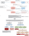
Figure 1
Figure 1
Flowchart to demonstrate the pathways to diagnosis of PCD as per the a) European Respiratory Society and b) American Thoracic Society consensus statements. It is important to note that not all patients will require all investigations. #: cystic fibrosis (CF) should be ruled out before performing nasal nitric oxide (nNO) measurement, as roughly one-third of CF patients can have nNO values below PCD diagnostic cut-offs. nNO measurements should only be performed with chemiluminescence analysers using standardised protocols at centres with specific expertise in nNO measurements. Some nNO analysers have not received approval from federal agencies worldwide (US Food and Drug Administration and Health Canada), which may have implications for clinical implementation. : genetic panel testing for mutations in >12 disease-associated PCD genes, including deletion/duplication analysis. +: as nNO levels can be significantly decreased by viral respiratory tract infections, a repeat nNO measurement, at least 2 weeks after the initial low value (expert opinion), is recommended to ensure the initial low value is not secondary to a viral process. A normal nNO value upon repeat testing is not suggestive of PCD, as nNO values remain consistently low in PCD. §: most forms of PCD resulting in normal nNO levels have normal or non-diagnostic electron microscopy studies. Thus, genetic testing is recommended in these cases. ƒ: or presence of variants of unknown significance. ##: additional corroborative testing may provide information on clinical prognosis, further disease understanding, and potential future therapeutic considerations. ¶¶: known disease-associated TEM ultrastructural defects include outer dynein arm (ODA) defects, ODA plus inner dynein arm (IDA) defects, IDA defect with microtubular disorganisation, and absent central pair, identified using established criteria. Of note, the presence of IDA defects alone is rarely diagnostic for PCD. ++: up to 30% of PCD cases can have normal ciliary ultrastructure on electron microscopy. Consider referral to PCD specialty centre if there is a strong clinical phenotype but all electron microscopy and genetic testing are negative. HSVMA: high-speed video microscopy analysis. Reproduced from [29] with permission.
Figure 2
Figure 2
TEM images of cross-sections of cilia from nasal brushing samples. a) Normal; b) outer and inner dynein arm defects; c) inner dynein arm defects plus microtubular disorganisation; d) central pair defect; and e) transposition defect.
Figure 3
Figure 3
IF staining in a healthy control where a) tubulin stains green to indicate presence of cilia on the cell surface, and b) staining with DNAH5 antibody for the outer dynein arm (ODA) in red is identical, indicating normal ODA structure; and in c) and d) a patient with DNAH11 mutations, where c) tubulin staining green for cilia is seen whereas in d) absence of staining for DNAH11 using red labelled antibody is evident.
Figure 4
Figure 4
3D electron tomography series demonstrating the clear difference between the healthy control sample and that from a patient with HYDIN mutation. a) Schematic interpretation of the central pair of a normal cilia highlighting the C2b protein that is absent in patients with HYDIN (red circle). b) 3D tomogram of healthy control showing normal appearance of C2b in the central complex and c) 3D tomogram from a patient with HYDIN showing absence of C2b.
Figure 5
Figure 5
Tomogram from a patient with DNAH11 mutation showing loss of ODA protein proximally in first image, and where the missing protein would be located in second image in red. The third image shows normal outer dynein arm structure in a patient with microtubular disorganisation (MTD).

References

    1. Reiter JF, Leroux MR. Genes and molecular pathways underpinning ciliopathies. Nat Rev Mol Cell Biol 2017; 18: 533–547. doi:10.1038/nrm.2017.60 - DOI - PMC - PubMed
    1. Singla V, Reiter JF. The primary cilium as the cell's antenna: signalling at a sensory organelle. Science 2006; 313: 629–633. doi:10.1126/science.1124534 - DOI - PubMed
    1. Spassky N, Meunier A. The development and functions of multiciliated epithelia. Nat Rev Mol Cell Biol 2017; 18: 423–436. doi:10.1038/nrm.2017.21 - DOI - PubMed
    1. Hildebrandt F, Benzing T, Katsanis N. Ciliopathies. N Engl J Med 2011; 364: 1533–1543. doi:10.1056/NEJMra1010172 - DOI - PMC - PubMed
    1. Goetz SC, Anderson KV. The primary cilium: a signalling centre during vertebrate development. Nat Rev Genet 2010; 11: 331–344. doi:10.1038/nrg2774 - DOI - PMC - PubMed

LinkOut - more resources