Case Reports
. 2020 Nov 28;16(2):237-240.
doi: 10.1016/j.radcr.2020.11.017. eCollection 2021 Feb.

Chronic tophaceous gout causing lumbar spinal stenosis

Affiliations
Case Reports

Chronic tophaceous gout causing lumbar spinal stenosis

Samantha Ayoub et al. Radiol Case Rep. .

Abstract

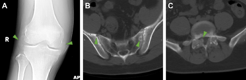
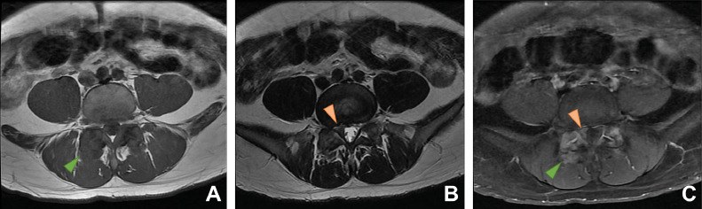
Gout is a common cause of inflammatory arthritis, typically affecting the joints of the appendicular skeleton. In this report, we present the relatively less common scenario of chronic tophaceous gout affecting the lumbar spine and pelvis, complicated by compressive neuropathy, and notable for its advanced initial presentation in a young patient. We review the pathophysiology underlying gout and discuss its clinical and laboratory presentation. We also use our case as an example to present the radiographic, CT, and MR imaging features of gout affecting the lumbar spine, which can often present a diagnostic dilemma. Finally, we discuss therapeutic options for gout resulting in spinal canal compromise, which include interventions not commonly performed for gout elsewhere in the body.

Keywords: Arthritis; Gout; Radiculopathy; Spine.


Figures

Fig 1
Fig. 1
Radiographic and CT findings of gout. (A) AP view of the knees demonstrates increased soft tissue density (arrowheads) along the lateral and medial aspects of the right knee joint corresponding to a clinically-evident 4 cm tophus. (B) Axial CT image at the level of the sacroiliac joints presented on bone window reveals subarticular erosions (arrowheads) along the sacroiliac joints. (C) Axial CT images at the level of the L5-S1 facet joints presented on bone window illustrates ground glass density along the superior and inferior articular processes and spinal laminae. On the right, there is notable extension into the right lateral epidural space (arrowhead).
Fig 2
Fig. 2
MR imaging features of spinal gout. Axial T1-weighted (A), T2-weighted (B), and post-contrast T1-weighted fat-suppressed (C) images of the spine at the L5-S1 level depict the gouty tophi as T1 isointense, T2 hypointense material with heterogeneous patchy enhancement (green arrowheads). On the right, the medial extension of the tophus associated with the facet joint combines with a disc bulge to produce impingement of the S1 nerve root in the lateral recess (orange arrowheads).

Similar articles

Cited by

References

    1. Golenbiewski J, Keenan RT. Moving the needle: improving the care of the gout patient. Rheumatol Ther. 2019;6(2):179–193. doi: 10.1007/s40744-019-0147-5. - DOI - PMC - PubMed
    1. Ryan MP, Monjazeb S, Goodwin BP, Group AR. Ulcerated tophaceous gout. Dermatol Online J. 2019;25(3) - PubMed
    1. Ragab G, Elshahaly M, Bardin T. Gout: An old disease in new perspective - A review. J Adv Res. 2017;8(5):495–511. doi: 10.1016/j.jare.2017.04.008. - DOI - PMC - PubMed
    1. Yoon J-W, Park K-B, Park H. Tophaceous gout of the spine causing neural compression. Korean J Spine. 2013;10(3):185–188. doi: 10.14245/kjs.2013.10.3.185. - DOI - PMC - PubMed
    1. Konatalapalli RM, Demarco PJ, Jelinek JS. Gout in the axial skeleton. J Rheumatol. 2009;36(3):609–613. doi: 10.3899/jrheum.080374. - DOI - PubMed

Publication types

LinkOut - more resources