

Acquired life-threatening uterine arteriovenous malformation treated by endovascular embolization
- PMID: 33304434
- PMCID: PMC7708760
- DOI: 10.1016/j.radcr.2020.11.013
Acquired life-threatening uterine arteriovenous malformation treated by endovascular embolization
Erratum in
-
Erratum regarding missing declaration of competing interest and patient consent statements in previously published articles.Radiol Case Rep. 2023 Jan 25;18(4):1643-1644. doi: 10.1016/j.radcr.2023.01.017. eCollection 2023 Apr. Radiol Case Rep. 2023. PMID: 36895588 Free PMC article.
Abstract
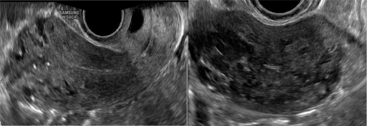
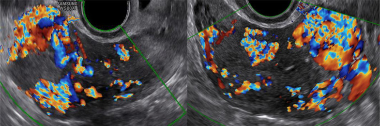
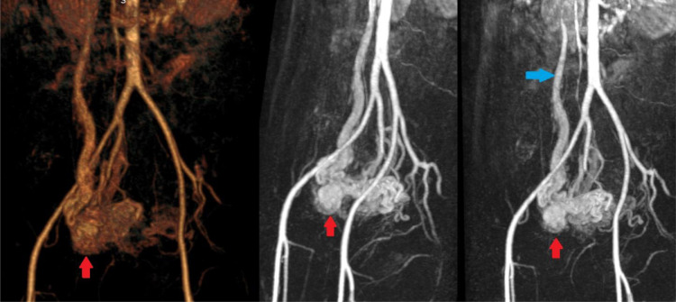
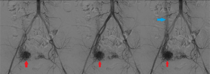
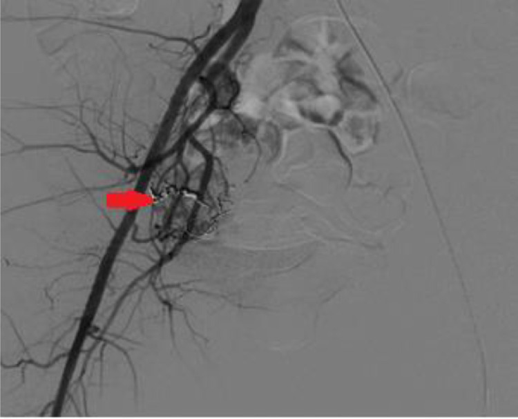
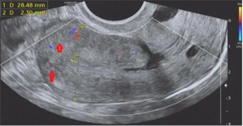
Uterine arteriovenous malformation (AVM) is a rare condition that may lead to a life-threatening state. The urgency of diagnosis and treatment for uterine AVM should be emphasized. This case report describes a 42-year-old woman with a vaginal hemorrhage. In the previous month, the patient also had a hemorrhage after induced abortion that required a bilateral artery suture hemostasis of the uterus. On ultrasound, there was a lesion suspected by acquired AVM. Magnetic Resonance Angiography and Digital Subtraction Angiography was indicated to confirm the diagnosis. The patient was successfully treated by uterine artery embolization. After 6 months, the re-examined result showed no lesion of AVM.
Keywords: Arteriovenous malformation; Ultrasound; Uterine artery; Uterine artery embolization; Uterus; Vaginal bleeding.
© 2020 The Authors. Published by Elsevier Inc. on behalf of University of Washington.
Figures
References
-
- Divya Sridhar, Robert L V. Diagnosis and treatment of uterine and pelvic arteriovenous malformations. 2018
-
- Thangam ma Kati Mada Annaiah SKS. Uterine arteriovenous malformations: clinical implications. 2015.
-
- Fleming H, Ostor A, Pickel H FD. Arteriovenous malformations of the uterus. Obs Gynaecol. 1989;73(2):209–213. - PubMed
-
- Polat P, Suma S, Kantarcy M, Alper F LA. Colour Doppler ultrasound in the evaluation of uterine vascular abnormalities. Radiographics. 2002;22:47–53. - PubMed
-
- Grivell R, Reid K MA. Uterine arteriovenous malformations: a review of the current literature. Obs Gynecol Surv. 2005;60(11):761–767. - PubMed
Publication types
LinkOut - more resources
Full Text Sources
Other Literature Sources
