

Thoracic endometriosis presenting as recurrent pleural effusions
- PMID: 33304436
- PMCID: PMC7708763
- DOI: 10.1016/j.radcr.2020.11.025
Thoracic endometriosis presenting as recurrent pleural effusions
Erratum in
-
Erratum regarding missing declaration of competing interest and patient consent statements in previously published articles.Radiol Case Rep. 2023 Jan 25;18(4):1643-1644. doi: 10.1016/j.radcr.2023.01.017. eCollection 2023 Apr. Radiol Case Rep. 2023. PMID: 36895588 Free PMC article.
Abstract
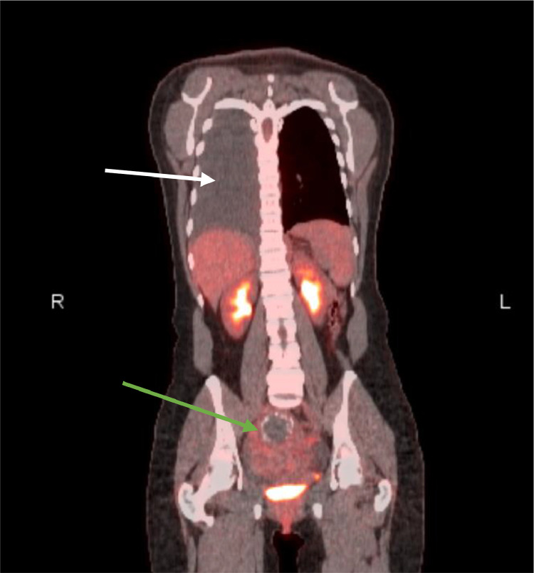
In rare instances, endometrial glandular tissue can implant in the thorax of women suffering from endometriosis. The presentation is variable depending on site of implant and can be a rare cause of hemothorax in women. A 28-year-old woman presented with shortness of breath and was found to have a significant right sided hemothorax. The hemothorax was drained but subsequently recurred, with shortness of breath increasing around the time of her menses. Considerable workup was performed and ultimately surgery was required to diagnose her with thoracic endometriosis. This case describes how thoracic endometriosis is a challenging diagnosis and may be under reported in the literature. However, there are key elements of the disease that can prevent delay in diagnosis to reduce pain and suffering.
Keywords: Catamenial; Fibroid; Hemothorax; Pleural effusion; Pneumothorax; Thoracic endometriosis.
© 2020 Published by Elsevier Inc. on behalf of University of Washington.
Figures


References
Publication types
LinkOut - more resources
Full Text Sources
Other Literature Sources
