Review
. 2020 Nov 21:18:3766-3773.
doi: 10.1016/j.csbj.2020.10.045. eCollection 2020.

Phytophthora zoospores: From perception of environmental signals to inoculum formation on the host-root surface

Affiliations
Review

Phytophthora zoospores: From perception of environmental signals to inoculum formation on the host-root surface

Ilaria Bassani et al. Comput Struct Biotechnol J. .

Abstract

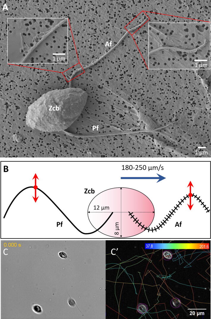
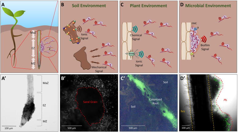
To explore moist soils and to target host plants, phytopathogenic Phytophthora species utilize the sensory and propulsion capabilities of the biflagellate unicellular zoospores they produce. Zoospore motion and interactions with the microenvironment are of primary importance for Phytophthora physiology. These are also of critical significance for plant pathology in early infection sequential events and their regulation: the directed zoospore migration toward the host, the local aggregation and adhesion at the host penetration site. In the soil, these early events preceding the root colonization are orchestrated by guidance factors, released from the soil particles in water films, or emitted within microbiota and by host plants. This signaling network is perceived by zoospores and results in coordinated behavior and preferential localization in the rhizosphere. Recent computational and structural studies suggest that rhizospheric ion and plant metabolite sensing is a key determinant in driving zoospore motion, orientation and aggregation. To reach their target, zoospores respond to various molecular, chemical and electrical stimuli. However, it is not yet clear how these signals are generated in local soil niches and which gene functions govern the sensing and subsequent responses of zoospores. Here we review studies on the soil, microbial and host-plant factors that drive zoospore motion, as well as the adaptations governing zoospore behavior. We propose several research directions that could be explored to characterize the role of zoospore microbial ecology in disease.

Keywords: Host-root; Microbiota; Motion; Perception; Phytophthora zoospore; Soil; Taxis.


Conflict of interest statement

The authors declare that they have no known competing financial interests or personal relationships that could have appeared to influence the work reported in this paper.

Figures

Fig. 1
Fig. 1
Structure and microswimmer traits of Phytophthora zoospores. (A) Micrograph of P. parasitica zoospore obtained using scanning electron microscopy (SEM). This shows the characteristic ellipsoidal zoospore cell body (Zcb) and the anterior and posterior flagella (Af and Pf, respectively). Tubular (left inset) and thinner (right inset) mastigonemes are found along the anterior flagellum, while the posterior flagellum is smooth. (B) Two-dimension schematic representation of the P. palmivora zoospore, including the two flagella beating with periodical waveforms in opposite directions and connected to the ellipsoidal cell body. The red arrows indicate the beating patterns of the flagella, while the blue arrow indicates the swimming direction of the zoospore. Cell body size and zoospore speed are obtained from Appiah et al. 2005 . Panels C and C′ show P. parasitica zoospores swimming in water (C) and the corresponding trajectory patterns delineated using the TrackMate plugin as per the procedure detailed in Galiana et al.. The trajectories indicate the randomness in swimming speed and direction of zoospores under no constraints. (C′) Correspondence between colors and mean speed (µm/s) is indicated in the scale at the top of panel C′. (For interpretation of the references to colour in this figure legend, the reader is referred to the web version of this article.)
Fig. 2
Fig. 2
Zoospore interactions with the surrounding environment. Panel A shows a schematic representation of a plant root being colonized by zoospores (Z). The zoomed longitudinal view highlights the different zones of the root tip (maturation zone (MaZ), elongation zone (EZ) and meristematic zone (MZ)), and illustrates the preferential aggregation of zoospores at the EZ as reported by Attard et al. . Panel A′ shows an EZ colonized by P. parasitica zoospores, 25 min after inoculation. Panels B, C and D give an overview of zoospore interactions with soil, plant and microbial environments, respectively. In Panel B, ionic signals emitted by charged soil particles and zoospore physical interactions with soil grains are represented. Panel B′ shows a fluorescence micrograph of a sand grain surrounded by zoospores (Z) that are exploring its surface. For cytoplasmic staining, zoospores were initially loaded for 10 min with 1 µM BCECF-AM (2′,7′ -bis-carboxyethyl-5(6′)-carboxyfluorescein acetoxymethyl ester). Panel C shows the ionic and chemical signals (e.g. root exudates) that are emitted or released by the plant root and subsequently attract zoospores. The fluorescence micrograph in C′ shows P. parasitica BCECF-stained zoospores having colonized a tomato root in the soil. The profile of fluorescence (green) illustrates the complete coverage of a tomato root by emerging Phytophthora mycelium (the part which can be visualized among soil elements), as the result of an extensive colonization by zoospores a 90 min soil exploration. The vast majority of zoospores had reached the root while very few were dispersed or still exploring the soil microenvironment. Panel D shows mixed biofilm formation on the root surface with incorporation of bacteria (B) and newly attracted zoospores, was well as extracellular matrix (ECM) formation. Panel D′ shows mixed biofilm formation on a tomato root surface. It illustrates the preferential colonization of the Phytophthora biofilm (Pb) rather than healthy root surface (hrs), by Pseudomonas species expressing Green Fluorescent Protein (GFP), 2 h post bacterial inoculation . (For interpretation of the references to colour in this figure legend, the reader is referred to the web version of this article.)

References

    1. Judelson H.S., Blanco F.A. The spores of Phytophthora: weapons of the plant destroyer. Nat Rev Microbiol. 2005;3:47–58. - PubMed
    1. Kamoun S., Furzer O., Jones J.D.G., Judelson H.S., Ali G.S. The top 10 oomycete pathogens in molecular plant pathology. Mol Plant Pathol. 2015;16:413–434. - PMC - PubMed
    1. Martin F.N., Blair J.E., Coffey M.D. A combined mitochondrial and nuclear multilocus phylogeny of the genus Phytophthora. Fungal Genet Biol. 2014;66:19–32. - PubMed
    1. Thompson S.E., Levin S., Rodriguez-Iturbe I. Rainfall and temperatures changes have confounding impacts on Phytophthora cinnamomi occurrence risk in the southwestern USA under climate change scenarios. Glob Chang Biol. 2014;20:1299–1312. - PubMed
    1. Cardillo E., Acedo A., Abad E. Topographic effects on dispersal patterns of Phytophthora cinnamomi at a stand scale in a Spanish heathland. PLoS ONE. 2018;13 - PMC - PubMed

LinkOut - more resources