. 2020 Oct;10(1):e12003.
doi: 10.1002/jev2.12003. Epub 2020 Oct 30.

EGFR-rich extracellular vesicles derived from highly metastatic nasopharyngeal carcinoma cells accelerate tumour metastasis through PI3K/AKT pathway-suppressed ROS

Affiliations

EGFR-rich extracellular vesicles derived from highly metastatic nasopharyngeal carcinoma cells accelerate tumour metastasis through PI3K/AKT pathway-suppressed ROS

Fei Li et al. J Extracell Vesicles. 2020 Oct.

Abstract

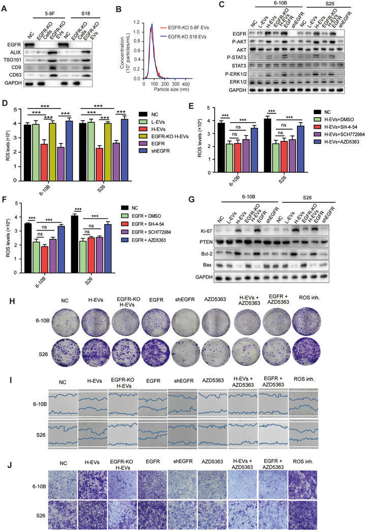
Nasopharyngeal carcinoma (NPC) is the most common cancer with high metastatic potential that occurs in the epithelial cells of the nasopharynx. Distant metastases are the primary cause for treatment failure and mortality of NPC patients. However, the underlying mechanism responsible for the initiation of tumour cell dissemination and tumour metastasis in NPC is not well understood. Here, we demonstrated that epidermal growth factor receptor (EGFR) was highly expressed in tumour tissues of NPC patients with distant metastases and was associated with a decrease in reactive oxygen species (ROS). We also revealed that extracellular vesicles (EVs) transfer occurred from highly to poorly metastatic NPC cells, mediating cell-cell communication and enhancing the metastatic potential of poorly metastatic NPC cells. Further experiments indicated that EVs derived from highly metastatic NPC cells induced the up-regulation of EGFR and down-regulation of ROS in low metastatic NPC cells. Mechanistically, EGFR-rich EVs-mediated EGFR overexpression down-regulated intracellular ROS levels through the PI3K/AKT pathway, thus promoting the metastatic potential of poorly metastatic NPC cells. Strikingly, treatment with EVs secreted from highly metastatic NPC cells was significantly associated with rapid NPC progression and shorter survival in xenografted mice. These findings not only improve our understanding of EVs-mediated NPC metastatic mechanism but also have important implications for the detection and treatment of NPC patients accompanied by aberrant EGFR-rich EVs transmission.

Keywords: epidermal growth factor receptor; extracellular vesicles; nasopharyngeal carcinoma; reactive oxygen species.


Conflict of interest statement

The authors report no conflict of interest.

Figures

FIGURE 1
FIGURE 1
ROS is reduced in NPC cells and patients with high metastatic potential and is correlated with an increase in EGFR. (a, b) Tumour single cell suspensions and tumour tissue sections of NPC patients with effective distant metastases (n = 16) and without distant metastases (n = 24) were incubated with DCFH‐DA probe and in situ intracellular ROS levels were analysed using (a) fluorescence microplate reader or (b) confocal microscope (Magnification × 200; Scale bar, 36.67 μm). (c) Immunohistochemical staining showing EGFR expression in tumour tissue of NPC patients with effective distant metastases (n = 16) and without distant metastases (n = 24) (Magnification × 400; Scale bar, 50 μm). (d) Intracellular ROS production of NPC cells 6–10B, 5–8F, S26 and S18, assessed using DCFH‐DA probe and recorded using confocal laser scanning microscope. (e) Levels of intracellular ROS production in NPC cells, quantified by DCFH‐DA probe and analysed using fluorescence microplate reader. (f) Western blot analysis of EGFR expression in NPC cells. GAPDH was used a loading control. (g) Quantitative PCR analysis of EGFR expression in NPC cells. Values were normalized to GAPDH. (h) A nonlinear correlation between intratumoral ROS levels and EGFR expression. The EGFR expression in tumour tissues of NPC patients (n = 40) was normalized to GAPDH. Experiments were performed in triplicate. Data are presented as mean ± SD (:P < 0.05; ::P < 0.01; :::P < 0.001)
FIGURE 2
FIGURE 2
EVs transfer from highly metastatic NPC cells to poorly metastatic NPC cells mediates cell‐cell communication. (a) Schematic representation of highly and poorly metastatic NPC cells co‐culture system. pmCherry‐CD63‐transfected highly metastatic NPC cells were seeded into the upper chamber and poorly metastatic NPC cells were seeded into the lower chamber. (b) Confocal microscopy observation of the internalization of EVs secreted from mCherry‐CD63‐labeled 5–8F or S18 (upper chamber) in 6–10B or S26 cells (lower chamber). Hoechst 33342 was used to stain cellular nuclei (blue fluorescence). Stereoscopic structure of cells was observed by differential interference contrast (DIC) (Magnification × 630; Scale bar, 10 μm). (c) Western blot analysis showing the presence of CD9, CD63, ALIX, TSG101, and EGFR and absence of GAPDH in EVs derived from the conditioned medium of NPC cells. (d) Representative nanoparticle tracking analysis plots showing the size distribution and total number of EVs isolated from the same volume of conditioned medium of 5–8F and S18 cells. (e) Confocal microscopy image showing the internalization of PKH26‐labeled EVs (red) isolated from the conditioned medium of highly metastatic NPC cells by 6–10B and S26 cells. Hoechst 33342 was used to stain the nuclei of cells (blue). Cell membrane was stained by Dio (green fluorescence). (Magnification × 630; Scale bar, 10 μm). Experiments were performed in triplicate
FIGURE 3
FIGURE 3
Uptake of EVs derived from highly metastatic NPC cells enhances NPC cells metastasis. (a) The effect of L‐EVs and H‐EVs on NPC cells clone formation. (b) Wound healing assay showing effects of L‐EVs and H‐EVs internalization on NPC cells migration. (c) Effect of L‐EVs and H‐EVs internalization on NPC cells invasion by Matrigel invasion assay. (d) Western blot for EMT markers E‐cadherin and Vimentin after treatment with L‐EVs and H‐EVs for 48 h. (e) Expression of proteins involved in EMT was confirmed by immunofluorescence after treatment with H‐EVs for 48 h. Stained cells were examined with E‐cadherin‐ and Vimentin‐specific antibodies and protein localization was recognized by FITC (green) and Alexa Fluor 555 (red) secondary antibodies. Hoechst 33342 was used to stain the nuclei of cells (blue) (Magnification × 630; Scale bar, 10 μm). Experiments were performed in triplicate
FIGURE 4
FIGURE 4
H‐EVs induces EGFR up‐regulation and ROS down‐regulation in low metastatic NPC cells. (a) Western blot analysis of EGFR expression in NPC cells following treatment with L‐EVs and H‐EVs. (b) EGFR expression by confocal microscope in 6–10B and S26 cells pretreated with H‐EVs. Stained cells were examined with EGFR‐specific antibodies, and protein expression was recognized by Alexa Fluor 555 (red) secondary antibodies. Hoechst 33342 was used to stain the nuclei of cells (blue). (Magnification × 630, Scale bar, 10 μm). (c) Detection of intracellular ROS in 6–10B and S26 cells (lower chamber) in co‐culture system following treatment with or without GW4869 in highly metastatic NPC cells (upper chamber). (d) Detection of intracellular ROS in NPC cells following treatment with L‐EVs or H‐EVs. (e) Expression of potential genes altered by H‐EVs treatment; numbers indicate quantity of genes in each DEG subset. (f) Heatmap displaying hierarchical clustering of genes in 6–10B and S26 cells in response to H‐EVs treatment. Gene expression values are displayed by applying progressively brighter shades of red (up‐regulated) or green (down‐regulated). (g) Column diagram represents the expression of genes correlated with tumor metastasis with H‐EVs treatment in 6–10B and S26 cells. (h) Western blot analysis for validation of the identified genes correlated with tumor metastasis after H‐EVs treatment in 6–10B and S26 cells. Experiments were performed in triplicate. Data represent the mean ± SD (:P < 0.05; ::P < 0.01; :::P < 0.001)
FIGURE 5
FIGURE 5
H‐EVs‐induced EGFR expression down‐regulates ROS via the PI3K/AKT pathway to promote metastasis of NPC cells. (a) Western blot analysis showing the presence of CD9, CD63, ALIX, and TSG101 and absence of GAPDH in EGFR‐KO EVs derived from the conditioned medium of EGFR‐KO 5–8F and S18 cells. (b) Representative NTA plots showing the size distribution and total number of EVs isolated from the same volume of conditioned medium of EGFR‐KO 5–8F and S18 cells. (c) Western blot analysis of phospho‐STAT3, STAT3, phospho‐AKT, AKT, phospho‐ERK1/2, and ERK1/2 in EVs‐treated, EGFR‐KO H‐EVs‐treated, EGFR overexpressed, and shEGFR transfected 6–10B and S26 cells. (d) Intracellular ROS levels in L‐EVs‐, H‐EVs‐ or EGFR‐KO H‐EVs‐treated, EGFR overexpressed, and shEGFR transfected 6–10B and S26 cells by fluorescence microplate reader. (e) Intracellular ROS levels in H‐EVs‐treated 6–10B and S26 cells pretreated with DMSO control, SH‐4‐54, SCH772984, or AZD5363. (f) Intracellular ROS levels in EGFR overexpressed 6–10B and S26 cells pretreated with DMSO control, SH‐4‐54, SCH772984, or AZD5363. (g) Western blot analysis of Ki‐67, PTEN, Bcl‐2, and Bax in L‐EVs‐, H‐EVs‐, or EGFR‐KO H‐EVs‐treated, EGFR overexpressed, and shEGFR 6–10B and S26 cells. (h) Clone formation capacity of H‐EVs‐treated, EGFR‐KO H‐EVs‐treated, EGFR overexpressed, ROS inhibited, shEGFR, AZD5363 treated, H‐EVs and AZD5363 co‐treated, and EGFR overexpression combined with AZD5363 treated 6–10B and S26 cells, respectively. (i) Wound healing assay showing cell migration of H‐EVs‐treated, EGFR‐KO H‐EVs‐treated, EGFR overexpressed, ROS inhibited, shEGFR,AZD5363 treated, H‐EVs and AZD5363 co‐treated, and EGFR overexpression combined with AZD5363 treated 6–10B and S26 cells. (j) Cell invasion of H‐EVs‐treated, EGFR‐KO H‐EVs‐treated, EGFR overexpressed, ROS inhibited, shEGFR, AZD5363 treated, H‐EVs and AZD5363 co‐treated, and EGFR overexpression combined with AZD5363 treated 6–10B and S26 cells using a Matrigel invasion assay. Experiments were performed in triplicate. Data are presented as mean ± SD (:P < 0.05; ::P < 0.01; :::P < 0.001)
FIGURE 6
FIGURE 6
H‐EVs treatment promotes the metastasis of NPC cells and causes shortened survival of NPC‐bearing mice. (a) Experimental design of cell transplantation with 6–10B and S26 cells by intraperitoneal injection (IP) and subcutaneous (SC) injection, respectively, and subsequent in vivo studies and NPC‐ascites sorting and monitoring. (b) Western blot analysis of EGFR, E‐cadherin, Vimentin, PTEN, Bcl‐2, and Bax in 6–10B and S26 ascitic cells developed from intraperitoneally injected cells following treatment with or without H‐EVs. (c) Intracellular ROS detection of 6–10B and S26 ascitic cells following intraperitoneally co‐injected H‐EVs. (d) Histological analysis of the lung and liver tissues obtained from 6–10B‐ and S26‐transplanted mice following treatment with or without H‐EVs (Magnification × 200; Scale bar, 10 μm). (e) Number of tumor nodules in the lung and liver metastases of 6–10B and S26 following treatment with or without H‐EVs. (f) Wet lung weight of 6–10B‐ and S26‐transplanted mice following treatment with or without H‐EVs. (g) Kaplan‐Meier survival curves for mice subcutaneously transplanted with 6–10B and S26 cells following treatment with or without H‐EVs. P values were determined by the log‐rank (Mantel‐Cox) test. (h) Schematic illustrating the transit of highly metastatic NPC cell‐derived EVs to poorly metastatic NPC cells to drive low metastatic potential cells toward a highly metastatic phenotype. All experiments were performed in triplicate. Data are presented as mean ± SD (:P < 0.05; ::P < 0.01; :::P < 0.001)

Similar articles

Cited by

References

    1. Barhoumi, T. , Fraulob‐Aquino, J. C. , Mian, M. O. R. , Ouerdm, S. , Idris‐Khodja, N. , Huo, K.‐G. , … Schiffrin, E. L. (2017). Matrix metalloproteinase‐2 knockout prevents angiotensin II‐induced vascular injury. Cardiovascular Research, 113(14), 1753–1762. - PMC - PubMed
    1. Bodega, G. , Alique, M. , Puebla, L. , Carracedo, J. , & Ramirez, R. M. (2019). Microvesicles: ROS scavengers and ROS producers. Journal of Extracellular Vesicles, 8(1), 1626654. - PMC - PubMed
    1. Cairns, R. A. , Harris, I. S , & Mak, T. W. (2011). Regulation of cancer cell metabolism. Nature Reviews Cancer, 11(2), 85–95. - PubMed
    1. Chan, K. C. , Hung, E. C. , Woo, J. K. , Chan, P. K. S. , Leung, S.‐F. , Lai, F. P. T. , … Lo, Y. M. D. (2013). Early detection of nasopharyngeal carcinoma by plasma Epstein‐Barr virus DNA analysis in a surveillance program. Cancer, 119(10), 1838–1844. - PubMed
    1. Chan, Y. K. , Zhang, H. , Liu, P. , Tsao, S.‐W. , Lung, M. L. , Mak, N.‐K. , … Yue, P. Y.‐K. (2015). Proteomic analysis of exosomes from nasopharyngeal carcinoma cell identifies intercellular transfer of angiogenic proteins. International Journal Of Cancer. Journal International du Cancer, 137(8), 1830–1841. - PubMed

Publication types