

Mitochondrial RNA in Alzheimer's Disease Circulating Extracellular Vesicles
- PMID: 33304899
- PMCID: PMC7701247
- DOI: 10.3389/fcell.2020.581882
Mitochondrial RNA in Alzheimer's Disease Circulating Extracellular Vesicles
Abstract
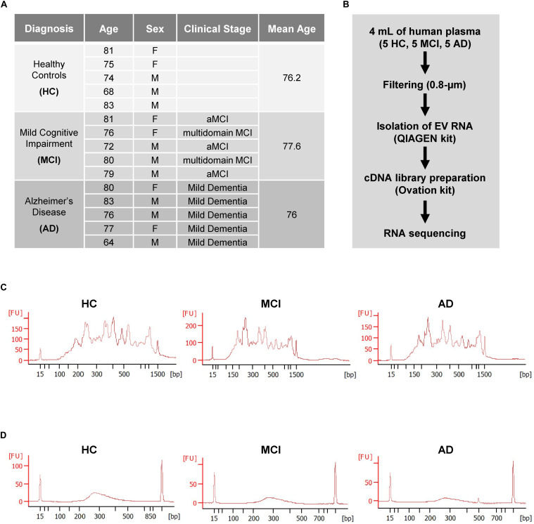
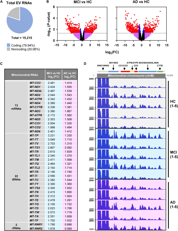
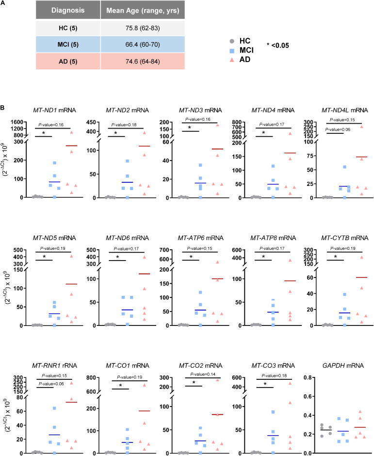
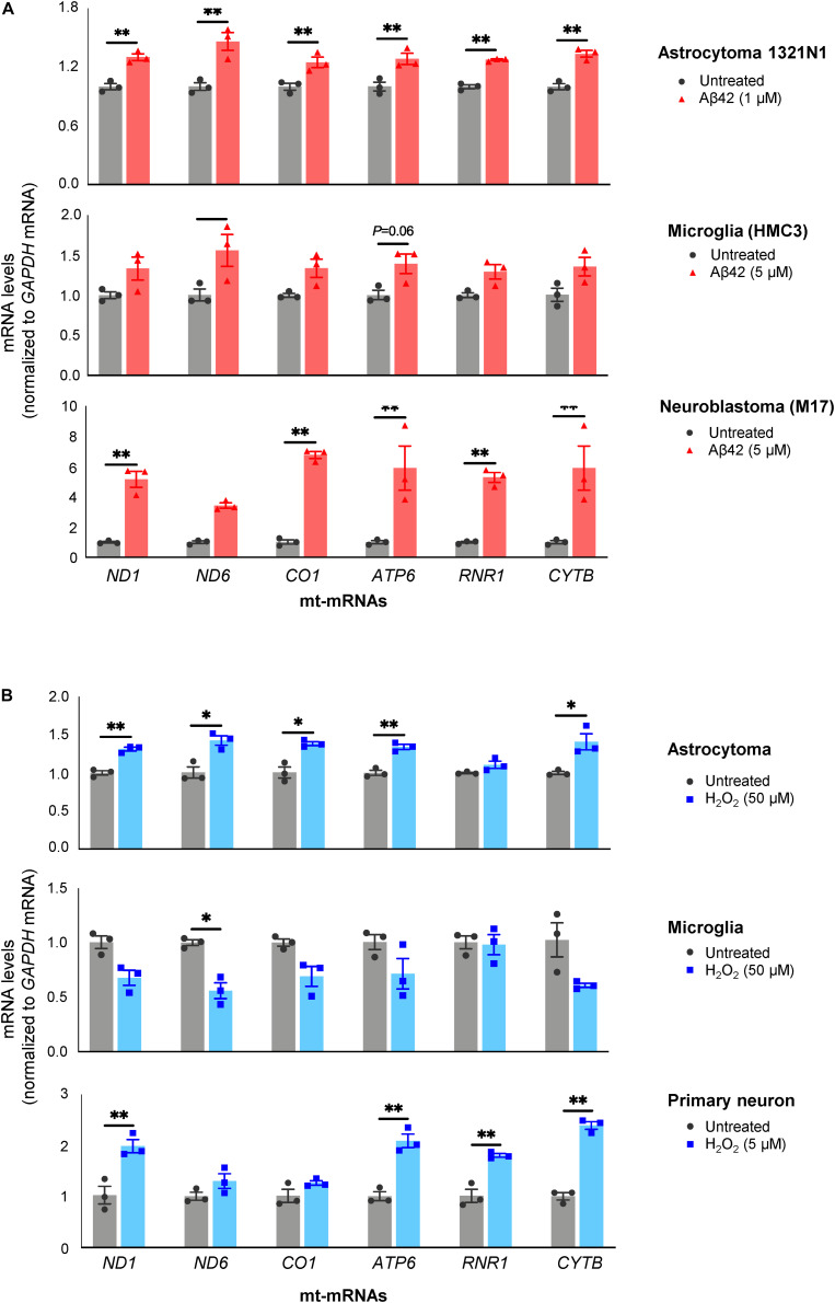
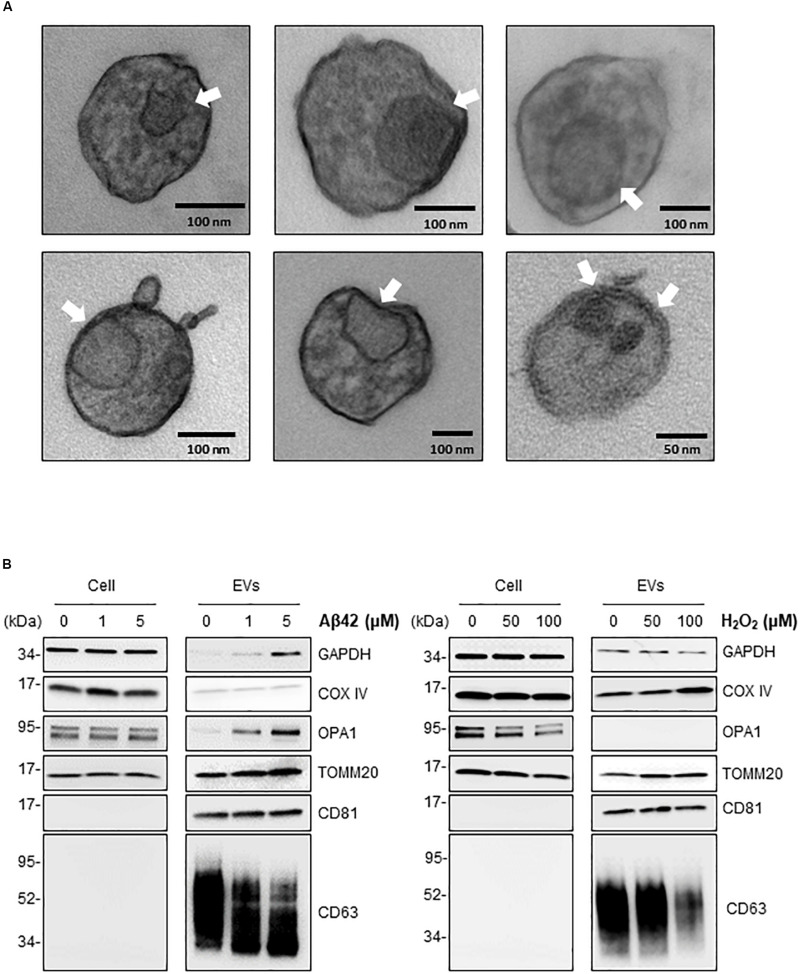
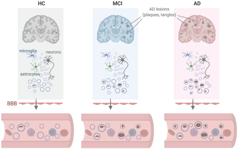
Alzheimer's disease (AD) is the most common type of dementia. Amyloid β (Aβ) plaques, tau-containing neurofibrillary tangles, and neuronal loss leading to brain atrophy are pathologic hallmarks of AD. Given the importance of early diagnosis, extensive efforts have been undertaken to identify diagnostic and prognostic biomarkers for AD. Circulating extracellular vesicles (EVs) provide a platform for "liquid biopsy" biomarkers for AD. Here, we characterized the RNA contents of plasma EVs of age-matched individuals who were cognitively normal (healthy controls (HC)) or had mild cognitive impairment (MCI) due to AD or had mild AD dementia (AD). Using RNA sequencing analysis, we found that mitochondrial (mt)-RNAs, including MT-ND1-6 mRNAs and other protein-coding and non-coding mt-RNAs, were strikingly elevated in plasma EVs of MCI and AD individuals compared with HC. EVs secreted from cultured astrocytes, microglia, and neurons after exposure to toxic conditions relevant to AD pathogenesis (Aβ aggregates and H2O2), contained mitochondrial structures (detected by electron microscopy) and mitochondrial RNA and protein. We propose that in the AD brain, toxicity-causing mitochondrial damage results in the packaging of mitochondrial components for export in EVs and further propose that mt-RNAs in plasma EVs can be diagnostic and prognostic biomarkers for MCI and AD.
Keywords: Alzheimer’s disease; biomarker; extracellular vesicles; mitochondrial RNA; mitochondrial protein.
Copyright © 2020 Kim, Meng, Perez de Acha, Mustapic, Cheng, Eren, Kundu, Piao, Munk, Wood, De, Noh, Delannoy, Cheng, Abdelmohsen, Kapogiannis and Gorospe.
Figures
Similar articles
-
Plasma neuronal exosomes serve as biomarkers of cognitive impairment in HIV infection and Alzheimer's disease.J Neurovirol. 2019 Oct;25(5):702-709. doi: 10.1007/s13365-018-0695-4. Epub 2019 Jan 4. J Neurovirol. 2019. PMID: 30610738 Free PMC article. Review.
-
Alzheimer's disease.Subcell Biochem. 2012;65:329-52. doi: 10.1007/978-94-007-5416-4_14. Subcell Biochem. 2012. PMID: 23225010 Review.
-
Multi-Omics Analysis of Microglial Extracellular Vesicles From Human Alzheimer's Disease Brain Tissue Reveals Disease-Associated Signatures.Front Pharmacol. 2021 Dec 2;12:766082. doi: 10.3389/fphar.2021.766082. eCollection 2021. Front Pharmacol. 2021. PMID: 34925024 Free PMC article.
-
Levels of Receptor for Advanced Glycation End Products and Glyoxalase-1 in the Total Circulating Extracellular Vesicles from Mild Cognitive Impairment and Different Stages of Alzheimer's Disease Patients.J Alzheimers Dis. 2021;84(1):227-237. doi: 10.3233/JAD-210441. J Alzheimers Dis. 2021. PMID: 34487040
-
miR-212 and miR-132 Are Downregulated in Neurally Derived Plasma Exosomes of Alzheimer's Patients.Front Neurosci. 2019 Nov 26;13:1208. doi: 10.3389/fnins.2019.01208. eCollection 2019. Front Neurosci. 2019. PMID: 31849573 Free PMC article.
Cited by
-
Extracellular vesicles: opening up a new perspective for the diagnosis and treatment of mitochondrial dysfunction.J Nanobiotechnology. 2024 Aug 14;22(1):487. doi: 10.1186/s12951-024-02750-8. J Nanobiotechnology. 2024. PMID: 39143493 Free PMC article. Review.
-
Plasma cell-free RNA profiling of Vietnamese Alzheimer's patients reveals a linkage with chronic inflammation and apoptosis: a pilot study.Front Mol Neurosci. 2023 Dec 21;16:1308610. doi: 10.3389/fnmol.2023.1308610. eCollection 2023. Front Mol Neurosci. 2023. PMID: 38178908 Free PMC article.
-
Role of Mitochondrial Nucleic Acid Sensing Pathways in Health and Patho-Physiology.Front Cell Dev Biol. 2022 Feb 11;10:796066. doi: 10.3389/fcell.2022.796066. eCollection 2022. Front Cell Dev Biol. 2022. PMID: 35223833 Free PMC article. Review.
-
Biomarker and therapeutic potential of peripheral extracellular vesicles in Alzheimer's disease.Adv Drug Deliv Rev. 2022 Nov;190:114486. doi: 10.1016/j.addr.2022.114486. Epub 2022 Aug 8. Adv Drug Deliv Rev. 2022. PMID: 35952829 Free PMC article. Review.
-
Extracellular vesicles in Alzheimer's disease.Arq Neuropsiquiatr. 2024 Mar;82(3):1-8. doi: 10.1055/s-0044-1779296. Epub 2024 Mar 11. Arq Neuropsiquiatr. 2024. PMID: 38467392 Free PMC article.
References
-
- Albert M. S., DeKosky S. T., Dickson D., Dubois B., Feldman H. H., Fox N. C., et al. (2011). The diagnosis of mild cognitive impairment due to Alzheimer’s disease: recommendations from the National Institute on Aging-Alzheimer’s Association workgroups on diagnostic guidelines for Alzheimer’s disease. Alzheimers Dement. 7 270–279. - PMC - PubMed
-
- Andaloussi S. E. L., Mäger I., Breakefield X. O., Wood M. J. (2013). Extracellular vesicles: biology and emerging therapeutic opportunities. Nat. Rev. Drug Discov. 12 347–357. - PubMed
-
- Athauda D., Gulyani S., Karnati H. K., Li Y., Tweedie D., Mustapic M., et al. (2019). Utility of neuronal-derived exosomes to examine molecular mechanisms that affect motor function in patients with parkinson disease: a secondary analysis of the Exenatide-PD Trial. JAMA Neurol. 76 420–429. - PMC - PubMed
-
- Beal M. F. (1998). Mitochondrial dysfunction in neurodegenerative diseases. Biochim. Biophys. Acta 1366 211–223. - PubMed
LinkOut - more resources
Full Text Sources
Molecular Biology Databases