Case Reports
. 2020 Nov 16:7:570572.
doi: 10.3389/fmed.2020.570572. eCollection 2020.

Phage Therapy as Adjuvant to Conservative Surgery and Antibiotics to Salvage Patients With Relapsing S. aureus Prosthetic Knee Infection

Affiliations
Case Reports

Phage Therapy as Adjuvant to Conservative Surgery and Antibiotics to Salvage Patients With Relapsing S. aureus Prosthetic Knee Infection

Tristan Ferry et al. Front Med (Lausanne). .

Abstract

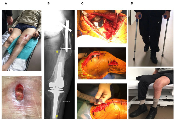
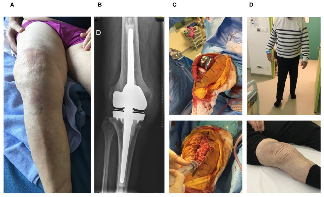
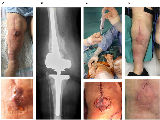
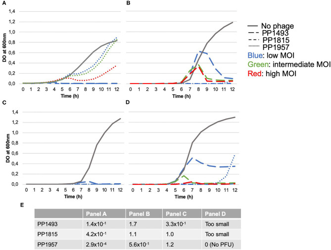
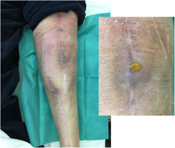
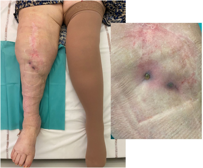
Objectives: To report the management of three consecutive patients with relapsing Staphylococcus aureus prosthetic knee infection (PKI) for whom explantation was not feasible who received a phage therapy during a "Debridement Antibiotics and Implant Retention" (DAIR) procedure followed by suppressive antimicrobial therapy. Methods: Each case was discussed individually in our reference center and with the French National Agency (ANSM). The lytic activity of three phages targeting S. aureus, which was produced with a controlled and reproducible process, was assessed before surgery (phagogram). A hospital pharmacist extemporaneously assembled the phage cocktail (1 ml of 1 × 10¹⁰ PFU/ml for each phage) as "magistral" preparation (final dilution 1 × 10⁹ PFU/ml), which was administered by the surgeon directly into the joint, after the DAIR procedure and joint closure (PhagoDAIR procedure). Results: Three elderly patients were treated with the PhagoDAIR procedure. Phagograms revealed a high susceptibility to at least two of the three phages. During surgery, all patients had poor local conditions including pus in contact to the implant. After a prolonged follow-up, mild discharge of synovial fluid persisted in two patients, for whom a subsequent DAIR was performed showing only mild synovial inflammation without bacterial persistence or super-infection. The outcome was finally favorable with a significant and impressive clinical improvement of the function. Conclusions: The PhagoDAIR procedure has the potential to be used as salvage for patients with relapsing S. aureus PKI, in combination with suppressive antibiotics to avoid considerable loss of function. This report provides preliminary data supporting the setup of a prospective multicentric clinical trial.

Keywords: S. aureus; bacteriophages; phage therapy; phagotherapy; prosthetic-joint infection.


Figures

Figure 1
Figure 1
The first patient is an 80-year-old man with past history of Parkinson disease, cardiac arrhythmia, hypertension, right hip prosthetic joint, and left hip fracture treated with osteosynthesis. A left knee prosthetic joint arthroplasty was performed in 2004. In 2014, the patient developed signs of septic arthritis of the left knee due to a methicillin-susceptible S. aureus. A one-stage exchange was performed with reimplantation of a hinged knee prosthesis with long cemented stem. The patient received intravenous daptomycin and rifampin orally, followed by cotrimoxazole and clindamycin for a total duration of 3 months. Unfortunately, a relapse occurred in April 2015 with persistence of the S. aureus, and pristinamycin (a streptogramin A+B antibiotic available in France) was prescribed as suppressive antimicrobial therapy. Despite pristinamycin treatment, the patient developed signs of left septic arthritis with severe pain, bedridden and large anterior fistula with purulent discharge (A). X-ray revealed a complex orthopedic situation with radiological signs of prosthesis loosening of the femoral stem (B). Open DAIR was performed showing poor soft tissue condition with pus in contact to the implant and the personalized cocktail of phages was injected in joint just after closure (C). The patient improved quickly, but at 3 months, a mild discharge of synoviual fluid persisted (Figure 5). A new DAIR was performed, revealing a drastic improvement of the local conditions, with only mild signs of non-specific synovitis. Multiple samples were performed for bacterial culture, but no recurrency/superinfection was diagnosed (cultures remained sterile, specific S. aureus PCR was negative). Cefalexin was then prescribed as suppressive therapy, and the outcome was favorable after 2 years and half of follow-up with no sign of infection, a negative C-reactive protein, and a pain-free walking [(D), Supplementary Video 1].
Figure 2
Figure 2
The second patient is an 84-year-old man with past history of dyslipidemia and right prosthetic-knee arthroplasty in 2006. A two-stage exchange was performed in 2007 for a S. epidermidis PKI. As the patient kept a painful knee, a one-stage exchange was performed in 2016, with implantation of a hinged cemented knee prosthesis with long stem. Cultures remained sterile. In 2019, the patient developed clinical signs of acute septic arthritis with fever. A DAIR without polyethylene exchange was performed and methicillin-susceptible S. aureus grew in perioperative samples and blood cultures. Endocarditis was excluded. The patient was treated using intravenous cefazolin and rifampin orally. Cefazolin was switched to ofloxacin 3 weeks after the DAIR and was combined with rifampin. Under this treatment, new signs of septic arthritis occurred 6 weeks after the DAIR, with local erythema, pain, and large joint effusion (A). A joint puncture was performed, but no pathogen was isolated in cultures, and the failure was attributed to S. aureus. X-ray showed no loosening of the prosthesis (B). Open DAIR was performed showing significant local inflammation and pus into the joint. The personalized cocktail of phages was administered after joint closure (C). The patient improved quickly, doxycycline was then prescribed as suppressive therapy, and the outcome was favorable at 7 months with no signs of infection, a negative C-reactive protein and pain-free walking [(D), Supplementary Video 2].
Figure 3
Figure 3
The third patient is an 83-year-old woman with past history of hypertension and lymphoedema. Right knee arthroplasty was performed in 2000. As prosthesis loosening occurred, a one-stage exchange was performed in 2018 with implantation of a hinged cemented knee prosthesis with long stem. In April 2019, a fistula occurred close to the tibial tuberosity, and in May 2019, the patient developed signs of PKI with fever. Open DAIR without polyethylene exchange was performed and methicillin-susceptible S. aureus grew in blood cultures, and in perioperative samples (with different phenotypes on agar for these latter, but with the same antibiogram. Determination of agr type by PCR showed that one strain belong to agr type I, and the other one to agr type II). Endocarditis was excluded. The patient received intravenous cloxacillin and rifampin orally. Unfortunately, the outcome was not favorable with occurrence of a large fistula with Bourgeon charnu (A). A joint puncture was performed, but no pathogen was isolated in cultures, and the failure was attributed to S. aureus. X-ray did not reveal prosthesis loosening (B). Open DAIR was performed showing catastrophic local condition with pus into the joint, and soft-tissue coverage with local flap was required. The personalized cocktail of phages was administered after joint closure, using a tube placed directly into the joint to preserve the flap (C). The patient improved quickly, but a mild discharge of synovial fluid persisted after 4 months (D). A new DAIR was performed, but no superinfection was diagnosed (cultures remained sterile, specific S. aureus PCR was negative). Doxycycline was then prescribed as suppressive therapy. At 11 months, a pain-free walking was observed, but the patient had persisting mild intermittent discharge of synovial fluid associated with a fistula and a C-reactive protein ≈20 mg/L (Figure 6, Supplementary Video 3), without any superinfection at the joint puncture performed at the end of the follow-up (cultures still sterile, and PCR still negative).
Figure 4
Figure 4
Phagograms performed using the kinetic assay (A–D) and the spot plaque assay (E). For the kinetic assay, the bacterium was incubated with or without bacteriophages, tested individually at three different initial concentrations to obtain low, intermediate, and high multiplicity of infection (MOI, ratio of phages/bacteria). (A) Corresponds to bacterial growth kinetic (Optical Density at 600 nm) obtained for the strain isolated on patient 1; (B) from patient 2; and (C,D) from the two different strains isolated from patient 3. Except for PP1957 on strain A (that only delayed bacterial growth whatever the MOI), all phages were able to inhibit bacterial growth at high MOI (A–D). Of note: (i) a slight delayed inhibition of the bacterial growth of strain B was observed with phage PP1493; (ii) only low MOI are represented concerning the phage activity on the strain C (blue lines), as intermediate and high MOI totally inhibited the bacterial growth; (iii) a partial growth inhibition of strain D (one of the two strains infecting patient 3) was observed with the phage PP1493 at low MOI, and a late growth of strain D appeared in presence of PP1957 at low MOI. The plaque test assay relies on the determination of the efficiency of plating score (EOP), calculated dividing the phage titer on the patient's strain by the phage titer on the reference strain (highly susceptible strain, used for phage amplification). The closer to 1 is the score is, the more efficient the phage is and likely active at low dose. Panel E showed the EOP scores of each strain and revealed that PP1493, PP1815, and PP1957 were active and very efficient on strains A, B, C from patients 1, 2, and 3 since EOP scores ranged from 1.4 × 10⁻¹ to 1.7 with the exception of PP1957 strain A, which showed a low efficiency, consistently with kinetic assay results. However, for strain D (second strain from patient 3), PFU formed with PP1493 and PP1815 could be observed but they were too small to be enumerated confidently. The minimum concentration of spotted phages leading to PFU was 5.5 × 10⁵ and 3.9 × 10⁴ PFU/ml, respectively. Partial lysis without PFU was observed for PP1957 (result concordant with the kinetic assay as only a late growth at the lowest dose was observed).
Figure 5
Figure 5
Local status of patient 1 showing, 3 months after the phagoDAIR procedure, the significant improvement of inflammatory signs of infection, with persistence of mild discharge of synovial fluid through the scar, for which a new DAIR was performed to exclude a superinfection. After the new DAIR, the outcome was favorable.
Figure 6
Figure 6
Local status of patient 3 (infected with two different S. aureus strains and for whom a soft-tissue coverage was required) showing 8 months after the phagoDAIR procedure a significant improvement of inflammatory signs of infection, but with fistula and discharge that persisted despite the phagoDAIR procedure.

Similar articles

Cited by

References

    1. Zimmerli W, Trampuz A, Ochsner PE. Prosthetic-joint infections. N Engl J Med. (2004) 351:1645–54. 10.1056/NEJMra040181 - DOI - PubMed
    1. Osmon DR, Berbari EF, Berendt AR, Lew D, Zimmerli W, Steckelberg JM, et al. Diagnosis and management of prosthetic joint infection: clinical practice guidelines by the infectious diseases society of America. Clin Infect Dis. (2013) 56:e1–25. 10.1093/cid/cis803 - DOI - PubMed
    1. Société de Pathologie Infectieuse de Langue Française (SPILF) Collège des Universitaires de Maladies Infectieuses et Tropicales (CMIT) Groupe de Pathologie Infectieuse Pédiatrique (GPIP) Société Française d'Anesthésie et de Réanimation (SFAR) Société Française de Chirurgie Orthopédique et Traumatologique (SOFCOT) Société Française d'Hygiène Hospitalière (SFHH) Société Française de Médecine Nucléaire (SFMN) et al. Recommendations for bone and joint prosthetic device infections in clinical practice (prosthesis, implants, osteosynthesis). société de pathologie infectieuse de langue française. Med Mal Infect. (2010) 40:185–211. 10.1016/j.medmal.2009.12.009 - DOI - PubMed
    1. Ariza J, Cobo J, Baraia-Etxaburu J, Benito N, Bori G, Cabo J, et al. . Executive summary of management of prosthetic joint infections. clinical practice guidelines by the Spanish society of infectious diseases and clinical microbiology (SEIMC). Enferm Infect Microbiol Clin. (2017) 35:189–95. 10.1016/j.eimce.2017.02.013 - DOI - PubMed
    1. Ferry T, Boucher F, Fevre C, Perpoint T, Chateau J, Petitjean C, et al. . Innovations for the treatment of a complex bone and joint infection due to XDR Pseudomonas aeruginosa including local application of a selected cocktail of bacteriophages. J Antimicrob Chemother. (2018) 73:2901–3. 10.1093/jac/dky263 - DOI - PubMed

Publication types

LinkOut - more resources