. 2020 Nov 16:7:547644.
doi: 10.3389/fvets.2020.547644. eCollection 2020.

Fibronectin Fragments and Inflammation During Canine Intervertebral Disc Disease

Affiliations

Fibronectin Fragments and Inflammation During Canine Intervertebral Disc Disease

Manuel Roland Schmidli et al. Front Vet Sci. .

Abstract

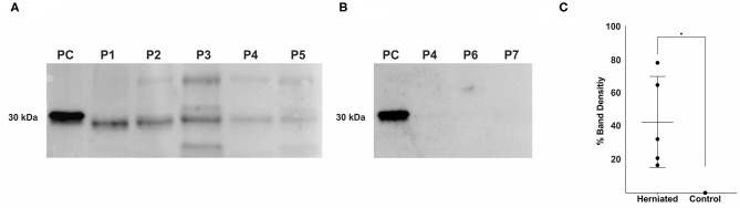
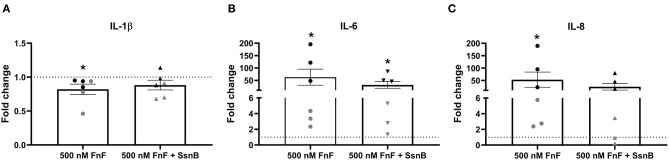
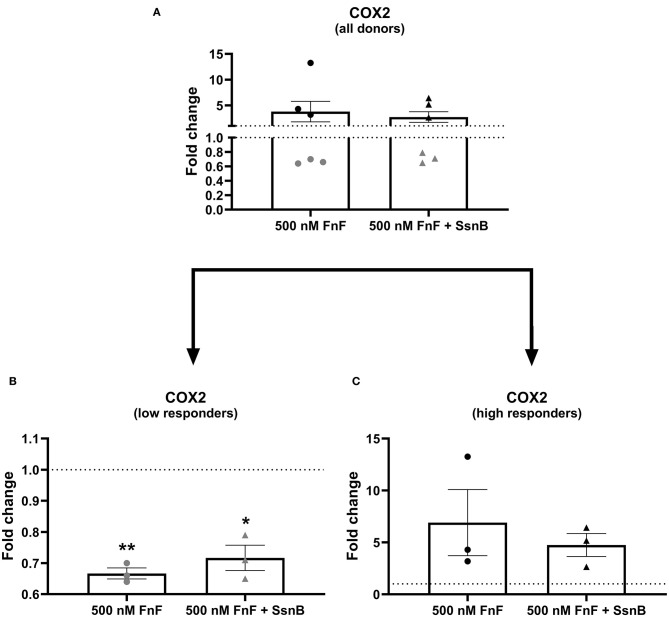
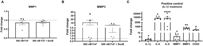
Background: Canine intervertebral disc disease (IVDD) represents a significant clinical problem in veterinary medicine, with similarities to the human pathology. Host-derived damage-associated molecular patterns like fibronectin fragments (FnF) that develop during tissue dysfunction may be of specific relevance to IVD pathologies by inducing an inflammatory response in resident cells. Aim: This project aimed to determine the presence and pathobiological role of FnF during IVD herniation in dogs, with a focus on inflammation. Methods: Herniated nucleus pulposus (NP) material from five dogs as well as non-herniated adjacent NP material from three dogs was collected during spinal surgery required due to acute IVD herniation. The presence of different types of FnF were determined by Western blot analysis. NP cells isolated from six herniated canine IVDs were then exposed to 30 kDa FnF. NP cell inflammation and catabolism was examined by investigating the expression of IL-1β, IL-6, IL-8, and COX-2, as well as MMP-1 and MMP-3 by qPCR (all targets) and ELISA (IL-6, PGE2). Results: Amongst multiple sized FnF (30, 35, 45, and >170kDa), N-terminal fragments at a size of ~30 kDa were most consistently expressed in all five herniated IVDs. Importantly, these fragments were exclusively present in herniated, but not in non-herniated IVDs. Exposure of canine NP cells to 500 nM 30 kDa FnF caused a significant upregulation of IL-6 (62.5 ± 79.9, p = 0.032) and IL-8 (53.0 ± 75.7, p = 0.031) on the gene level, whereas IL-6 protein analysis was inconclusive. Donor-donor variation was observed in response to FnF treatment, whereby this phenomenon was most evident for COX-2, with three donors demonstrating a significant downregulation (0.67 ± 0.03, p = 0.003) and three donors showing upregulation (6.9 ± 5.5, p = 0.21). Co-treatment with Sparstolonin B, a TRL-2/TRL-4 antagonist, showed no statistical difference to FnF treatment alone in all tested target genes. Conclusion: Given the presence of the 30 kDa FnF in canine herniated IVDs and the proinflammatory effect of 30 kDa FnF on NP cells, we concluded that the accumulation of FnF may be involved in the pathogenesis of canine IVDD. These results correspond to the findings in humans with IVDD.

Keywords: DAMPs; IVDD; NP cells; fibronectin fragments; intervertebral disc; neuroinflammation.


Figures

Figure 1
Figure 1
Detection of fibronectin fragments in herniated and non-herniated canine nucleus pulposus material. FnF were detected using a mouse monoclonal antibody specific for the N-terminal end of fibronectin. (A) Lane 1: 1 × 10⁻⁴ μg, 30 kDa proteolytic fragments from human plasma fibronectin (F9911, Sigma-Aldrich); Lane 2: Patient 1; Lane 3: Patient 2; Lane 4: Patient 3; Lane 5: Patient 4; Lane 6, Patient 5; (B) Lane 1: 1 × 10⁻⁴ μg, 30 kDa proteolytic fragments from human plasma fibronectin (F9911, Sigma-Aldrich); Lane 2: Patient 4; Lane 3: Patient 6; Lane 4: Patient 7; (for patient description see Table 1); (C) Density values of the bands between 27 and 29.2 kDa of all donors. Black points represent the individual bands of the corresponding donors (A,B) and their respective signal intensity in relation to the positive control group (Band 1: 30 kDa FnF). Data is presented as mean and 95% CI. *denotes statistically significant differences with p < 0.05.
Figure 2
Figure 2
Gene expression of IL-1β (A), IL-6 (B), and IL-8 (C) after 18 h exposure to 30 kDa fibronectin fragments (500 nM) alone and in combination with the TRL-2/TRL-4 inhibitor Sparstolonin B. Values were normalized to the reference gene (GAPDH) and are shown as fold change compared to untreated control (for 500 nM FnF) or Sparstolonin B alone (for 500 nm FnF + SsnB). *Denotes statistically significant differences with p < 0.05. Black symbols indicate high responders (n = 3) and gray symbols indicate low responders (n = 3) (n = 6 total). Points demonstrate NP cells treated with 500 nM 30 kDa FnF. Triangles demonstrate NP cells treated with a combination of FnF and Sparstolonin B.
Figure 3
Figure 3
Gene expression of COX-2 after 18 h exposure to 30 kDa fibronectin fragments (500 nM) alone and in combination with the TRL-2/TRL-4 inhibitor Sparstolonin B. All donors (A) and split according to low responders (B) and high responders (C). mRNA was quantified by real time RT-qPCR (RT-qPCR). Values of COX-2 were normalized to the reference gene (GAPDH) and presented as the fold change compared to untreated control (FnF stimulation alone) or NP cells treated with Sparstolonin B alone. *Denotes statistically significant differences with p < 0.05. Due to significant donor variation, low and high responders are displayed in a separate graph (B,C). Points demonstrate NP cells treated with 500 nM 30 kDa FnF. Triangles demonstrate NP cells treated with a combination of FnF and Sparstolonin B. The color black indicates high responders and the color gray indicates low responders. *Denotes statistically significant differences with p < 0.05 and **p < 0.01.
Figure 4
Figure 4
Gene expression of MMP-1 (A) and MMP-3 (B) after 18 h exposure to 30 kDa fibronectin fragments (500nM) alone and in combination with the TRL-2/TRL-4 inhibitor Sparstolonin B. Gene expression of IL-1β, IL-6, IL-8, COX-2, MMP-1, and MMP-3 after 8h exposure to canine IL-1β (C). mRNA was quantified by real time RT-qPCR (RT-qPCR). Values of MMP-1 and MMP-3 were normalized to the reference gene (GAPDH) and presented as the fold change compared to untreated control (FnF stimulation alone) or NP cells treated with Sparstolonin B alone (A,B). Values of IL-1β, IL-6, IL-8, MMP-1, MMP-3, and COX-2 were normalized to the reference gene (GAPDH) and presented as the fold change compared between IL-1β treatment and untreated controls (C). *Denotes statistically significant differences with p < 0.05. Points demonstrate NP-cells treated with 500nM 30kDa FnF. Triangles demonstrate NP cells treated with a combination of FnF and Sparstolonin B. The color black indicates high responders and the color gray indicates low responders. *Denotes statistically significant differences with p < 0.05, **p < 0.01 and ****p < 0.0001.

References

    1. Modic MT. Degenerative disc disease and back pain. Magn Reson Imaging Clin N Am. (1999) 7:481–91. - PubMed
    1. Webb AA. Potential sources of neck and back pain in clinical conditions of dogs and cats: a review. Vet J. (2003) 165:193–213. 10.1016/S1090-0233(02)00249-6 - DOI - PubMed
    1. Pai S, Sundaram LJ. Low back pain: an economic assessment in the United States. Orthop Clin North Am. (2004) 35:1–5. 10.1016/S0030-5898(03)00101-9 - DOI - PubMed
    1. Thompson K, Moore S, Tang S, Wiet M, Purmessur D. The chondrodystrophic dog: a clinically relevant intermediate-sized animal model for the study of intervertebral disc-associated spinal pain. JOR Spine. (2018) 1:e1011. 10.1002/jsp2.1011 - DOI - PMC - PubMed
    1. Wuertz K, Haglund L. Inflammatory mediators in intervertebral disk degeneration and discogenic pain. Global Spine J. (2013) 3:175–84. 10.1055/s-0033-1347299 - DOI - PMC - PubMed